. 2022 May 13;20(1):212.
doi: 10.1186/s12967-022-03416-5.

VCP interaction with HMGB1 promotes hepatocellular carcinoma progression by activating the PI3K/AKT/mTOR pathway

Affiliations

VCP interaction with HMGB1 promotes hepatocellular carcinoma progression by activating the PI3K/AKT/mTOR pathway

Zhangya Pu et al. J Transl Med. .

Abstract

Background: Hepatocellular carcinoma (HCC) is the most common pathological type of liver cancer. Valosin-containing protein (VCP) is a member of the AAA-ATPase family associated with multiple molecular functions and involved in tumor metastasis and prognosis. However, the role of VCP in HCC progression is still unclear.

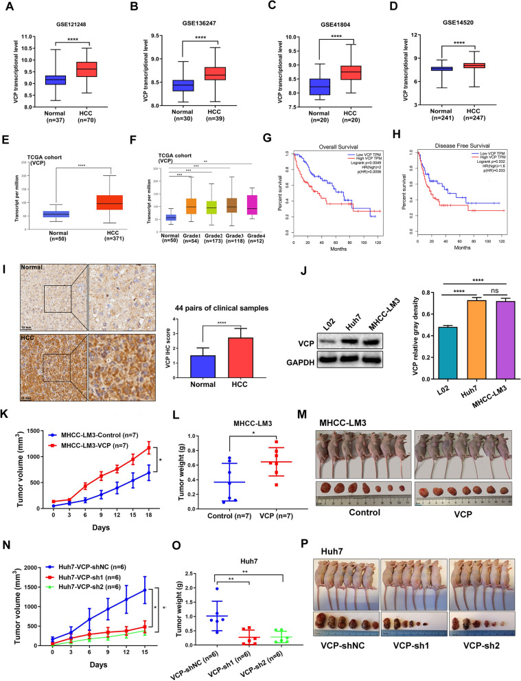
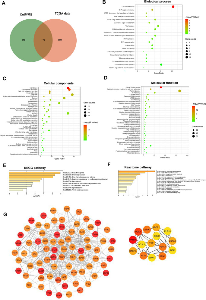
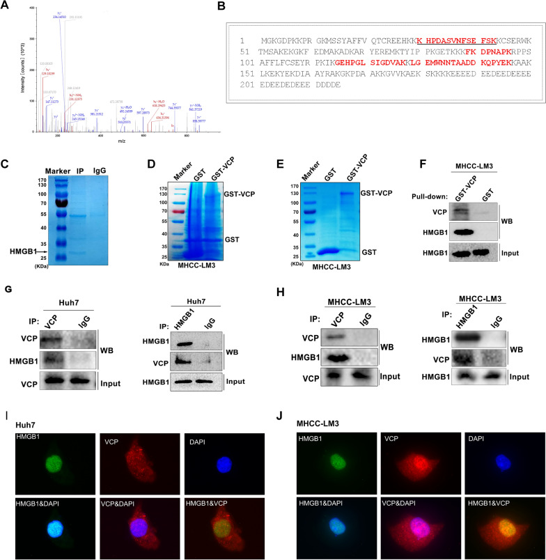
Methods: We examined the expression of VCP in HCC using the RNA sequencing and microarray data from public databases and measured it in clinical samples and cell lines by western blot, and immunohistochemistry (IHC). We also evaluated the correlation between VCP and clinical features. The VCP-interacting proteins were identified by co-immunoprecipitation combined with mass spectrometry (CoIP/MS). The underlying molecular mechanisms were investigated using in vitro and in vivo models of HCC.

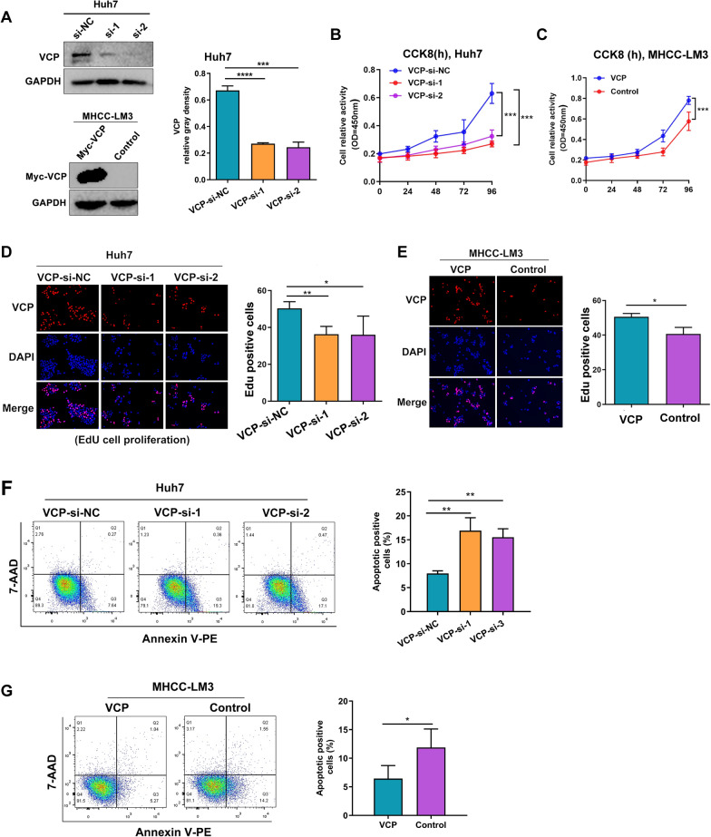
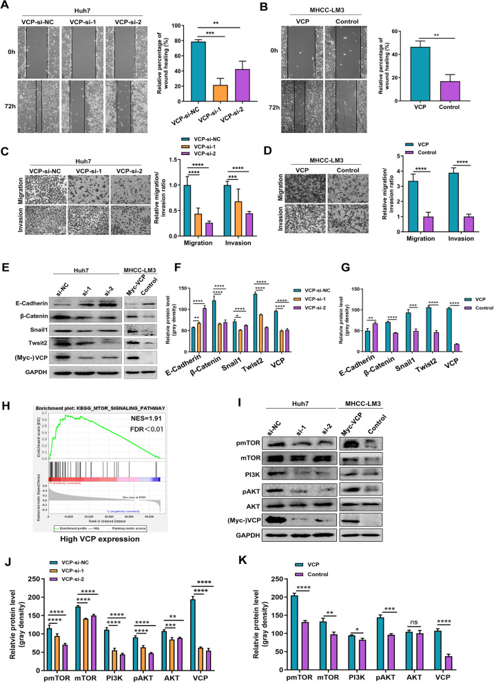
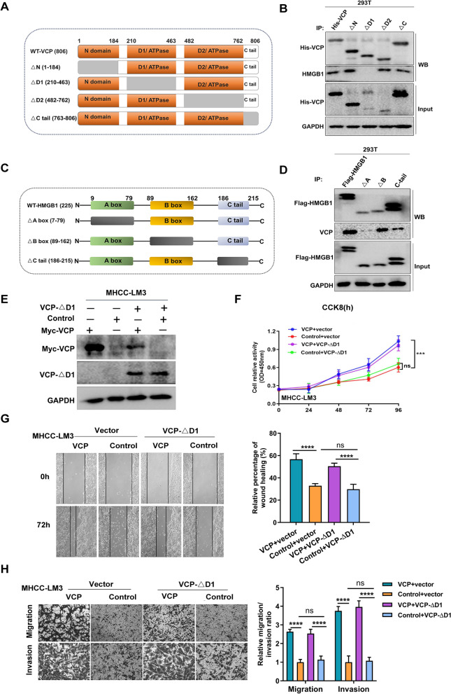
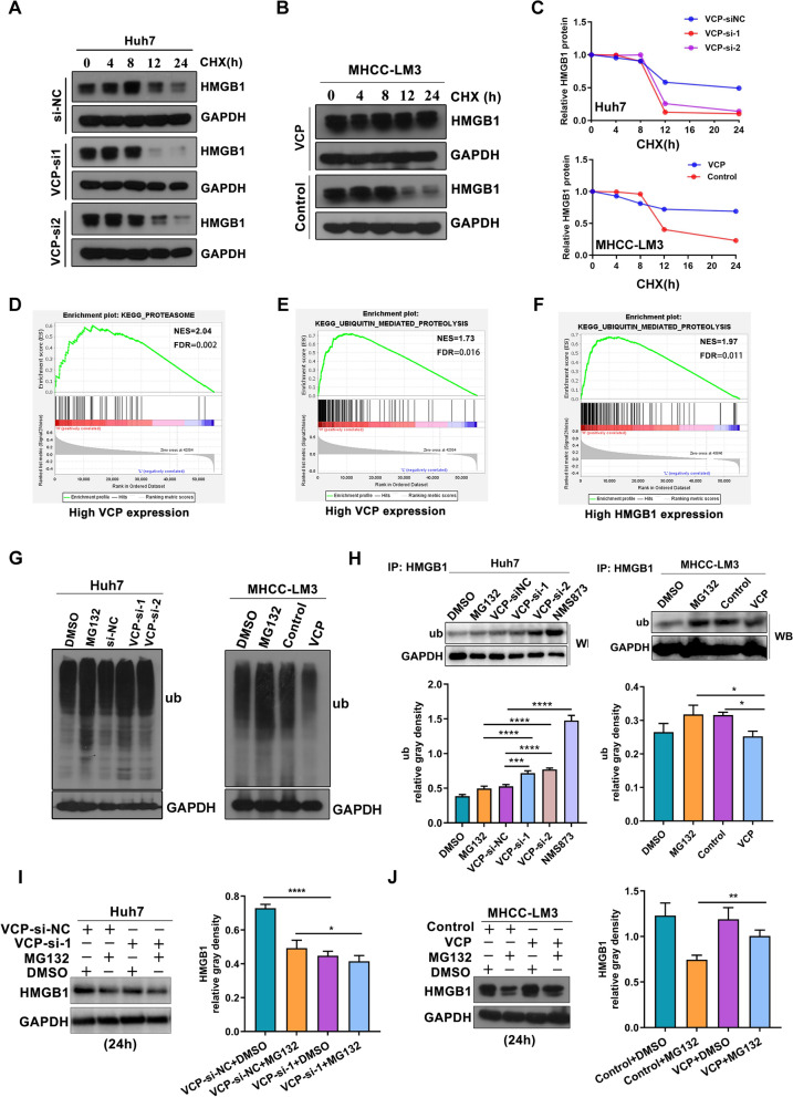
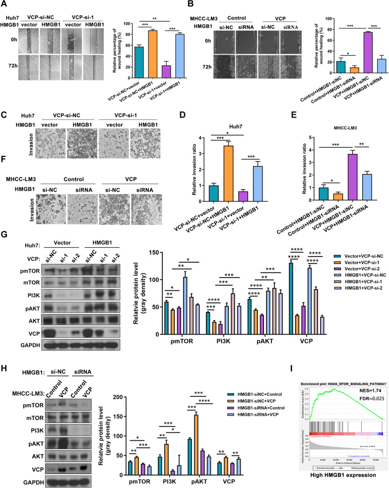
Results: We found that VCP expression is significantly increased in tumor tissues and is associated with advanced TNM stages and poorer prognosis in HCC patients. In vitro analyses revealed that VCP overexpression promoted HCC cell proliferation, migration, and invasion via PI3K/AKT/mTOR pathway activation. Conversely, VCP knockdown resulted in the reverse phenotypes. In vivo studies indicated that up-regulated VCP expression accelerated tumor growth in a subcutaneous HCC model. The D1 domain of VCP and A box of HMGB1 were identified as the critical regions for their interaction, and D1 area was required for the tumor-promoting effects induced by VCP expression. VCP enhanced the protein stability of HMGB1 by decreasing its degradation via ubiquitin-proteasome process. Inhibition of HMGB1 markedly attenuated VCP-mediated HCC progression and downstream activation of PI3K/AKT/mTOR signals.

Conclusion: Collectively, these findings demonstrate that VCP is a potential prognostic biomarker in HCC and exhibits oncogenic roles via PI3K/AKT/mTOR pathway activation. HMGB1 played an essential role in VCP-mediated HCC progression, indicating that VCP and HMGB1 are potential therapeutic targets in human HCC.

Keywords: HMGB1; Hepatocellular carcinoma; PI3K/AKT/mTOR pathway; Tumor progression; VCP.


Conflict of interest statement

Dr. Duda received consultant fees from Bayer, BMS, Simcere, Sophia Biosciences, and Surface Oncology and has received research grants from Bayer, Merrimack, Exelixis, and BMS. No reagents or support from these companies were used for this study. All the other authors declare that they have no competing interests.

Figures

Fig.1
Fig.1
Elevated VCP expression is associated with poor prognosis and promotes HCC growth in vivo. AD The mRNA expression of VCP was elevated in HCC samples compared with nontumorous in GEO datasets including GSE121248, GSE136247, GSE41804, and GSE14520. E, F The HCC cohort from TCGA database presented the increase of VCP in tumor samples and various disease grades. G, H The clinical significance of VCP expression in overall and disease-free survivals in HCC patients was evaluated in the TCGA cohort. I The expression of VCP in HCC and normal tissues from 44 pairs of clinical samples was detected by IHC. The representative images and the statistical result of IHC scores were displayed. J The protein expression of VCP in human HCC cell lines (Huh7 and MHCC-LM3) and immortalized hepatic cell line (L02) was determined by western blot. The statistical analysis was further analyzed. K, N The nude mice were injected with MHCC-LM3 cells with stable VCP overexpression and Huh7 cells with stable VCP depletion, respectively, in the right flank followed by monitoring growth for three weeks. The tumor volumes were measured every 3 days. L, O The differences in tumor weight in various groups when mice were sacrificed. M, P The representative morphology of tumors in nude mice. TCGA: The Cancer Genome Atlas. GEO: Gene Expression Omnibus. IHC: immunohistochemistry. All *P < 0.05, **P < 0.01, ***P < 0.001, and ****P < 0.0001
Fig. 2
Fig. 2
The effect of VCP on HCC cell proliferation and apoptosis in vitro. A The efficacy verification of VCP-siRNA and exogenous VCP transfection in HCC cells. B, C CCK8 assay was performed to measure the cell viability. D, E The EdU experiment was also used to detect cell ability of proliferation induced by different expressed levels of VCP. F, G The apoptotic level of HCC cells was determined by flow cytometry. All *P < 0.05, **P < 0.01, and ***P < 0.001
Fig. 3
Fig. 3
VCP expression promotes migration and invasion in HCC in vitro. A, B the expression of VCP was silenced in Huh7 cells treated with siRNAs and exogenously overexpressed in MHCC-LM3 cells. Wound-healing assay demonstrated cell movement capacity. C, D The transwell system was utilized to detect the migration and invasion abilities of HCC cells. The representative images and the corresponding statistical results were shown. EG The protein expression of EMT-related markers was determined by western blot and the relative protein density was analyzed. H The GSEA analysis in the HCC cohort from TCGA database based on the VCP transcriptional expression showed the up-regulated PI3K/AKT/mTOR pathway. The median value of VCP transcriptional level was regarded as the cut-off to divide HCC patients into high- or low-VCP expression groups. IK Proteins extracted from Huh7 and MHCC-LM3 cells in various groups were subjected to western blot to detect the markers within the mTOR pathway. Meanwhile, the relative protein gray density was summarized. EMT epithelial-mesenchymal transition, GSEA gene set enrichment analysis, FDR false discovery rate, NES normalized enrichment score, EMT epithelial-mesenchymal transformation, ns no significance. All *P < 0.05, **P < 0.01, ***P < 0.001, ****P < 0.0001
Fig. 4
Fig. 4
The GO and pathway enrichment, and protein to protein (PPI) network analysis of VCP-interacting proteins. A The Venn diagram showed 79 VCP-interacting proteins obtained by overlapping the candidates of CoIP/MS and the profile of VCP co-expressed genes in the HCC cohort from TCGA database. BD The GO enriched terms of top 20 biological processes, cell components, and molecular function, respectively. E, F The enriched pathways through KEGG and Reactome database, separately. G The PPI network of 79 VCP-interacting proteins was analyzed in STRING database and further visualized by Cytoscape software (version 3.8.2). A total of 15 Hub genes were determined by CytoHubba plug-in. The colors of nodes from red to yellow indicated the overall combined scores of each molecule interacting with others from high to low. GO gene ontology, KEGG Kyoto Encyclopedia of Genes and Genomes
Fig. 5
Fig. 5
VCP interacts with HMGB1 in HCC cells. A The representative MS/MS spectrum of HMGB1 and the amino acid sequence was identified as KHPDASVNFSEFSK from mass differences in the y and b fragment ions series. B The protein sequence of HMGB1, the matched peptides are labeled in red bold letters. C MHCC-LM3 cells were treated with exogenous VCP and immunoprecipitation was performed. The HMGB1 protein shown by the arrow was observed by Coomassie staining after running SDS-PAGE. DF GST pull-down assay indicated that VCP could be fused (D) and purified (E) in vitro, and the purified GST-tagged VCP pulls down HMGB1 in MHCC-LM3 cells (F). G, H The CoIP was operated to validate the VCP-HMGB1 interaction in Huh7 and MHCC-LM3 cells. I, J The colocalization of VCP and HMGB1 in HCC cells was observed by confocal microscopy. SDS-PAGE sodium dodecyl sulfate–polyacrylamide gel electrophoresis, MS/MS tandem mass spectrometry, CoIP co-immunoprecipitation
Fig. 6
Fig. 6
The D1 domain is the key region for VCP exhibiting biological function in HCC. A The scheme of VCP mutants. The WT VCP including 806 amino acids was divided into four domains (△N, △D1, △D2, and △C tail). The gray area indicated the deleted region. B 293T cells were transfected with His-tagged full-length VCP and its mutants with deletion of various regions. Cell lysates were immunoprecipitated with anti-His antibody. And western blot was used to detect the expression of His probe and HMGB1. C The scheme of HMGB1. The full length of HMGB1 included 215 amino acids and was divided into three regions including A box, B box, and C-tail. The box with gray color indicates the deleted domain of HMGB1. D 293 T cells were transfected with WT HMGB1 and its various mutants with the expression of the Flag probe. Cell lysates were collected for immunoprecipitation with anti-Flag antibody. Then the expression of Flag probe and VCP were determined by western blot. E The protein expression of WT VCP and D1 mutant in MHCC-LM3 cells. F The ability of cell proliferation was measured by CCK8 assay in MHCC-LM3 cells with transfection of WT VCP and D1 mutant, separately. G The movement capacity of MHCC-LM3 cells with transfection of WT VCP or D1 mutant was evaluated by wound-healing assay. H Transwell system indicates the cell's ability of invasion and migration in various groups. The representative images and the corresponding statistical results were shown. WT: wild type. All ***P < 0.001, ****P < 0.0001, and ns no significance
Fig. 7
Fig. 7
VCP increases the protein stability of HMGB1. AC Western blot was performed to evaluate the half-life of HMGB1 protein in HCC cells. All groups were treated with cycloheximide (CHX, 50 μg/mL), a classic protein synthesis inhibitor, from 0 to 24 h. DF The GSEA analysis indicated the enriched pathways in HCC patients of high VCP and HMGB1 expressing groups from TCGA database, respectively. The median value of VCP and HMGB1 transcriptional levels in the HCC cohort was regarded as the cut-off. G The total cellular levels of ubiquitylated proteins in HCC cells was determined by western blot. Both Huh7 and MHCC-LM3 cells were treated with MG132 (20 μM, 6 h), which is a general proteasome inhibitor. H The ubiquitylated HMGB1 protein was detected by western blot after HMGB1 immunoprecipitation in HCC cells. Cells were treated with MG132 (20 μM, 6 h) or the VCP inhibitor NMS873 (10 μM, 6 h). I, J The Huh7 cells with VCP depletion and MHCC-LM3 cells with ectopic VCP overexpression were incubated with MG132 (20 μM) for 24 h. The whole-cell lysates were collected and separated by SDS-PAGE. Proteins were detected by indicated antibodies. All *P < 0.05, **P < 0.01, and ****P < 0.0001
Fig. 8
Fig. 8
HMGB1 is critical for VCP to activate P13K/AKT/mTOR pathway in HCC cells. A, B The wound-healing assay was operated to test the moveable ability of Huh7 and MHCC-LM3 cells with indicated treatment. CF The transwell system was used to evaluate the cell’s invasive capacity. G, H The influence of HMGB1 expression on VCP triggering the PI3K/AKT/mTOR pathway was assessed by western blot. The representative results and quantitative analyzed data were shown. I The GSEA analysis showed that the mTOR pathway was enriched in HCC patients with high expression of HMGB1 from TCGA database. The median value of HMGB1 transcriptional expression in the HCC cohort was adopted to classify patients into high and low HMGB1 expressed groups. All *P < 0.05, **P < 0.01, ***P < 0.001, and ****P < 0.0001

References

    1. Forner A, Reig M, Bruix J. Hepatocellular carcinoma. The Lancet. 2018;391(10127):1301–1314. doi: 10.1016/S0140-6736(18)30010-2. - DOI - PubMed
    1. Yang YF, Zhang MF, Tian QH, Fu J, Yang X, Zhang CZ, et al. SPAG5 interacts with CEP55 and exerts oncogenic activities via PI3K/AKT pathway in hepatocellular carcinoma. Mol Cancer. 2018;17(1):117. doi: 10.1186/s12943-018-0872-3. - DOI - PMC - PubMed
    1. Bray F, Ferlay J, Soerjomataram I, Siegel RL, Torre LA, Jemal A. Global cancer statistics 2018: GLOBOCAN estimates of incidence and mortality worldwide for 36 cancers in 185 countries. CA Cancer J Clin. 2018;68(6):394–424. doi: 10.3322/caac.21492. - DOI - PubMed
    1. Llovet JM, Zucman-Rossi J, Pikarsky E, Sangro B, Schwartz M, Sherman M, et al. Hepatocellular carcinoma. Nat Rev Dis Primers. 2016;2:16018. doi: 10.1038/nrdp.2016.18. - DOI - PubMed
    1. Fu S, Wang J, Hu X, Zhou RR, Fu Y, Tang D, et al. Crosstalk between hepatitis B virus X and high-mobility group box 1 facilitates autophagy in hepatocytes. Mol Oncol. 2018;12(3):322–338. doi: 10.1002/1878-0261.12165. - DOI - PMC - PubMed

MeSH terms