. 2022 Apr 20;23(9):4528.
doi: 10.3390/ijms23094528.

Alleviation of Endoplasmic Reticulum Stress Enhances Human Corneal Epithelial Cell Viability under Hyperosmotic Conditions

Affiliations

Alleviation of Endoplasmic Reticulum Stress Enhances Human Corneal Epithelial Cell Viability under Hyperosmotic Conditions

Damien Guindolet et al. Int J Mol Sci. .

Abstract

Tear hyperosmolarity plays an essential role in the initiation and progression of dry-eye disease. Under a hyperosmotic environment, corneal epithelial cells experience perturbations in endoplasmic reticulum function that can lead to proinflammatory signaling and apoptosis. In this study, we investigated the effect of tauroursodeoxycholic acid (TUDCA), a chemical chaperone known to protect against endoplasmic reticulum stress, on corneal epithelial cells exposed to hyperosmotic conditions. We found that the expression of the genes involved in the activation of the unfolded protein response and the pro-apoptotic transcription factor DDIT3 were markedly upregulated in patients with Sjögren's dry-eye disease and in a human model of corneal epithelial differentiation following treatment with hyperosmotic saline. Experiments in vitro demonstrated that TUDCA prevented hyperosmotically induced cell death by reducing nuclear DNA fragmentation and caspase-3 activation. TUDCA supplementation also led to the transcriptional repression of CXCL8 and IL5, two inflammatory mediators associated with dry-eye pathogenesis. These studies highlight the role of hyperosmotic conditions in promoting endoplasmic reticulum stress in the cornea and identify TUDCA as a potential therapeutic agent for the treatment of dry-eye disease.

Keywords: corneal epithelium; dry eye; endoplasmic reticulum stress; hyperosmotic stress; tauroursodeoxycholic acid; unfolded protein response.


Conflict of interest statement

The authors declare no conflict of interest. The funders had no role in the design of the study; in the collection, analyses, or interpretation of data; in the writing of the manuscript, or in the decision to publish the results.

Figures

Figure 1
Figure 1
Expression of indicators of cells undergoing ER stress in conjunctival epithelium of patients with Sjögren’s syndrome and control subjects. Conjunctival impression cytology samples were collected from the superior and temporal bulbar conjunctiva. Cells in the filters were immunostained with antibodies against HSPA5 (a) or DDIT3 (b), and counterstained with DAPI. Scale bars: 20 µm.
Figure 2
Figure 2
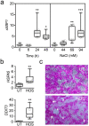
Hyperosmolarity induces the UPR in stratified human corneal epithelial cells. (a) Cell cultures exposed to sodium chloride were evaluated for time- and concentration-dependent changes in sXBP1 expression. qPCR experiments demonstrated significant upregulation of sXBP1 following addition of sodium chloride (94 mM) for 24–48 h and using 69–94 mM sodium chloride (for 24 h) (n = 7 independent experiments). (b) HSPA5 and DDIT3 mRNA were significantly upregulated by qPCR following exposure to HOS (94 mM NaCl, 24 h) compared to untreated (UT) cells (n = 6 independent experiments). (c) HOS led to the formation of areas with substantial cell loss (arrowheads) and the penetration of rose bengal into damaged cells with altered morphology (arrows). The box-and-whisker plots show the 25th and 75th percentiles (boxes), the median, and the minimum and maximum data values (whiskers). Significance was determined using the Kruskal–Wallis test followed by Dunn’s multiple comparison (a) or Mann–Whitney test (b). *, p < 0.05; **, p < 0.01; ***, p < 0.001.
Figure 3
Figure 3
TUDCA reduces hyperosmotically induced cell death. (a) Crystal violet staining of stratified human corneal epithelial cells: untreated (UT), treated with 1 mM TUDCA alone (TUDCA), or exposed to HOS (94 mM NaCl, 24 h) with or without addition of TUDCA. Scale bar: 10 µm. (b) Quantitative assessment of the cellular area covered by crystal violet in (a) normalized-to-UT-control (n = 3 independent experiments in triplicate). (c) Quantitative assessment of cell viability using the MTS assay (n = 3 independent experiments in triplicate). The box-and-whisker plots show the 25th and 75th percentiles (boxes), the median, and the minimum and maximum data values (whiskers). Significance was determined using the Kruskal–Wallis test followed by Dunn’s multiple comparison. ****, p < 0.0001.
Figure 4
Figure 4
TUDCA reduces hyperosmotically induced apoptotic events. (a) DAPI staining showing increased chromatin condensation and fragmentation (arrowhead in the inset) in stratified human corneal epithelial cells exposed to HOS (94 mM NaCl, 24 h) compared to control cells (UT, TUDCA alone) or cells exposed to HOS in the presence of TUDCA. UT, untreated cells. Scale bar: 50 µm. (b) Upper panel: Western blotting analysis showing cleaved caspase-3 (cCasp3) and ß-actin. Molecular weights of standard proteins are shown to the left. Lower panel: Quantification of the relative levels of cCasp3 protein in cell culture (n = 3 independent experiments in duplicate or triplicate). The box-and-whisker plots show the 25th and 75th percentiles (boxes), the median, and the minimum and maximum data values (whiskers). Significance was determined using the Kruskal–Wallis test followed by Dunn’s multiple comparison. *, p < 0.05.
Figure 5
Figure 5
TUDCA downregulates CXCL8 and IL5 under hyperosmotic conditions. Stratified human corneal epithelial cells were exposed to HOS (94 mM NaCl, 24 h) with or without addition of 1 mM TUDCA. (a) Relative transcript abundance of genes encoding proteins involved in the inflammatory response in cells exposed to HOS. The expression of genes was normalized using the comparative ∆∆CT method. (b) Scatterplot comparing the expression of genes in cells exposed to HOS with or without TUDCA (n = 3 independent experiments). The red dots indicate statistically significant downregulation of the immune regulatory gene. The corresponding quantitative graphs are shown to the right. The data are presented as floating bars (min. to max.) with line at the median. Significance was determined using the unpaired t test with a Holm–Sidak’s multiple comparison. *, p < 0.05.

References

    1. Lemp M.A. Advances in understanding and managing dry eye disease. Am. J. Ophthalmol. 2008;146:350–356. doi: 10.1016/j.ajo.2008.05.016. - DOI - PubMed
    1. Schaumberg D.A., Dana R., Buring J.E., Sullivan D.A. Prevalence of dry eye disease among US men: Estimates from the Physicians’ Health Studies. Arch. Ophthalmol. 2009;127:763–768. doi: 10.1001/archophthalmol.2009.103. - DOI - PMC - PubMed
    1. Schaumberg D.A., Sullivan D.A., Buring J.E., Dana M.R. Prevalence of dry eye syndrome among US women. Am. J. Ophthalmol. 2003;136:318–326. doi: 10.1016/S0002-9394(03)00218-6. - DOI - PubMed
    1. Craig J.P., Nelson J.D., Azar D.T., Belmonte C., Bron A.J., Chauhan S.K., de Paiva C.S., Gomes J.A.P., Hammitt K.M., Jones L., et al. TFOS DEWS II Report Executive Summary. Ocul. Surf. 2017;15:802–812. doi: 10.1016/j.jtos.2017.08.003. - DOI - PubMed
    1. Coursey T.G., Tukler Henriksson J., Barbosa F.L., de Paiva C.S., Pflugfelder S.C. Interferon-γ-Induced Unfolded Protein Response in Conjunctival Goblet Cells as a Cause of Mucin Deficiency in Sjögren Syndrome. Am. J. Pathol. 2016;186:1547–1558. doi: 10.1016/j.ajpath.2016.02.004. - DOI - PMC - PubMed

Substances