. 2022 Apr 20;23(9):4544.
doi: 10.3390/ijms23094544.

Probing and Manipulating the Lateral Pressure Profile in Lipid Bilayers Using Membrane-Active Peptides-A Solid-State 19F NMR Study

Affiliations

Probing and Manipulating the Lateral Pressure Profile in Lipid Bilayers Using Membrane-Active Peptides-A Solid-State 19F NMR Study

Stephan L Grage et al. Int J Mol Sci. .

Abstract

The lateral pressure profile constitutes an important physical property of lipid bilayers, influencing the binding, insertion, and function of membrane-active peptides, such as antimicrobial peptides. In this study, we demonstrate that the lateral pressure profile can be manipulated using the peptides residing in different regions of the bilayer. A 19F-labeled analogue of the amphiphilic peptide PGLa was used to probe the lateral pressure at different depths in the membrane. To evaluate the lateral pressure profile, we measured the orientation of this helical peptide with respect to the membrane using solid-state 19F-NMR, which is indicative of its degree of insertion into the bilayer. Using this experimental approach, we observed that the depth of insertion of the probe peptide changed in the presence of additional peptides and, furthermore, correlated with their location in the membrane. In this way, we obtained a tool to manipulate, as well as to probe, the lateral pressure profile in membranes.

Keywords: lateral pressure profile; lipid bilayer; membrane protein; membrane-active amphiphilic peptide; peptide crowding; solid-state 19F nuclear magnetic resonance.


Conflict of interest statement

The authors declare no conflict of interest. The funders had no role in the design of the study; in the collection, analyses, or interpretation of data; in the writing of the manuscript, or in the decision to publish the results.

Figures

Figure A1
Figure A1
Antimicrobial activity of pairs of the antimicrobial peptide PGLa, labeled with 4-CF3-phenylglycine in position 13, with one of the crowder peptides Gramicidin S (GS), Gramicidin A (GA), MAP, or Magainin-2 (MAG2) were examined with respect to their antimicrobial activity against Gram-negative E. coli (a) and Gram-positive M. luteus (b), using an agar diffusion assay. Inhibition of growth after incubation of the agar plates at 37 °C overnight is visible as a clear zone around the wells, in which 50 µL of 200 µg/mL peptide solution of the indicated peptide was placed. The synergistically-enhanced activity of PGLa and MAG2 is visible as bulges in the boundary of the inhibition zone (marked by an arrow).
Figure A2
Figure A2
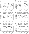
The agreement of the solid-state 19F-NMR spectra (see Figure 2) with the characteristic spectrum of one of the respective alignment states of PGLa was quantified using an averaged correlation product, as defined in the text. The averaged correlation products denoting the agreement with an S-state are shown as a dotted line, with a T-state as a dashed line, and with an I-state as a solid line. Samples at a PGLa:lipid ratio of 0.5:100 (left) and 2.5:100 (right) were studied at temperatures from 15 °C to 55 °C, varied in steps of 5 °C. The second crowder peptide was added, resulting into a total peptide:lipid ratio of 5:100. Several crowders were tested: (a) reference spectra without crowder, (b) with gramicidin S (GS), (c) with gramicidin A (GA), (d) with MSI-103, (e) with MAP, (f) with magainin-2 (MAG2).
Figure A3
Figure A3
The relative dimensions of the peptides used in this study: PGLa, gramicidin S (GS), gramicidin A (GA, shown as a head-to-head dimer), MSI-103, MAP, and magainin 2 (MAG2). The peptides are shown with their van der Waals interaction surface in top view (top row) and side views (bottom rows). Polar residues are colored in blue, with hydrophobic residues in yellow. The membrane headgroup region and hydrophobic core are indicated by blue and yellow-orange background, respectively.
Figure 1
Figure 1
(a) Crowding in different regions within the lipid bilayer was created using a series of peptides: the cyclic β-hairpin gramicidin S (GS) was previously found aligned parallel to the surface in DMPC bilayers for most temperatures [31,32], the β-helix gramicidin A (GA) is known to span the bilayer as a dimer [33], the amphiphilic helices of the model peptides MSI-103 and MAP were tilted into the membrane as a T-state at the peptide:lipid molar ratio of this study [36], and the antimicrobial peptide magainin 2 (MAG2), also forming an amphiphilic helix, was found in a surface-parallel or marginally tilted orientation [27,28]. (b) In the absence of additional peptides, PGLa assumes an alignment parallel to the membrane surface (S-state) at low peptide:lipid molar ratios (top left) and tilts obliquely into the membrane (T-state) at high peptide:lipid molar ratios (top right). When exposed to these different crowders, PGLa was not influenced by the rather small peptide GS, but responded to crowding by the other peptides in a manner depending on their location in the membrane. PGLa stayed in an S-state in the presence of transmembrane GA, assumed a T-state when mixed with MSI-103 or MAP, which effectively acted like additional PGLa peptide, and inserted in the presence of surface-bound MAG2.
Figure 2
Figure 2
(a) The influence of selected crowder peptides (denoted above the spectra), which mimic membrane proteins with different structures and locations, on the orientation of the antimicrobial peptide PGLa, labeled with CF3-Phg in position 13, was examined on the basis of solid-state 19F-NMR spectra. The spectra are indicative of the insertion of PGLa into the membrane in the presence of the crowder. Spectra fingerprints of S-, T-, and I-state are colored in red, blue, and green, respectively. In case of the coexistence of several spectral fingerprints, the pattern is highlighted that caused the highest level of agreement with the respective pattern of the pure PGLa sample in the data analysis. Spectra were acquired of samples at a PGLa:lipid molar ratio of 0.5:100 (left column within each set) and 2.5:100 (right column in each set), and at temperatures from 15 °C (lowest spectrum) to 55 °C, varied in steps of 5 °C. The gel-to-fluid phase transition temperature of DMPC is indicated by “Tm”. A second peptide was added, to result in a total peptide:lipid molar ratio of 5:100. Several crowders were tested: (a) reference spectra without crowder, (b) with gramicidin S (GS), (c) with gramicidin A (GA), (d) with MSI-103, (e) with MAP, (f) with magainin 2 (MAG2).
Figure 3
Figure 3
The insertion states of PGLa in DMPC bilayers alone (top) and in the presence of the crowder peptides GS, GA, MSI-103, MAP, or MAG2 were determined from the solid-state 19F-NMR spectra of Figure 2, for temperatures from 15 °C to 55 °C, acquired in steps of 5 °C. The spectra were compared with spectra typical for each state (see SI for details), and the state with the best agreement is denoted with S (peptide aligned on the surface, S-state), T (peptide obliquely tilted into the membrane, T-state), or I (peptide inserted in a transmembrane alignment, I-state) for each temperature. Each peptide combination was prepared in two peptide:lipid molar ratios of PGLa:DMPC 0.5:100 and PGLa:DMPC 2.5:100. The crowder peptide was added, to result into a total peptide:lipid molar ratio of 5:100. Tm denotes the melting temperature characteristic for DMPC.

Similar articles

Cited by

References

    1. Cantor R.S. Lateral pressures in cell membranes: A mechanism for modulation of protein function. J. Phys. Chem. B. 1997;101:1723–1725. doi: 10.1021/jp963911x. - DOI
    1. Marsh D. Lateral pressure profile, spontaneous curvature frustration, and the incorporation and conformation of proteins in membranes. Biophys. J. 2007;93:3884–3899. doi: 10.1529/biophysj.107.107938. - DOI - PMC - PubMed
    1. Perozo E., Cortes D.M., Sompornpisut P., Kloda A., Martinac B. Open channel structure of MscL and the gating mechanism of mechanosensitive channels. Nature. 2002;418:942–948. doi: 10.1038/nature00992. - DOI - PubMed
    1. Perozo E., Kloda A., Cortes D.M., Martinac B. Physical principles inderlying the transduction of bilayer deformation forces during mechanosensitive channel gating. Nat. Struct. Biol. 2002;9:696–703. doi: 10.1038/nsb827. - DOI - PubMed
    1. Grage S.L., Keleshian A.M., Turdzeladze T., Battle A.R., Tay W.C., May R.P., Holt S.A., Contera S.A., Moulin M., Pal P., et al. Bilayer-mediated clustering and functional interaction of MscL channels. Biophys. J. 2011;100:1252–1260. doi: 10.1016/j.bpj.2011.01.023. - DOI - PMC - PubMed

MeSH terms

LinkOut - more resources