. 2022 Apr 20;23(9):4556.
doi: 10.3390/ijms23094556.

Prognostic Significance of GPR55 mRNA Expression in Colon Cancer

Affiliations

Prognostic Significance of GPR55 mRNA Expression in Colon Cancer

Hager Tarek H Ismail et al. Int J Mol Sci. .

Abstract

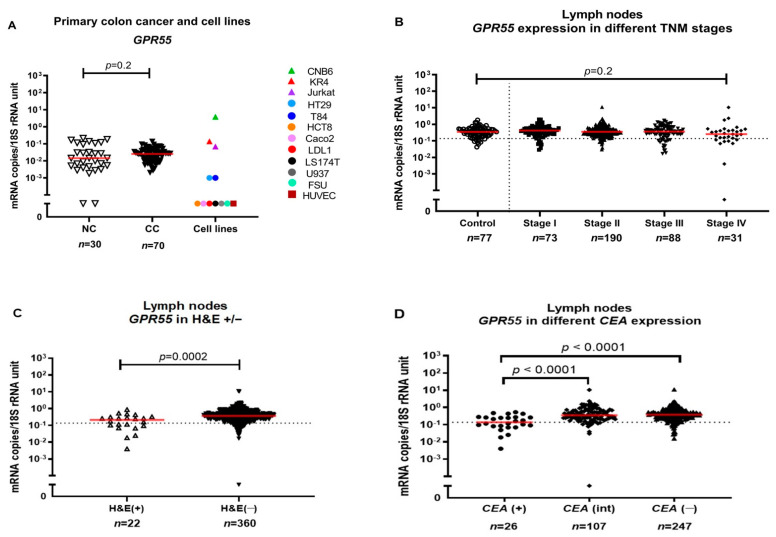
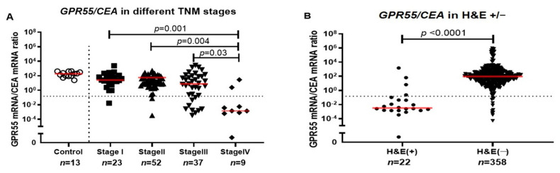
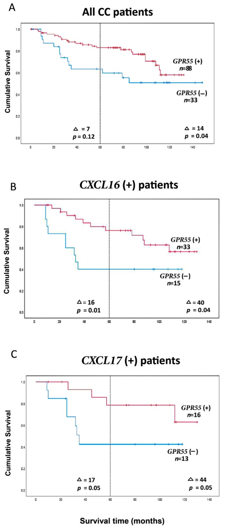
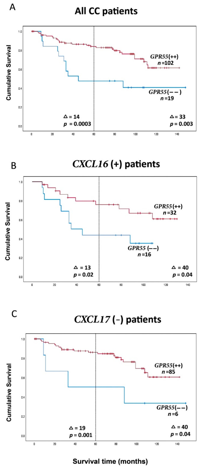
G protein-coupled receptor 55 (GPR55) probably plays a role in innate immunity and tumor immunosurveillance through its effect on immune cells, such as T cells and NK cells. In this study, the prognostic value of GPR55 in colon cancer (CC) was investigated. mRNA expression levels of GPR55 were determined in 382 regional lymph nodes of 121 CC patients with 12 years observation time after curative surgery. The same clinical material had previously been analyzed for expression levels of CEA, CXCL16, CXCL17, GPR35 V2/3 and LGR5 mRNAs. Clinical cutoffs of 0.1365 copies/18S rRNA unit for GPR55 and 0.1481 for the GPR55/CEA ratio were applied to differentiate between the high- and low-GPR55 expression groups. Kaplan-Meier survival analysis and Cox regression risk analysis were used to determine prognostic value. Improved discrimination between the two groups was achieved by combining GPR55 with CEA, CXCL16 or CXCL17 compared with GPR55 alone. The best result was obtained using the GPR55/CEA ratio, with an increased mean survival time of 14 and 33 months at 5 and 12 years observation time, respectively (p = 0.0003 and p = 0.003) for the high-GPR55/CEA group. The explanation for the observed improvement is most likely that GPR55 is a marker for T cells and B cells in lymph nodes, whereas CEA, CXCL16 and CXCL17, are markers for tumor cells of epithelial origin.

Keywords: CEA; CXCL16; CXCL17; GPR55; colon cancer; prognosis; qRT-PCR; regional lymph nodes.


Conflict of interest statement

The authors declare no conflict of interest.

Figures

Figure 1
Figure 1
(A) GPR55 mRNA expression levels in resected normal colon tissues (NC), primary colon cancer tissues (CC) and in a panel of colon cancer cell lines: DLD1, LS174T, HT29, T84, HCT8 and Caco2, primary foreskin fibroblast cells (FSU), an endothelial cell line (HUVEC), a T cell line (Jurkat), two B cell lines (CNB6 and KR4) and a monocyte cell line (U937). (B) GPR55 mRNA expression levels in lymph nodes from non-cancerous disease patients (Control) and from colon cancer patients of different TNM stages (Stage I–IV). (C) GPR55 mRNA expression levels in metastatic (H&E(+)) lymph nodes and non-metastatic (H&E(−)) lymph nodes. (D) GPR55 mRNA expression levels in lymph nodes from CC patients divided into three groups according to their CEA mRNA levels: CEA(+) = CEA mRNA levels >3.67 copies/18S rRNA unit; CEA(int) = intermediate CEA mRNA levels, that is, 0.013–3.67 copies/18S rRNA unit; and CEA(−) = CEA mRNA levels <0.013 copies/18S rRNA unit. Red horizontal lines indicate median values. Dashed horizontal lines indicate clinical cutoff values of 0.1365 mRNA copies/18S rRNA unit for GPR55. n = number of lymph nodes. p-values were calculated by Kruskal–Wallis non-parametric ANOVA, followed by post hoc Dunn’s test for multiple comparisons in (B,D) and by two-tailed Mann–Whitney test for comparison between expression levels in (A,C).
Figure 2
Figure 2
(A) GPR55/CEA mRNA ratio in lymph nodes from non-cancerous disease patients (control) and colon cancer patients in different TNM stages (Stage I–IV). (B) GPR55/CEA mRNA ratio in metastatic (H&E(+)) and non-metastatic (H&E(−)) lymph nodes. Red horizontal lines indicate median values. Dashed horizontal line shows clinical cutoff equal to 0.1481 GPR55 mRNA/CEA mRNA ratio and (n) number of lymph nodes. p-Values were calculated by Kruskal–Wallis non-parametric ANOVA, followed by post hoc Dunn’s test for multiple comparisons in (A) and by two-tailed Mann–Whitney test for comparison between expression levels in (B).
Figure 3
Figure 3
(A) Kaplan–Meier cumulative survival curves for all 121 CC patients. Each patient is represented by the lymph node with the lowest GPR55 mRNA value. The cutoff level between the two groups was 0.1365 GPR55 mRNA copies/18S rRNA unit. (B) Kaplan–Meier cumulative survival curves for GPR55(−) and GPR55(+) patients. Analysis is restricted to patients with CXCL16 mRNA levels in their highest lymph node > 7.2 mRNA copies/18S rRNA unit. The cutoff level between the two groups was 0.1365 GPR55 mRNA copies/18S rRNA unit. The number of patients was 48. (C) Kaplan–Meier cumulative survival curves for GPR55(−) and GPR55(+) patients. Analysis is restricted to patients with CXCL17 mRNA levels in their highest lymph node > 0.0014 mRNA copies/18S rRNA unit. The cutoff level between the two groups was 0.1365 GPR55 mRNA copies/18S rRNA unit. The number of patients was 29. Patients were followed for 12 years. Differences in disease-free survival time after surgery between the two groups are given as a ∆-value in months and statistical significance as p-values; n = number of patients in the respective group.
Figure 4
Figure 4
(A) Kaplan–Meier cumulative survival curves for GPR55(−−) and GPR55(++) patients. All 121 CC patients are included. Each patient is represented by the lymph node with the lowest GPR55 mRNA value. A GPR55 mRNA/CEA mRNA ratio of 0.1481 was used to divide the patients into two groups. (B) Kaplan–Meier cumulative survival curves for GPR55(−−) and GPR55(++) patients. The analysis is restricted to patients with CXCL16 mRNA levels in the highest lymph node > 7.2 mRNA copies/18S rRNA unit. The number of patients was 48. A GPR55 mRNA/CEA mRNA ratio of 0.1481 was used to divide the patients into two groups. (C) Kaplan–Meier cumulative survival curves for GPR55(−−) and GPR55(++) patients. The analysis is restricted to the CXCL17 mRNA levels in the highest lymph node < 0.0014 mRNA copies/18S rRNA unit. The number of patients was 91. A GPR55 mRNA/CEA mRNA ratio of 0.1481 was used to divide the patients into two groups. The patients were followed for 12 years. Differences in disease-free survival time after surgery between the two groups are given as a ∆-value in months and statistical significance as p-values; n = number of patients in the respective group.

References

    1. Sriram K., Insel P.A. G Protein-coupled receptors as targets for approved drugs: How many targets and how many drugs? Mol. Pharmacol. 2018;93:251–258. doi: 10.1124/mol.117.111062. - DOI - PMC - PubMed
    1. Fredriksson R., Lagerström M.C., Lundin L.G., Schiöth H.B. The G-protein-coupled receptors in the human genome form five main families. Phylogenetic analysis, paralogon groups, and fingerprints. Mol. Pharmacol. 2003;63:1256–1272. doi: 10.1124/mol.63.6.1256. - DOI - PubMed
    1. Uhlén M., Fagerberg L., Hallström B.M., Lindskog C., Oksvold P., Mardinoglu A., Sivertsson Å., Kampf C., Sjöstedt E., Asplund A., et al. Tissue-based map of the human proteome. Science. 2015;347:1260419. doi: 10.1126/science.1260419. - DOI - PubMed
    1. Insel P.A., Zhang L., Murray F., Yokouchi H., Zambon A.C. Cyclic AMP is both a pro-apoptotic and anti-apoptotic second messenger. Acta Physiol. 2012;204:277–287. doi: 10.1111/j.1748-1716.2011.02273.x. - DOI - PMC - PubMed
    1. Chiurchiu V., Lanuti M., De Bardi M., Battistini L., Maccarrone M. The differential characterization of GPR55 receptor in human peripheral blood reveals a distinctive expression in monocytes and NK cells and a proinflammatory role in these innate cells. Int. Immunol. 2015;27:153–160. doi: 10.1093/intimm/dxu097. - DOI - PubMed

LinkOut - more resources