Review
. 2022 Apr 20;23(9):4565.
doi: 10.3390/ijms23094565.

Demystifying the Neuroprotective Role of Neuropeptides in Parkinson's Disease: A Newfangled and Eloquent Therapeutic Perspective

Affiliations
Review

Demystifying the Neuroprotective Role of Neuropeptides in Parkinson's Disease: A Newfangled and Eloquent Therapeutic Perspective

Tapan Behl et al. Int J Mol Sci. .

Abstract

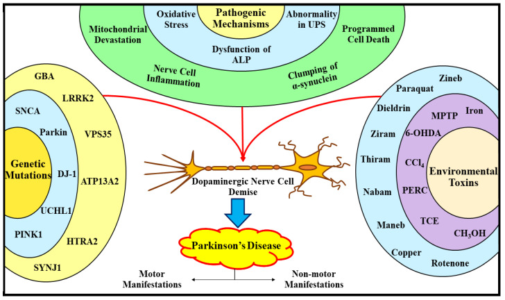
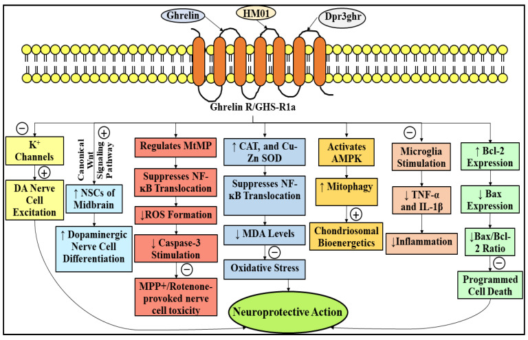
Parkinson's disease (PD) refers to one of the eminently grievous, preponderant, tortuous nerve-cell-devastating ailments that markedly impacts the dopaminergic (DArgic) nerve cells of the midbrain region, namely the substantia nigra pars compacta (SN-PC). Even though the exact etiopathology of the ailment is yet indefinite, the existing corroborations have suggested that aging, genetic predisposition, and environmental toxins tremendously influence the PD advancement. Additionally, pathophysiological mechanisms entailed in PD advancement encompass the clumping of α-synuclein inside the lewy bodies (LBs) and lewy neurites, oxidative stress, apoptosis, neuronal-inflammation, and abnormalities in the operation of mitochondria, autophagy lysosomal pathway (ALP), and ubiquitin-proteasome system (UPS). The ongoing therapeutic approaches can merely mitigate the PD-associated manifestations, but until now, no therapeutic candidate has been depicted to fully arrest the disease advancement. Neuropeptides (NPs) are little, protein-comprehending additional messenger substances that are typically produced and liberated by nerve cells within the entire nervous system. Numerous NPs, for instance, substance P (SP), ghrelin, neuropeptide Y (NPY), neurotensin, pituitary adenylate cyclase-activating polypeptide (PACAP), nesfatin-1, and somatostatin, have been displayed to exhibit consequential neuroprotection in both in vivo and in vitro PD models via suppressing apoptosis, cytotoxicity, oxidative stress, inflammation, autophagy, neuronal toxicity, microglia stimulation, attenuating disease-associated manifestations, and stimulating chondriosomal bioenergetics. The current scrutiny is an effort to illuminate the neuroprotective action of NPs in various PD-experiencing models. The authors carried out a methodical inspection of the published work procured through reputable online portals like PubMed, MEDLINE, EMBASE, and Frontier, by employing specific keywords in the subject of our article. Additionally, the manuscript concentrates on representing the pathways concerned in bringing neuroprotective action of NPs in PD. In sum, NPs exert substantial neuroprotection through regulating paramount pathways indulged in PD advancement, and consequently, might be a newfangled and eloquent perspective in PD therapy.

Keywords: Parkinson’s disease; ghrelin; neuropeptide Y; neuropeptides; neuroprotective action; neurotensin; pituitary adenylate cyclase-activating polypeptide; substance P.


Conflict of interest statement

The authors declare no conflict of interest.

Figures

Figure 1
Figure 1
The diagram illustrates the expansion in the incidence rate of Parkinson’s disease with aging, and also outlines the motor and nonmotor manifestations associated with Parkinson’s disease. Aging is reckoned as the critical parameter actively engaged in the evolution of PD. The incidence rate of PD escalates with aging, i.e., people ranging under 30–40 years rarely experience PD, 2% of people ranging above 60–70 years experience PD, and 5% of people ranging above 80–90 years experience PD. The motor manifestations of the disease comprehends rigor, postural deformities, tremor, and bradykinesia. On the other hand, the nonmotor manifestations associated with the disease comprehends urinary dysfunction, sexual abnormalities, sleep disturbances, psychosis, dementia, anxiety, apathy, depression, constipation, and erectile dysfunction. Howbeit, the nonmotor manifestations are comparably less noticeable than motor manifestations. PD, Parkinson’s disease; ↑, increasing.
Figure 2
Figure 2
Portraying the active participation of genetic mutations, environmental toxins, and pathogenic processes in the progression/evolvement of Parkinson’s disease. The trio, namely genetic mutations (SNCA, Parkin, DJ-1, UCHL1, PINK1, GBA, LRRK2, VPS35, ATP13A2, HTRA2, and SYNJ1), subjection to environmental toxins (MPTP, 6-OHDA, CH3OH, PERC, TCE, CCl4, zineb, paraquat, rotenone, dieldrin, ziram, thiram, nabam, maneb, copper, and iron), and pathogenic mechanisms (oxidative stress, dysfunction of ALP, abnormality in UPS, mitochondrial devastation, nerve cell inflammation, clumping of α-synuclein, and programmed cell death), give rise to dopaminergic nerve cell demise, which as a result culminates in the progression/evolvement of PD. SNCA, α-synuclein; Parkin, Parkin RBR E3 ubiquitin–protein ligase; DJ-1, protein deglycase; UCHL1, ubiquitin carboxy (C)-terminal hydrolase L1; PINK1, PTEN-induced kinase 1; GBA, glucocerebrosidase; LRRK2, leucine-rich repeat kinase 2; VPS35, vacuolar protein sorting 35; ATP13A2, neuronal P-type adenosine triphosphate (ATP)ase gene; HTRA2, high temperature requirement A2; SYNJ1, synaptojanin 1; MPTP, 1-methyl-4-phenyl-1,2,3,6-tetrahydropyridine; 6-OHDA, 6-hydroxy DA; CH3OH, methanol; PERC, perchloroethylene; TCE, trichloroethylene; CCl4, carbon tetrachloride; ALP, autophagy lysosomal pathway; UPS, ubiquitin-proteasome system; PD, Parkinson’s disease.
Figure 3
Figure 3
Highlighting the neuropeptides that contribute to significant nerve cell protection in Parkinson’s disease, encompassing substance P, ghrelin, neuropeptide Y, neurotensin, PACAP, nesfatin-1, and somatostatin. PACAP, pituitary adenylate cyclase-activating polypeptide.
Figure 4
Figure 4
Highlighting the neuroprotective role of Substance P in Parkinson’s disease. SP upon interaction with NK1 receptor, decreases Ca2+ entry, caspase-3 stimulation, ROS formation, and modulates MtMP, which in turn inhibit programmed cell death and cytotoxicity, and thereby protects MES23.5 cells from MPP+-prompted neurotoxicity. Septide, an analog of SP, upon interaction with NK1 receptor culminates in the suppression of programmed cell death pathways and stimulation of PKB/Akt signaling pathway, thereby protects nerve cells against 6-OHDA-prompted neurotoxicity. Further, senktide, upon interaction with NK3 receptor, reinstated the temporal order memory in the 6-OHDA-lesioned hemiparkinsonian rat model. In addition, the three NK1 receptor antagonists, namely NAT, L-733060, and LY303870, upon introduction, decrease cellular demise provoked by 6-OHDA subjection, upgrade motor operations, and decrease levodopa-precipitated dyskinesia. Finally, by virtue of these mechanisms, SP markedly contributes to neuroprotective action in PD. Subtraction symbol indicates inhibitory/suppressing action, while addition symbol indicates stimulatory action. SP, Substance P; NAT, N-acetyl-L-tryptophan; LY303870, lanepitant; NK, neurokinin; NK1, neurokinin 1; NK3, neurokinin 3; Ca2+, calcium ions; ROS, reactive oxygen species; MtMP, mitochondrial membrane potential; MES23.5, DArgic nerve cell line; MPP+, 1-methyl-4-phenylpyridinium ion; PKB/Akt, protein kinase B signaling pathway; 6-OHDA, 6-hydroxy DA; PD, Parkinson’s disease; ↓, decreasing or reducing.
Figure 5
Figure 5
Highlighting the neuroprotective role of ghrelin in Parkinson’s disease. Ghrelin, along with its analogs, namely HM01 and Dpr3ghr, markedly render nerve cell protective actions against MPTP, MPP+, and rotenone-provoked neuronal destruction, via suppressing oxidative stress, programmed cell death, and de-escalating inflammation. Ghrelin, following its interaction with ghrelinR/GHS-R1a, suppresses K+ channels, which, as a consequence, contributes to DA nerve cell excitation. In addition, ghrelin, by means of the canonical Wnt signaling pathway, increases the number or amount of NSCs of the midbrain and in vitro and ex vivo DArgic nerve cell differentiation. Moreover, ghrelin treatment regulates MtMP, suppresses mitochondrial complex-I operation, de-escalates ROS formation and caspase-3 stimulation, and eventually restrains nerve cells from MPP+ or rotenone-instigated detrimental repercussions. Additionally, ghrelin escalates the levels of antioxidant biocatalysts, namely CAT and Cu-Zn SOD, de-escalates the MDA levels, and suppresses the NF-κB translocation, and thereby suppresses the oxidative stress via ceasing the lipid peroxidation and generation of ROS, and finally contributes to nerve cell protective action. Moreover, ghrelin possesses its nerve cell protective action via activating AMPK and escalating the mitophagy, which subsequently culminates in the amplification in the chondriosomal bioenergetics. Furthermore, therapy with the assistance of ghrelin culminates in the suppression of microglia stimulation, which, as a result, de-escalates inflammation (via de-escalating the levels of TNF-α and IL-1β), and finally culminates into nerve cell protective action. Apart from this, the two, namely ghrelin and Dpr3ghr, suppress programmed cell death and possess substantial nerve cell protective action by elevating Bcl-2 expression, and declining Bax expression and the Bax/Bcl-2 ratio. Subtraction symbol indicates inhibitory/suppressing action, while addition symbol indicates stimulatory action. GhrelinR, ghrelin receptor; GHS-R1a, growth hormone secretagogue receptor 1a; K+, potassium; DA, dopamine; NSCs, neural stem cells; DArgic, dopaminergic; MtMP, mitochondrial membrane potential; ROS, reactive oxygen species; MPP+, 1-methyl-4-phenylpyridinium ion; CAT, catalase; Cu-Zn SOD, copper-zincsuperoxide dismutase; NF-κB, Nuclear Factor kappa-light-chain-enhancer of activated B cells; MDA, malondialdehyde; AMPK, 5′ adenosine monophosphate-activated protein kinase; TNF-α,tumor necrosis factor-alpha; IL-1β, interleukin-1β;Bcl-2,B-cell lymphoma-2; Bax, Bcl-2-associated X protein; MPTP, 1-methyl-4-phenyl-1,2,3,6-tetrahydropyridine; ↑, increasing; ↓, decreasing.
Figure 6
Figure 6
Highlighting the neuroprotective role of neuropeptide Y in Parkinson’s disease. NPY exhibits its significant nerve cell protective action upon interacting with DA-forming cells by means of Y2R. NPY stimulates the two, namely the Akt signaling pathway and the MAPK signaling pathway and contributes to the enhanced viability of DA-forming nerve cells of the nigral area of the encephalon. In addition, NPY exerts its nerve cell protective action by suppressing the microglia, which in turn suppresses the NO formation and IL-1β liberation, and finally results in the suppression of inflammation. Furthermore, NPY, via stimulating the PI3K-XBP1s-Bip/GRP78 signaling pathway, suppresses the ER stress-provoked nerve cell demise, and ultimately contributes to neuroprotection. Moreover, therapy with the assistance of NPY de-escalated the stimulation/operation of caspase-3 and caspase-4, which consequently suppressed the programmed cell death and ER stress-triggered nerve cell demise and contributed to nerve cell protection in PD. Subtraction symbol indicates inhibitory/suppressing action, while addition symbol indicates stimulatory action. NPY, neuropeptide Y; Y2R,Y2 receptor; DArgic, dopaminergic; SN, substantia nigra; MAPK, mitogen-activated protein kinase; NO, nitric oxide; IL-1β, interleukin-1β; PI3K, phosphoinositide 3-kinase; XBP1s, spliced form of X-box binding protein 1; BiP, binding immunoglobulin protein; GRP78, 78-kDa glucose-regulated protein; ER stress, endoplasmic reticulum stress; DA, dopamine; PD, Parkinson’s disease; ↑, increasing; ↓, decreasing.
Figure 7
Figure 7
Highlighting the neuroprotective role of pituitary adenylate cyclase-activating polypeptide in Parkinson’s disease. The duo, namely experimental animal and cellular PD models illustrate that PACAP therapy renders significant nerve cell protective action against rotenone, MPTP, and 1-methyl-6,7-dihydroxy-1,2,3,4-tetrahydroisochinolin-provoked deleterious repercussions through suppressing inflammation, autophagy, and cellular demise. The prior therapy of SH-SY5Y cells with PACAP (1–38) safeguarded these cells against inflammatory-mediated detrimental effects via decreasing the liberation of inflammatory mediators. PACAP regulates the sequestosome-1/p62 protein concentrations and elevates the LC3-II formation, and thereby suppresses the autophagic-operation. PACAP safeguarded against rotenone-precipitated cellular demise via carrying out the stimulation of PKA signaling process, as well as the two downstream signals, viz., p38 MAPK and ERK. In addition, intravenously introduced PACAP27 safeguarded against MPTP-instigated nerve cell demise via altering the DArgic and cholinergic synaptic conveyance, by way of elevating the D2R operation and the KATP subunits expression within the striatal region of the basal nuclei. Further, therapy with the assistance of PACAP/agonists of PACAP receptors elevated the BDNF and p-CREB expression, and suppressed the caspase-3 expression, and, as a consequence, decreased the 1-methyl-6,7-dihydroxy-1,2,3,4-tetrahydroisochinolin-prompted SH-SY5Y cells toxicity. Apart from this, PACAP has the tendency to considerably suppress the LPS-prompted microglia stimulation, which in turn suppresses the formation and liberation of NO and TNF-α, and finally suppresses the inflammation. Subtraction symbol indicates inhibitory/suppressing action, while addition symbol indicates stimulatory action. PACAP, pituitary adenylate cyclase-activating polypeptide; VIP, vasoactive intestinal polypeptide; PAC1, PACAP type I receptor; VPAC1, VIP receptor 1; VPAC2, VIP receptor 2; NO, nitric oxide; TNF-α, tumor necrosis factor-alpha; IL-1β, interleukin-1β; IL-6, interleukin-6; ROS, reactive oxygen species; MMP-9, matrix metallopeptidase 9; LC3-II, microtubule-associated protein light chain 3-phosphatidylethanolamine conjugate; PKA, protein kinase A; MAPK, mitogen-activated protein kinase; ERK, extracellular signal-regulated kinase; D2R, DA 2 receptors; KATP, ATP-sensitive potassium channel; DArgic, dopaminergic; MPTP, 1-methyl-4-phenyl-1,2,3,6-tetrahydropyridine; BDNF, brain-derived neurotrophic factor; p-CREB, phosphorylated cAMP-response element binding protein; SH-SY5Y, human neuroblastoma cells; LPS, lipopolysaccharide; PD, Parkinson’s disease; ↑, increasing; ↓, decreasing.

Similar articles

Cited by

References

    1. Palacios-Sánchez L., Nupan M.T., Botero-Meneses J.S. James Parkinson and His Essay on “Shaking Palsy”, Two Hundred Years Later. Arq. Neuro-Psiquiatr. 2017;75:671–672. doi: 10.1590/0004-282x20170108. - DOI - PubMed
    1. Hayes M.T. Parkinson’s Disease and Parkinsonism. Am. J. Med. 2019;132:802–807. doi: 10.1016/j.amjmed.2019.03.001. - DOI - PubMed
    1. Dawson T.M. Parkinson’s Disease: Clinical Manifestations and Treatment. Int. Rev. Psychiatry. 2000;12:263–269. doi: 10.1080/09540260020002488. - DOI
    1. Surmeier D.J. Determinants of Dopaminergic Neuron Loss in Parkinson’s Disease. FEBS J. 2018;285:3657–3668. doi: 10.1111/febs.14607. - DOI - PMC - PubMed
    1. Muangpaisan W., Hori H., Brayne C. Systematic Review of the Prevalence and Incidence of Parkinson’s Disease in Asia. J. Epidemiol. 2009;19:281–293. doi: 10.2188/jea.JE20081034. - DOI - PMC - PubMed

MeSH terms

Substances