. 2022 Apr 21;23(9):4597.
doi: 10.3390/ijms23094597.

JAK2 Inhibitor, Fedratinib, Inhibits P-gp Activity and Co-Treatment Induces Cytotoxicity in Antimitotic Drug-Treated P-gp Overexpressing Resistant KBV20C Cancer Cells

Affiliations

JAK2 Inhibitor, Fedratinib, Inhibits P-gp Activity and Co-Treatment Induces Cytotoxicity in Antimitotic Drug-Treated P-gp Overexpressing Resistant KBV20C Cancer Cells

Yunmoon Oh et al. Int J Mol Sci. .

Abstract

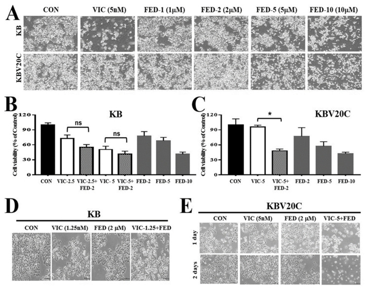
P-glycoprotein (P-gp) overexpression is one of the major mechanisms of multidrug resistance (MDR). Previously, co-treatment with Janus kinase 2 (JAK2) inhibitors sensitized P-gp-overexpressing drug-resistant cancer cells. In this study, we assessed the cytotoxic effects of JAK2 inhibitor, fedratinib, on drug-resistant KBV20C cancer cells. We found that co-treatment with fedratinib at low doses induced cytotoxicity in KBV20C cells treated with vincristine (VIC). However, fedratinib-induced cytotoxicity was little effect on VIC-treated sensitive KB parent cells, suggesting that these effects are specific to resistant cancer cells. Fluorescence-activated cell sorting (FACS), Western blotting, and annexin V analyses were used to further investigate fedratinib's mechanism of action in VIC-treated KBV20C cells. We found that fedratinib reduced cell viability, increased G2 arrest, and upregulated apoptosis when used as a co-treatment with VIC. G2 phase arrest and apoptosis in VIC-fedratinib-co-treated cells resulted from the upregulation of p21 and the DNA damaging marker pH2AX. Compared with dimethyl sulfoxide (DMSO)-treated cells, fedratinib-treated KBV20C cells showed two-fold higher P-gp-inhibitory activity, indicating that VIC-fedratinib sensitization is dependent on the activity of fedratinib. Similar to VIC, fedratinib co-treatment with other antimitotic drugs (i.e., eribulin, vinorelbine, and vinblastine) showed increased cytotoxicity in KBV20C cells. Furthermore, VIC-fedratinib had similar cytotoxic effects to co-treatment with other JAK2 inhibitors (i.e., VIC-CEP-33779 or VIC-NVP-BSK805) at the same dose; similar cytotoxic mechanisms (i.e., early apoptosis) were observed between treatments, suggesting that co-treatment with JAK2 inhibitors is generally cytotoxic to P-gp-overexpressing resistant cancer cells. Given that fedratinib is FDA-approved, our findings support its application in the co-treatment of P-gp-overexpressing cancer patients showing MDR.

Keywords: JAK2; P-gp; cancer; co-treatment; drug-resistance; fedratinib.


Conflict of interest statement

The Authors declare no conflicts of interest regarding this study.

Figures

Figure 1
Figure 1
Co-treatment with fedratinib specifically increases the sensitization of drug-resistant KBV20C cancer cells to VIC treatment. (A) Parent sensitive KB cells and drug-resistant KBV20C were grown on 60 mm diameter dishes and treated with 5 nM vincristine (VIC), 1 µM fedratinib (FED-1), 2 µM fedratinib (FED-2), 5 µM fedratinib (FED-5), 10 µM fedratinib (FED-10), or 0.1% DMSO (CON). After 1 day, all cells were observed using an inverted microscope at ×40 magnification. (B,C) Parent sensitive KB cells and drug-resistant KBV20C were plated on 96-well plates and grown to 30–40% confluence. The cells were then treated for 48 h with 2.5 nM vincristine (VIC-2.5), 5 nM vincristine (VIC-5), 2 µM fedratinib (FED-2), 5 µM fedratinib (FED-5), 10 µM fedratinib (FED-10), 10 µM fedratinib (FED-10), 2.5 nM VIC with 2 µM fedratinib (VIC-2.5 + FED-2), 5 nM VIC with 2 µM fedratinib (VIC-5 + FED-2), or 0.1% DMSO (CON). Cell viability assay was performed as described in the Materials and Methods. The data are presented as the mean ±SD from two independent experiments repeated in triplicate. For co-treatments, significantly different at * p < 0.05 compared with the corresponding control. The “ns” is an abbreviation of “not significant”. (D,E) Parent sensitive KB cells and drug-resistant KBV20C were grown on 60 mm-diameter dishes and treated with 1.25 nM vincristine (VIC-1.25), 5 nM vincristine (VIC-5), 2 µM fedratinib (FED), 1.25 nM VIC with 2 µM fedratinib (VIC-1.25 + FED), 5 nM VIC with 2 µM fedratinib (VIC-5 + FED), or 0.1% DMSO (CON). After 1 day, all cells were observed using an inverted microscope at ×100 magnification.
Figure 2
Figure 2
DNA damage signal induces apoptosis and autophagy via G2 arrest in VIC–fedratinib co-treated KBV20C cells. (A) KBV20C cells were grown on 60 mm-diameter dishes and treated with 5 nM vincristine (VIC), 2 µM fedratinib (FED), 5 nM VIC with 2 µM fedratinib (VIC + FED), or 0.1% DMSO (CON). After 24 h, annexin V analyses were performed as described in Materials and Methods. (B,C) KBV20C cells were plated on 60 mm-diameter dishes and treated with 5 nM vincristine (VIC), 1 µM fedratinib (FED-1), 2 µM fedratinib (FED-2), 5 nM VIC with 1 µM fedratinib (VIC + FED-1), 5 nM VIC with 2 µM fedratinib (VIC + FED-2), or 0.1% DMSO (CON). After 24 h, western blot analysis was performed using antibodies against C-PARP, α-LC3B, and GAPDH. (D) KBV20C cells were plated on 60 mm-diameter dishes and treated with 5 nM vincristine (VIC), 1 µM fedratinib (FED-1), 2 µM fedratinib (FED-2), 5 nM VIC with 1 µM fedratinib (VIC + FED-1), 5 nM VIC with 2 µM fedratinib (VIC + FED-2), or 0.1% DMSO (CON). After 24 h, FACS analyses were performed as described in Materials and Methods. (E) KBV20C cells were plated on 60 mm-diameter dishes and treated with 5 nM vincristine (VIC), 1 µM fedratinib (FED-1), 2 µM fedratinib (FED-2), 5 nM VIC with 1 µM fedratinib (VIC + FED-1), 5 nM VIC with 2 µM fedratinib (VIC + FED-2), or 0.1% DMSO (CON). After 24 h, western blot analysis was performed using antibodies against Cyclin D1, CDK2, CDK1, p21, pH2AX, and GAPDH.
Figure 3
Figure 3
Fedratinib with P-gp inhibitory activity increases the sensitization of KBV20C cells treated to other anti-mitotic drugs. (A) KBV20C cells were grown on 60 mm-diameter dishes and treated with 10 µM verapamil, 2.5 µM aripiprazole, 5 µM reserpine, 2 µM fedratinib (FED), or 0.1% DMSO (CON). After 1 h, all cells were stained with rhodamine for 3 h and examined by using FACS analysis, as described in Section 4, Materials and Methods. (B,C) KBV20C cells were grown on 60 mm diameter dishes and treated with 30 nM eribulin, 0.1 µg/mL vinorelbine, 5 nM vinblastine, 2 µM fedratinib (FED), 30 nM eribulin with 2 µM fedratinib (Eribulin + FED), 0.1 µg/mL vinorelbine with 2 µM fedratinib (Vinorelbine + FED), 5 nM vinblastine with 1 µM fedratinib (Vinblastine + FED), or 0.1% DMSO (CON). After 1 day, all cells were observed using an inverted microscope at x40 or x100 magnification.
Figure 4
Figure 4
JAK2 inhibitors (fedratinib, CEP-33779, and NVP-BSK805) sensitize VIC-treated KBV20C cells via similar mechanisms of action. (A) KBV20C cells were plated on 60 mm diameter dishes and treated with 5 nM vincristine (VIC), 2 µM fedratinib (FED), 10 µM verapamil, 5 nM VIC with 2 µM fedratinib (VIC + FED), 5 nM VIC with 10 µM verapamil (VIC+VER), or 0.1% DMSO (CON). After 1 day, all cells were observed using an inverted microscope at ×100 magnification. (B,C) KBV20C cells were plated on 60 mm diameter dishes and treated with 5 nM vincristine (VIC), 1 µM fedratinib (FED-1), 2 µM fedratinib (FED-2), 2 µM CEP-33779 (CEP), 2 µM NVP-BSK805 (NVP), 5 nM VIC with 1 µM fedratinib (VIC + FED-1), 5 nM VIC with 2 µM fedratinib (VIC + FED-2), 5 nM VIC with 2 µM CEP-33779 (VIC + CEP), 5 nM VIC with 2 µM NVP-BSK805 (VIC + NVP), or 0.1% DMSO (CON). After 24 h, microscopic observation at x100 magnification (B) or annexin V analyses (C) were performed as described in Section 4, Materials and Methods.
Figure 5
Figure 5
Fedratinib did not change P-gp protein levels. (A,B) KBV20C cells were plated on 60 mm diameter dishes and treated with 2 µM fedratinib (FED-2) or 0.1% DMSO (CON). After 4 h or 24 h, Western blot analysis was performed using antibodies against (A) P-gp (Antibody 1 from Cell signaling company), (B) P-gp (Antibody 2 from Santia Cruz company), and GAPDH. (C) Drug-resistant KBV20C were grown on 60 mm diameter dishes and treated with 5 nM vincristine (VIC), 0.5 µM fedratinib (FED-0.5), 1 µM fedratinib (FED-1), 2 µM fedratinib (FED-2), 4 µM fedratinib (FED-4), 6 µM fedratinib (FED-6), 5 nM VIC with 0.5 µM fedratinib (VIC-5 + FED-0.5), 5 nM VIC with 1 µM fedratinib (VIC-5 + FED-1), 5 nM VIC with 2 µM fedratinib (VIC-5 + FED-2), 5 nM VIC with 4 µM fedratinib (VIC-5 + FED-4), 5 nM VIC with 6 µM fedratinib (VIC-5 + FED-6), or 0.1% DMSO (CON). After 2 days, all cells were observed using an inverted microscope at x40 magnification. (D) Drug-resistant KBV20C were plated on 96-well plates and grown to 30–40% confluence. The cells were then treated with 5 nM vincristine (VIC-5), 0.5 µM fedratinib (FED-0.5), 1 µM fedratinib (FED-1), 2 µM fedratinib (FED-2), 3 µM fedratinib (FED-3), 5 µM fedratinib (FED-5), 5 nM VIC with 0.5 µM fedratinib (VIC-5 + FED-0.5), 5 nM VIC with 1 µM fedratinib (VIC-5 + FED-1), 5 nM VIC with 2 µM fedratinib (VIC-5 + FED-2), 5 nM VIC with 3 µM fedratinib (VIC-5 + FED-3), 5 nM VIC with 5 µM fedratinib (VIC-5+FED-5), or 0.1% DMSO (CON). Cell viability assay was performed as described in Section 4, Materials and Methods. The data are presented as the mean ±SD from two independent experiments repeated in triplicate. For co-treatments, significantly different at * p < 0.05 compared with the corresponding control. The “ns” is an abbreviation of “not significant”.

References

    1. Jordan M.A., Wilson L. Microtubules as a target for anticancer drugs. Nat. Rev. Cancer. 2004;4:253–265. doi: 10.1038/nrc1317. - DOI - PubMed
    1. Kim J.H., Yoo H.I., Kang H.S., Ro J., Yoon S. Salinomycin sensitizes antimitotic drugs-treated cancer cells by increasing apoptosis via the prevention of G2 arrest. Biochem. Biophys. Res. Commun. 2012;418:98–103. doi: 10.1016/j.bbrc.2011.12.141. - DOI - PubMed
    1. McGrogan B.T., Gilmartin B., Carney D.N., McCann A. Taxanes, microtubules and chemoresistant breast cancer. Biochim. Biophys. Acta. 2008;1785:96–132. doi: 10.1016/j.bbcan.2007.10.004. - DOI - PubMed
    1. Chen Z., Shi T., Zhang L., Zhu P., Deng M., Huang C., Hu T., Jiang L., Li J. Mammalian drug efflux transporters of the ATP binding cassette (ABC) family in multidrug resistance: A review of the past decade. Cancer Lett. 2016;370:153–164. doi: 10.1016/j.canlet.2015.10.010. - DOI - PubMed
    1. Shukla S., Wu C.P., Ambudkar S.V. Development of inhibitors of ATP-binding cassette drug transporters: Present status and challenges. Expert Opin. Drug Metab. Toxicol. 2008;4:205–223. doi: 10.1517/17425255.4.2.205. - DOI - PubMed

MeSH terms