

A Reliable Approach for Revealing Molecular Targets in Secondary Ion Mass Spectrometry
- PMID: 35563005
- PMCID: PMC9103194
- DOI: 10.3390/ijms23094615
A Reliable Approach for Revealing Molecular Targets in Secondary Ion Mass Spectrometry
Abstract
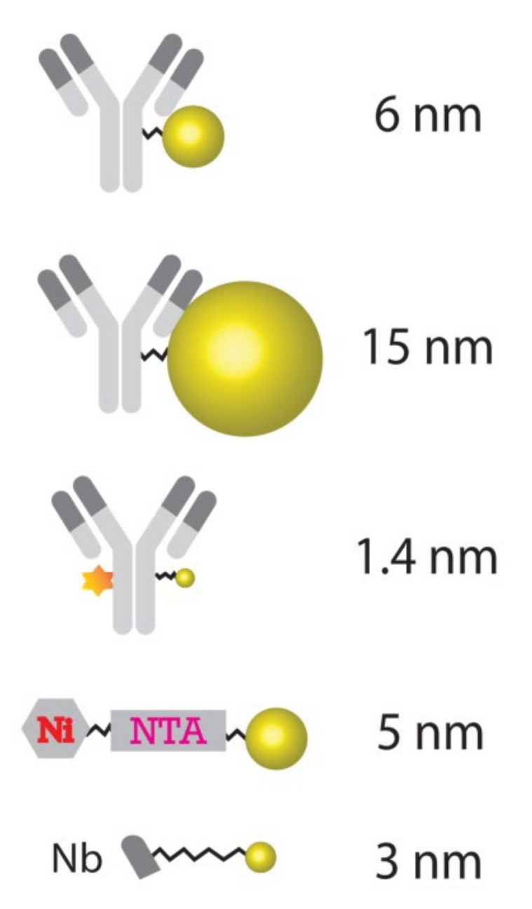
Nano secondary ion mass spectrometry (nanoSIMS) imaging is a rapidly growing field in biological sciences, which enables investigators to describe the chemical composition of cells and tissues with high resolution. One of the major challenges of nanoSIMS is to identify specific molecules or organelles, as these are not immediately recognizable in nanoSIMS and need to be revealed by SIMS-compatible probes. Few laboratories have generated such probes, and none are commercially available. To address this, we performed a systematic study of probes initially developed for electron microscopy. Relying on nanoscale SIMS, we found that antibodies coupled to 6 nm gold particles are surprisingly efficient in terms of labeling specificity while offering a reliable detection threshold. These tools enabled accurate visualization and sample analysis and were easily employed in correlating SIMS with other imaging approaches, such as fluorescence microscopy. We conclude that antibodies conjugated to moderately sized gold particles are promising tools for SIMS imaging.
Keywords: immunogold labeling; mass spectrometry imaging; nanoSIMS; protein turnover.
Conflict of interest statement
The authors declare no conflict of interest.
Figures

Similar articles
-
Elemental mapping of Neuromelanin organelles of human Substantia Nigra: correlative ultrastructural and chemical analysis by analytical transmission electron microscopy and nano-secondary ion mass spectrometry.J Neurochem. 2016 Jul;138(2):339-53. doi: 10.1111/jnc.13648. Epub 2016 Jun 2. J Neurochem. 2016. PMID: 27121280
-
Gold-Conjugated Nanobodies for Targeted Imaging Using High-Resolution Secondary Ion Mass Spectrometry.Nanomaterials (Basel). 2021 Jul 10;11(7):1797. doi: 10.3390/nano11071797. Nanomaterials (Basel). 2021. PMID: 34361183 Free PMC article.
-
Correlative High-Resolution Imaging of Iron Uptake in Lung Macrophages.Anal Chem. 2022 Sep 20;94(37):12798-12806. doi: 10.1021/acs.analchem.2c02675. Epub 2022 Sep 7. Anal Chem. 2022. PMID: 36070604 Free PMC article.
-
Progress in analytical imaging of the cell by dynamic secondary ion mass spectrometry (SIMS microscopy).Biochim Biophys Acta. 2005 Aug 5;1724(3):228-38. doi: 10.1016/j.bbagen.2005.05.013. Biochim Biophys Acta. 2005. PMID: 15982822 Review.
-
Highest resolution chemical imaging based on secondary ion mass spectrometry performed on the helium ion microscope.Rep Prog Phys. 2021 Sep 15;84(10). doi: 10.1088/1361-6633/ac1e32. Rep Prog Phys. 2021. PMID: 34404033 Review.
Cited by
-
Stable isotope labeling and ultra-high-resolution NanoSIMS imaging reveal alpha-synuclein-induced changes in neuronal metabolism in vivo.Acta Neuropathol Commun. 2023 Sep 29;11(1):157. doi: 10.1186/s40478-023-01608-8. Acta Neuropathol Commun. 2023. PMID: 37770947 Free PMC article.
-
Application of metal stable isotopes labeling and elemental mass spectrometry for biomacromolecule profiling.Biophys Rep. 2025 Apr 30;11(2):112-128. doi: 10.52601/bpr.2024.240039. Biophys Rep. 2025. PMID: 40308936 Free PMC article.
References
-
- Swales J.G., Hamm G., Clench M.R., Goodwin R.J.A. Mass Spectrometry Imaging and Its Application in Pharmaceutical Research and Development: A Concise Review. Int. J. Mass Spectrom. 2019;437:99–112. doi: 10.1016/j.ijms.2018.02.007. - DOI
MeSH terms
Substances
Grants and funding
LinkOut - more resources
Full Text Sources
