Review
. 2022 Apr 22;23(9):4635.
doi: 10.3390/ijms23094635.

Molecular and Signaling Mechanisms for Docosahexaenoic Acid-Derived Neurodevelopment and Neuroprotection

Affiliations
Review

Molecular and Signaling Mechanisms for Docosahexaenoic Acid-Derived Neurodevelopment and Neuroprotection

Hee-Yong Kim et al. Int J Mol Sci. .

Abstract

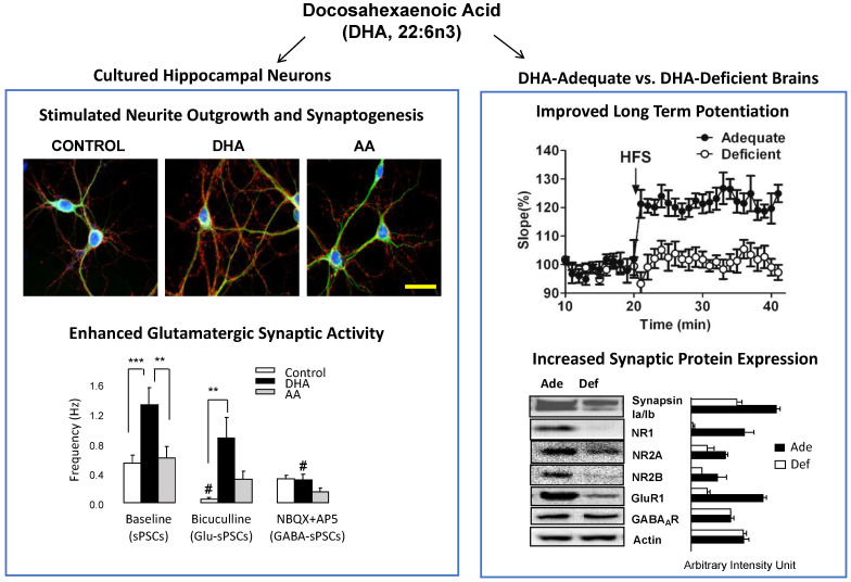
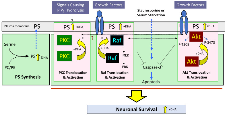
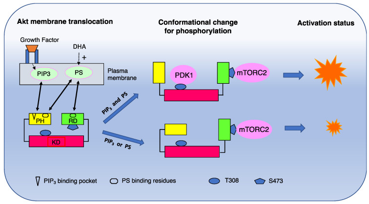
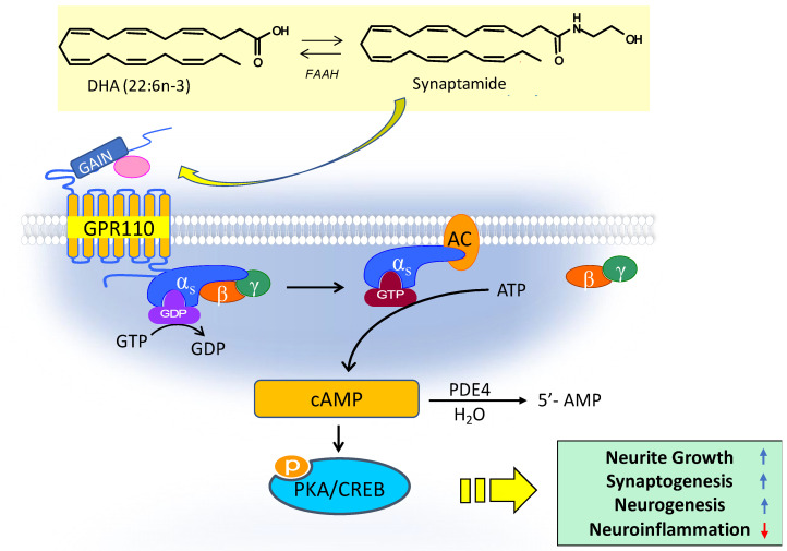
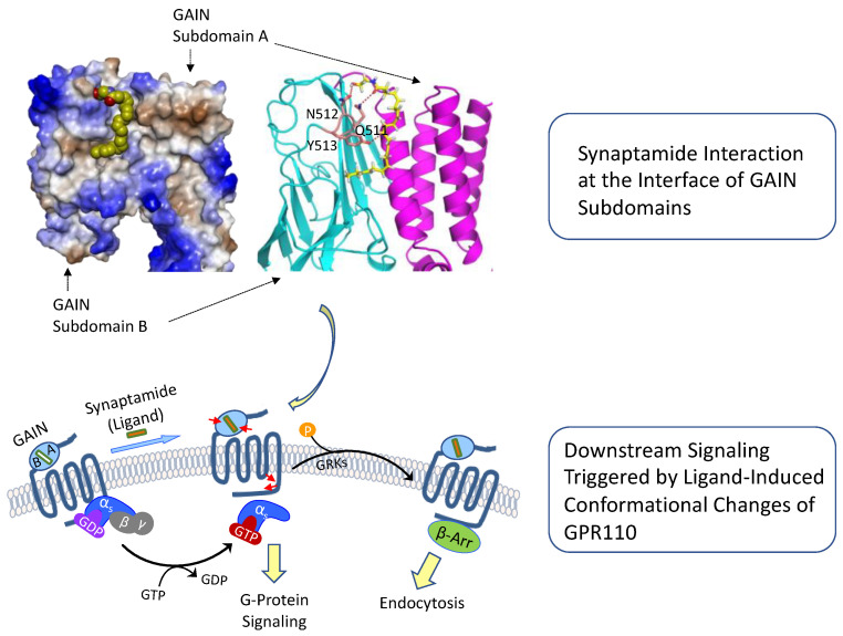
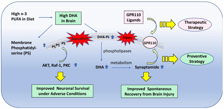
The neurodevelopmental and neuroprotective actions of docosahexaenoic acid (DHA) are mediated by mechanisms involving membrane- and metabolite-related signal transduction. A key characteristic in the membrane-mediated action of DHA results from the stimulated synthesis of neuronal phosphatidylserine (PS). The resulting DHA-PS-rich membrane domains facilitate the translocation and activation of kinases such as Raf-1, protein kinase C (PKC), and Akt. The activation of these signaling pathways promotes neuronal development and survival. DHA is also metabolized in neural tissues to bioactive mediators. Neuroprotectin D1, a docosatriene synthesized by the lipoxygenase activity, has an anti-inflammatory property, and elovanoids formed from DHA elongation products exhibit antioxidant effects in the retina. Synaptamide, an endocannabinoid-like lipid mediator synthesized from DHA in the brain, promotes neurogenesis and synaptogenesis and exerts anti-inflammatory effects. It binds to the GAIN domain of the GPR110 (ADGRF1) receptor, triggers the cAMP/protein kinase A (PKA) signaling pathway, and activates the cAMP-response element binding protein (CREB). The DHA status in the brain influences not only the PS-dependent signal transduction but also the metabolite formation and expression of pre- and post-synaptic proteins that are downstream of the CREB and affect neurotransmission. The combined actions of these processes contribute to the neurodevelopmental and neuroprotective effects of DHA.

Keywords: ADGRF1; Akt; GPR110; N-docosahexaenoylethanolamine; N-docosahexaenoylphosphatidylethanolamine; PKA; cAMP; docosahexaenoic acid; phosphatidylserine; synaptamide; synaptic membrane proteins.


Conflict of interest statement

The authors declare no conflict of interest.

Figures

Figure 1
Figure 1
Neurodevelopmental effects of DHA. DHA among fatty acids uniquely promotes neurite growth, synaptogenesis, and glutamatergic synaptic activity in cultured hippocampal neurons. DHA-adequate mouse brains show improved LTP and increased synaptic protein expression compared to DHA-deficient brains. Markers used for the staining of hippocampal neurons include DAPI (for nuclei, blue), synapsin1 (a presynaptic marker protein, red), and MAP2 (a neuronal marker protein, green). AA, arachidonic acid. Adapted from reference [4].
Figure 2
Figure 2
Neurodevelopmental and neuroprotective actions resulting from the membrane incorporation and metabolism of DHA. DHA increases PS in the neuronal membrane as DHA-containing phospholipids are the best substrate for PS biosynthetic activity. Susceptibility to neuronal cell death is reduced by increasing the PS level. Certain metabolites of free DHA can also exert neuroprotective effects. DHA, docosahexaenoic acid; PC, phosphatidylcholine; PE, phosphatidylethanolamine; PS, phsphatidylserine. Adapted from references [6,17].
Figure 3
Figure 3
Membrane PS-dependent signaling mechanisms modulated by DHA that mediate neuronal survival under adverse conditions. By increasing PS in the neuronal membrane, DHA facilitates the translocation and activation of PKC, Raf-1, and Akt, positively influencing neuronal survival. PKC, protein kinase C; ERK, mitogen-activated kinase (MAPK); MEK, MAPK kinase. PC, phosphatidylcholine; PE, phosphatidylethanolamine; PS, phsphatidylserine. Adapted from reference [12].
Figure 4
Figure 4
Molecular mechanism for PS-facilitated Akt activation. Upon stimulation of a growth factor receptor, cytosolic Akt is recruited to the plasma membrane though interacting with PIP3 and PS. The membrane interaction causes conformational changes of Akt, exposing T308 and S473 for phosphorylation by PDK1 and mTORC2, respectively. While S473 in the regulatory domain can be exposed by interacting with either PS or PIP3, both lipids are required for the conformational change of the PH domain to expose T308 in the kinase domain for full activation of Akt. DHA, docosahexaenoic acid; PS, phsphatidylserine; PH, pleckstrin homology domain; RD, regulatory domain; KD, kinase domain. Adapted from reference [25].
Figure 5
Figure 5
Synaptamide-activated GPR110 signaling. By binding to GPR110, synaptamide activates Gαs protein; increases cAMP; promotes neurogenesis, neurite growth, and synaptogenesis; and inhibits neuroinflammation. DHA, docosahexaenoic acid; FAAH, fatty acid amide hydrolase; GAIN, GPCR autoproteolysis domain; GTP, guanosine-5′-triphosphate; GDP, guanosine-5′-diphosphate; ATP, adenosine-5′-triphosphate; AC, adenylyl cyclase; CREB, cAMP response element-binding protein. Adapted from references [15,49].
Figure 6
Figure 6
Activation of GPR110 and downstream signaling through synaptamide binding to the GAIN domain. Synaptamide is shown with a space-filling or stick-and-ball representation. Synaptamide binds to the interface between two subdomains, causing conformational changes in the GAIN and intracellular domains of GPR110 (depicted by red arrows) that trigger downstream signaling events. Adapted from reference [56]. GAIN, GPCR autoproteolysis domain; GTP, guanosine-5′-triphosphate; GDP, guanosine-5′-diphosphate.
Figure 7
Figure 7
Preventive and therapeutic strategies for neuroprotection based on DHA-derived bioactivity. PUFA, polyunsaturated fatty acids; DHA, docosahexaenoic acid; DHA-PL, DHA-containing phospholipids. PKC, protein kinase C.

Similar articles

Cited by

References

    1. Salem N., Litman B., Kim H.-Y., Gawrisch K. Mechanisms of action of docosahexaenoic acid in the nervous system. Lipids. 2001;36:945–959. doi: 10.1007/s11745-001-0805-6. - DOI - PubMed
    1. Green P., Glozman S., Kamensky B., Yavin S. Developmental changes in rat brain membrane lipids and fatty acids: The preferential prenatal accumulation of docosahexaenoic acid. J. Lipid Res. 1999;40:960–966. doi: 10.1016/S0022-2275(20)32132-5. - DOI - PubMed
    1. Calderon F., Kim H.-Y. Docosahexaenoic acid promotes neurite growth in hippocampal neurons. J. Neurochem. 2004;90:1540. doi: 10.1111/j.1471-4159.2004.02808.x. - DOI - PubMed
    1. Cao D.H., Kevala K., Kim J., Moon H.S., Jun S.B., Lovinger D., Kim H.-Y. Docosahexaenoic acid promotes hippocampal neuronal development and synaptic function. J. Neurochem. 2009;111:510–521. doi: 10.1111/j.1471-4159.2009.06335.x. - DOI - PMC - PubMed
    1. Kawakita E., Hashimoto M., Shido O. Docosahexaenoic acid promotes neurogenesis in vitro and in vivo. Neuroscience. 2006;139:991–997. doi: 10.1016/j.neuroscience.2006.01.021. - DOI - PubMed

MeSH terms

Substances