. 2022 Apr 23;23(9):4692.
doi: 10.3390/ijms23094692.

Comparison of Physicochemical, Mechanical, and (Micro-)Biological Properties of Sintered Scaffolds Based on Natural- and Synthetic Hydroxyapatite Supplemented with Selected Dopants

Affiliations

Comparison of Physicochemical, Mechanical, and (Micro-)Biological Properties of Sintered Scaffolds Based on Natural- and Synthetic Hydroxyapatite Supplemented with Selected Dopants

Andrzej Hudecki et al. Int J Mol Sci. .

Abstract

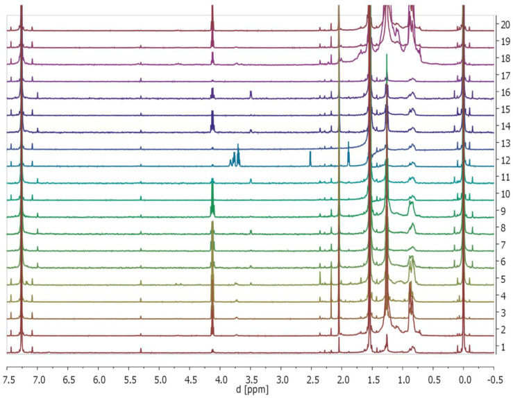
The specific combinations of materials and dopants presented in this work have not been previously described. The main goal of the presented work was to prepare and compare the different properties of newly developed composite materials manufactured by sintering. The synthetic- (SHAP) or natural- (NHAP) hydroxyapatite serves as a matrix and was doped with: (i) organic: multiwalled carbon nanotubes (MWCNT), fullerenes C60, (ii) inorganic: Cu nanowires. Research undertaken was aimed at seeking novel candidates for bone replacement biomaterials based on hydroxyapatite-the main inorganic component of bone, because bone reconstructive surgery is currently mostly carried out with the use of autografts; titanium or other non-hydroxyapatite -based materials. The physicomechanical properties of the developed biomaterials were tested by Scanning Electron Microscopy (SEM), Dielectric Spectroscopy (BSD), Nuclear Magnetic Resonance (NMR), and Differential Scanning Calorimetry (DSC), as well as microhardness using Vickers method. The results showed that despite obtaining porous sinters. The highest microhardness was achieved for composite materials based on NHAP. Based on NMR spectroscopy, residue organic substances could be observed in NHAP composites, probably due to the organic structures that make up the tooth. Microbiology investigations showed that the selected samples exhibit bacteriostatic properties against Gram-positive reference bacterial strain S. epidermidis (ATCC 12228); however, the property was much less pronounced against Gram-negative reference strain E. coli (ATCC 25922). Both NHAP and SHAP, as well as their doped derivates, displayed in good general compatibility, with the exception of Cu-nanowire doped derivates.

Keywords: composites; fullerenes; hydroxyapatite; implants; multiwalled carbon nanotubes.


Conflict of interest statement

The authors declare no conflict of interest. The funders had no role in the design of the study; in the collection, analyses, or interpretation of data; in the writing of the manuscript, or in the decision to publish the results.

Figures

Figure 1
Figure 1
Assessment of the surface-nanostructure of the obtained biomaterials by SEM. (A) I and II (upper panels) show representative examples of ultrastructural features of SHAP. III-IV (lower panels) show representative examples of ultrastructural features of NHAP. Micrographs I and III show tested biomaterials under 300× magnification, whereas micrographs II and IV show tested biomaterials under 3000× magnification. (B) Upper panel shows NHAP and SHAP under 50,000× magnification, whereas the composite lower panel shows 50,000× micrographs of SHAP doped with the indicated (left side) additives, while the concentrations of the additives are shown at the top of the panel. Cu: copper nanowires.
Figure 1
Figure 1
Assessment of the surface-nanostructure of the obtained biomaterials by SEM. (A) I and II (upper panels) show representative examples of ultrastructural features of SHAP. III-IV (lower panels) show representative examples of ultrastructural features of NHAP. Micrographs I and III show tested biomaterials under 300× magnification, whereas micrographs II and IV show tested biomaterials under 3000× magnification. (B) Upper panel shows NHAP and SHAP under 50,000× magnification, whereas the composite lower panel shows 50,000× micrographs of SHAP doped with the indicated (left side) additives, while the concentrations of the additives are shown at the top of the panel. Cu: copper nanowires.
Figure 2
Figure 2
Assessment of microhardness and of dielectric properties of the obtained biomaterials. (A) The dielectric properties of NHSP and NHAP were assessed by Dielectric Spectroscopy (BSD). The measurement of AC conductivity of samples prepared from natural and synthetic hydroxyapatite was performed at a constant temperature of 37 °C. Randomly chosen example of the data is presented. (B) The assessment of microhardness was performed by the Vickers method. During the test, a load of 300 gf was used. No dop.: no dopants; Cu: indicates the percentage content of copper nanowires; MW: multiwalled carbon nanotubes at various concentrations; Ful.: fullerenes at the indicated concentrations. For all doped NHAP derivates, the differences in microhardness were highly statistically significant (p < 0.0001) when compared to non-doped NHAP. For SHAP supplemented with dopants, differences in microhardness were less pronounced, and in some instances, they were statistically non-significant. A statistically significant difference was found between non-doped SHAP and SHAP doped with copper nanowires (Cu) (p < 0.001). However, no statistically significant difference was found between SHAP without dopants and SHAP variants supplemented with MWCNT (MW) at concentrations of 1% and 3%, and between SHAP variants supplemented with fullerene (Ful) at concentrations of 3% and 5%. The microhardness of SHAP supplemented with 5% MW, 1% Ful, as well as with all concentrations of copper nanowires (Cu) were statistically different. A comparative analysis was performed for each material separately between different concentrations of dopant materials, and no statistically significant differences in microhardness were observed between the different concentrations except for NHAP doped with Cu 3% vs. NHAP doped with Cu 1% and SHAP between the same concentrations of MW dopants and in the case of Ful between concentrations of 5% vs. 3%. When microhardness of NHAP and SHAP was compared (between non-doped biomaterials and between biomaterials supplemented with similar dopants and at similar concentrations), the differences in microhardness were statistically significant (p < 0.001).
Figure 3
Figure 3
Assessment of thermal properties of the obtained biomaterials using thermogravimetric analysis. The thermogravimetric measurement of natural hydroxyapatite (NHAP). The black line represents the mass loss of the powder, while the blue line is the first derivative of mass loss. From the derivative peaks, one can obtain temperatures of most intensive mass losses. The data represents single sample tests. Randomly-chosen example of the data is presented.
Figure 4
Figure 4
Monitoring of the potential release of selected organic compounds from tested composites to the solvent (CDCl3) of the obtained biomaterials by 1H NMR. The displayed NMR-spectra (numbered from bottom to top) are as follows: 1: NHAP; 2: NHAP/FUL/1; 3: NHAP/FUL/3; 4: NHAP/FUL/5; 5: NHAP/CU/1; 6: NHAP/CU/3; 7: NHAP/CU/5; 8: NHAP/MWCNT/1; 9: NHAP/MWCNT/3; 10: NHAP/MWCNT/5; 11: SHAP; 12: SHAP/FUL/1; 13: SHAP/FUL/3; 14: SHAP/FUL/5; 15: SHAP/CU/1; 16: SHAP/CU/3; 17: SHAP/CU/5; 18: SHAP/MWCNT/1; 19: SHAPMW/CNT/3; 20: SHAP/MWCNT/5 (please see the main text for peak descriptions). Randomly-chosen example of the data is presented.
Figure 5
Figure 5
Assessment of antibacterial properties of the obtained biomaterials. We have assessed the adherence and survival of our composites on our biomaterials and the potential effects of dopants on two reference bacterial strains: (A) S. epidermidis (Gram-positive) and (B) E. coli (Gram-negative). M1, M3, and M5 represent, respectively, SHAP-doped with −1%, −3%, and 5% MWCNT. F1, F3, and F5 represent SHAP composites doped respectively with −1%, −3%, and −5% of Fullerenes C60. Cu1, Cu3, Cu5, represent SHAP doped with −1%, −3%, and −5% of copper nanowires, respectively. TCPS represents a positive control.
Figure 6
Figure 6
Assessment of the biocompatibility of SHAP-based biomaterials and their doped derivates. MW: doped with multiwalled carbon nanotubes; Ful: doped with fullerenes; Cu: doped with copper nanowires. The bars represent the percentage of cell viability as a percentage of control, that is, cells treated with PBS: medium only, where PBS was not with prior contact with any biomaterials. “*” indicates statistically-significant difference (p < 0.05) as compared to control (cells in 50% medium; 50% PBS).
Figure 7
Figure 7
Schematic representation of materials used for the preparation of SHAP-, NHAP-based biomaterials, with the indication of the dopants.

References

    1. Bray F., Ferlay J., Soerjomataram I., Siegel R.L., Torre L.A., Jemal A. Global cancer statistics 2018: GLOBOCAN estimates of incidence and mortality worldwide for 36 cancers in 185 countries. CA Cancer J. Clin. 2018;68:394–424. doi: 10.3322/caac.21492. - DOI - PubMed
    1. Hashemi M., Bahari G., Markowski J., Małecki A., Łos M.J., Ghavami S. Association of PDCD6 polymorphisms with the risk of cancer: Evidence from a meta-analysis. Oncotarget. 2018;9:24857–24868. doi: 10.18632/oncotarget.25324. - DOI - PMC - PubMed
    1. Hashemi M., Bahari G., Tabasi F., Markowski J., Małecki A., Ghavami S., Łos M.J. LAPTM4B gene polymorphism augments the risk of cancer: Evidence from an updated meta-analysis. J. Cell. Mol. Med. 2018;22:6396–6400. doi: 10.1111/jcmm.13896. - DOI - PMC - PubMed
    1. Hashemi M., Fazaeli A., Ghavami S., Eskandari E., Arbabi F., Mashhadi M.A., Taheri M., Chaabane W., Jain M.V., Łos M.J. Functional Polymorphisms of FAS and FASL Gene and Risk of Breast Cancer—Pilot Study of 134 Cases. PLoS ONE. 2013;8:e53075. doi: 10.1371/journal.pone.0053075. - DOI - PMC - PubMed
    1. Hombach-Klonisch S., Kalantari F., Medapati M.R., Natarajan S., Krishnan S.N., Kumar-Kanojia A., Thanasupawat T., Begum F., Xu F.Y., Hatch G.M., et al. HMGA 2 as a functional antagonist of PARP 1 inhibitors in tumor cells. Mol. Oncol. 2019;13:153–170. doi: 10.1002/1878-0261.12390. - DOI - PMC - PubMed

LinkOut - more resources