. 2022 Apr 24;23(9):4708.
doi: 10.3390/ijms23094708.

Evaluation of a Gel Containing a Propionibacterium Extract in an In Vivo Model of Wound Healing

Affiliations

Evaluation of a Gel Containing a Propionibacterium Extract in an In Vivo Model of Wound Healing

Campolo M et al. Int J Mol Sci. .

Abstract

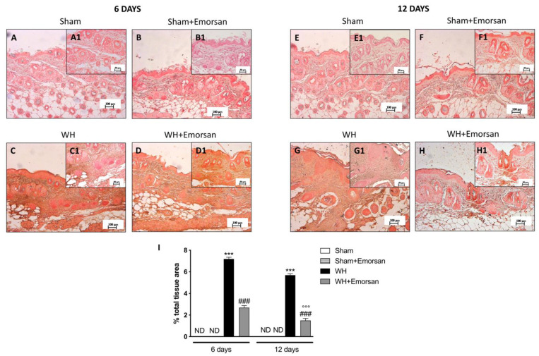
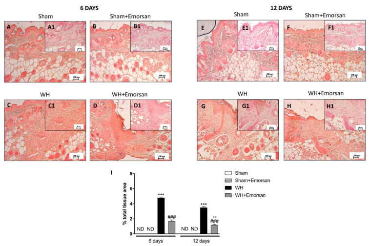
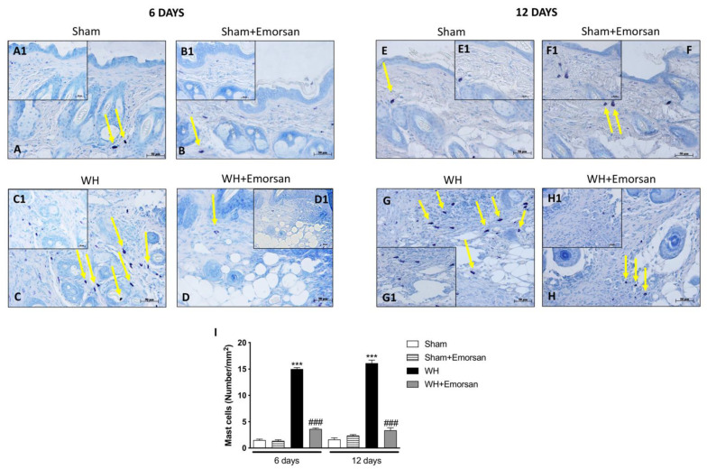
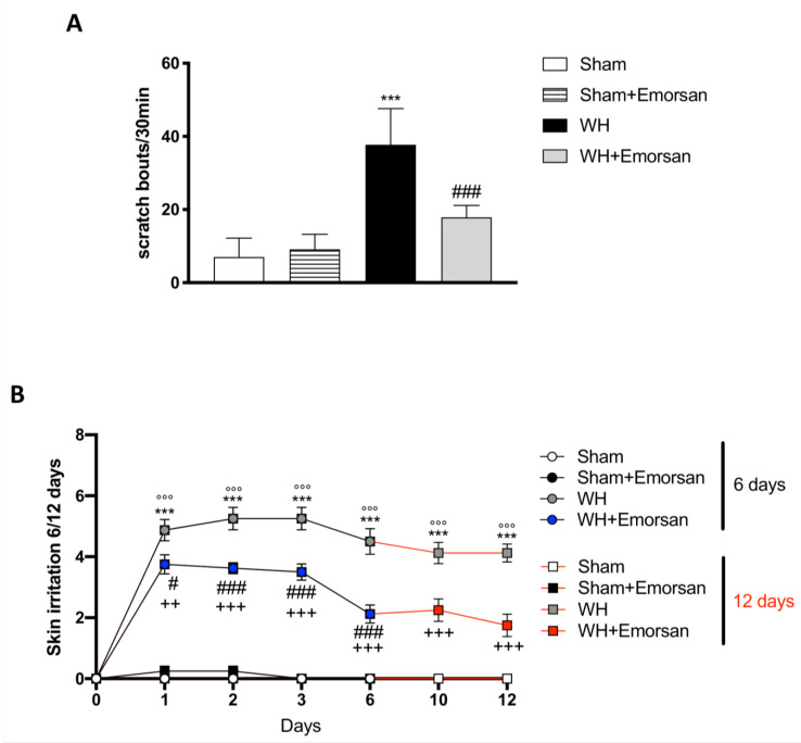
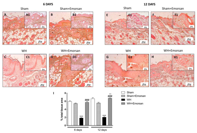
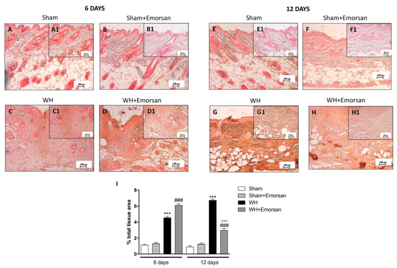
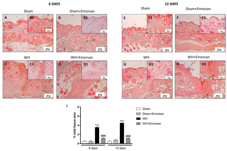
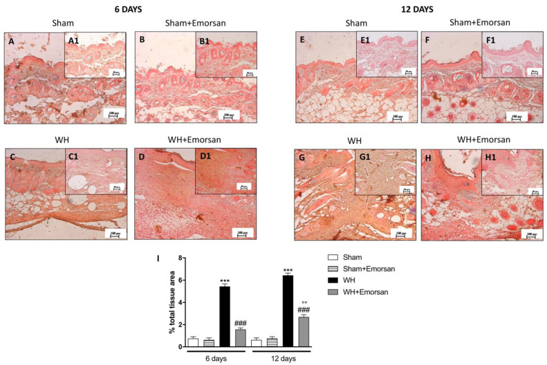
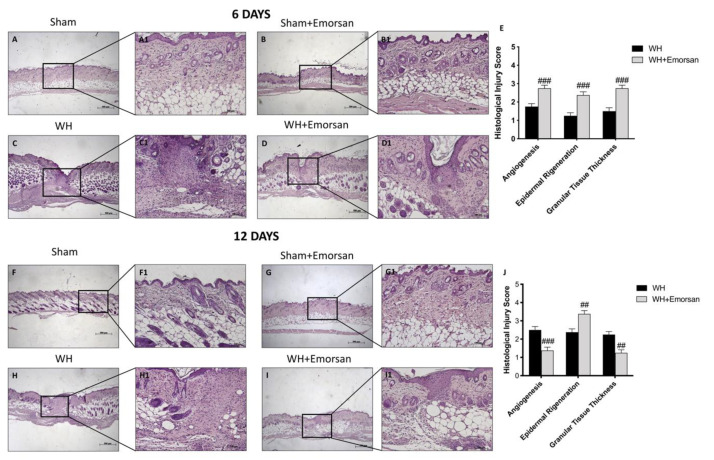
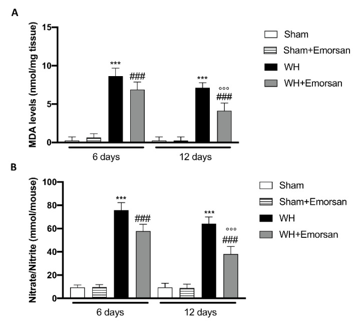
Inappropriate wound healing (WH) management can cause significant comorbidities, especially in patients affected by chronic and metabolic diseases, such as diabetes. WH involves several different, partially overlapping processes, including hemostasis, inflammation, cell proliferation, and remodeling. Oxidative stress in WH contributes to WH impairment because of the overexpression of radical oxygen species (ROS) and nitrogen species (RNS). This study aimed to evaluate the in vitro antioxidative action of a gel containing a Propionibacterium extract (Emorsan® Gel) and assess its skin re-epithelialization properties in a mouse model of WH. The scavenging effects of the bacterial extract were assessed in vitro through the ABTS and DPPH assays and in L-929 murine fibroblasts. The effects of the Emorsan® Gel were studied in vivo in a murine model of WH. After WH induction, mice were treated daily with vehicle or Emorsan® Gel for 6 or 12 days. According to the in vitro tests, the Propionibacterium extract exerted an inhibitory effect on ROS and RNS, consequently leading to the reduction in malondialdehyde (MDA) and nitrite levels. Before proceeding with the in vivo study, the Emorsan® Gel was verified to be unabsorbed. Therefore, the observed effects could be ascribed to a local action. The results obtained in vivo showed that through local reduction of oxidative stress and inflammation (IL-1β, TNF-α), the Emorsan® Gel significantly reduced the infiltration of mast cells into the injured wound, leading to the amelioration of symptoms such as itch and skin irritation. Therefore, the Emorsan® Gel improved the speed and percentage of wound area closure by improving the tissue remodeling process, prompting vascular-endothelial growth factor (VEGF) and transforming growth factor (TGF)- β production and reducing the expression of adhesion molecules. Emorsan® Gel, by its ability to inhibit free radicals, could reduce local inflammation and oxidative stress, thus enhancing the speed of wound healing.

Keywords: Emorsan® Gel; Propionibacterium extract; anti-inflammatory effect; antioxidant effect; bacterial lysate; skin care; topical treatment; wound healing.


Conflict of interest statement

The authors declare no conflict of interest.

Figures

Figure 1
Figure 1
Antioxidant effect of Propionibacterium extract in L-929 culture. Pre-treatment with Propionibacterium extract, in a concentration-dependent manner, considerably prevented H2O2 cytotoxicity (A) and reduced MDA (B) and NO2 (C) levels in the cell lysate, following H2O2 stimulation. Data are representative of at least three independent experiments. Values are provided as means ± SEM. One-way ANOVA test. *** p < 0.001 vs. CTR; # p < 0.05 vs. H2O2; ## p < 0.01 vs. H2O2; ### p < 0.001 vs. H2O2.
Figure 2
Figure 2
Effect of Emorsan® Gel treatment on TNF-α expression after WH. Epidermal tissues collected from the WH-vehicle group revealed positive immunostaining for TNF-α (C,C1,G,G1,I), compared to the sham groups (A,A1,B,B1,E,E1,F,F1,I). Emorsan® Gel topical treatment strongly reduced such positive immunostaining at both 6 days (D,D1,I) and 12 days (H,H1,I) after WH induction. Data are representative of at least three independent experiments. Values are means ± SEM. One-Way ANOVA test. *** p < 0.001 vs. corresponding Sham; ### p < 0.001 vs. corresponding WH; °°° p < 0.001 vs. WH+Emorsan 6 days; ND: not detectable.
Figure 3
Figure 3
Effect of Emorsan® Gel treatment on IL-1β expression after WH. Epidermal tissues collected from WH-vehicle injured mice showed positive IL-1β immunostaining (C,C1,G,G1,I), compared to the sham-treated mice 6 days and 12 days after WH induction (A,A1,B,B1,E,E1,F,F1,I). Emorsan® topical administration reduced IL-1β expression in a significant way (D,D1,H,H1,I). Data are representative of at least three independent experiments. Values are means ± SEM. One-Way ANOVA test. *** p < 0.001 vs. corresponding Sham; ### p < 0.001 vs. corresponding WH; °° p < 0.01 vs. WH+Emorsan 6 days; ND: not detectable.
Figure 4
Figure 4
Effect of Emorsan®Gel on mast cells in epidermal tissues after WH induction. A high degree of mast cells was found in mice with WH (C,C1,G,G1,I) compared to control animals (A,A1,B,B1,E,E1,F,F1,I). However, a decreased number of mast cells was identified in mice topically treated with Emorsan® at both 6 days (D,D1,I) and 12 days (H,H1,I). Data are representative of at least three independent experiments. Values are means ± SEM. One-Way ANOVA test. *** p < 0.001 vs. corresponding Sham; ### p < 0.001 vs. corresponding WH.
Figure 5
Figure 5
Effect of Emorsan® Gel administration on itch-induced scratching and skin irritation. An increase in scratch bouts was detected in WH mice compared to sham animals (A); conversely, Emorsan® Gel treatment was effective in reducing itch-induced scratching (A). Moreover, Emorsan® Gel administration decreased skin irritation until 6 days and 12 days after WH induction (B). Data are representative of at least three independent experiments. Values are means ± SEM. One-Way ANOVA test. *** p < 0.001 vs. Sham 6 days; °°° p < 0.001 vs. Sham 12 days; # p < 0.05 vs. WH 6 days; ### p < 0.001 vs. WH 6 days; ++ p < 0.01 vs. WH 12 days; +++ p < 0.001 vs. WH 12 days.
Figure 6
Figure 6
Effects of Emorsan®Gel on TGF-β levels in the skin after 6 and 12 days from WH induction. The epidermal tissues collected from WH-injured animals showed positive immunostaining for TGF-β (C,C1,G,G1,I), compared to the sham groups (A,A1,B,B1,E,E1,F,F1,I). Emorsan® Gel topical administration notably reduced TGF-β levels in the skin after 6 and 12 days from WH induction (D,D1,H,H1,I). Data are representative of at least three independent experiments. Values are means ± SEM. One-Way ANOVA test. *** p < 0.001 vs. corresponding Sham; ### p < 0.001 vs. corresponding WH; °° p < 0.01 vs. WH+Emorsan 6 days.
Figure 7
Figure 7
Effect of Emorsan® Gel treatment on VEGF expression after WH. Mice of the vehicle group showed a marked positive immunostaining for VEGF after 6 days of WH induction (C,C1,I) compared to sham mice both on day 6 and day 12 after WH induction (A,A1, B,B1,E,E1,F,F1, I). In the vehicle group, such positive immunostaining was even more intense 12 days after WH induction (G,G1,I). Contrarily, Emorsan®-treated mice revealed a modulation of VEGF levels, which were increased 6 days after WH induction (D,D1,I), and decreased 12 days after the start of the experiment (H,H1,I). Data are representative of at least three independent experiments. Values are means ± SEM. One-Way ANOVA test. *** p < 0.001 vs. corresponding Sham; ### p < 0.001 vs. corresponding WH; °°° p < 0.001 vs. WH+Emorsan 6 days.
Figure 8
Figure 8
Effect of Emorsan® Gel treatment on ICAM-1 expression after WH. Epidermal tissues obtained from WH-injured animals exhibited positive immunostaining for ICAM-1 (C,C1,G,G1,I), compared to the sham-treated animals (A,A1,B,B1,E,E1,F,F1,I). Emorsan® Gel administered topically reduced this expression in both times (D,D1,H,H1,I). Data are representative of at least three independent experiments. Values are means ± SEM. One-Way ANOVA test. *** p < 0.001 vs. corresponding Sham; ### p < 0.001 vs. corresponding WH.
Figure 9
Figure 9
Effect of Emorsan® Gel treatment on P-Selectin expression after WH. The intensity of P-selectin positive staining was markedly increased in the tissue section obtained from the WH group (C,C1,G,G1,I) compared to control animals (A,A1,B,B1,E,E1,F,F1,I). Mice topically treated with Emorsan® Gel expressed a diminished expression of P-selectin positive staining (D,D1,H,H1,I). Data are representative of at least three independent experiments. Values are means ± SEM. One-Way ANOVA test. *** p < 0.001 vs. corresponding Sham; ### p < 0.001 vs. corresponding WH; °° p < 0.01 vs. WH+Emorsan 6 days.
Figure 10
Figure 10
Effect of Emorsan® Gel treatment on histological parameters after WH induction. WH-injured epidermis showed tissue damage, edema, and neutrophilic infiltration at both 6 days (C,C1,score E) and 12 days (H,H1,score J) compared to the sham mice (A,A1,score E,F,F1,score J). compared to the sham animals (B,(B1) histological score G,(G1), score J) at both 6 and 12 days. Emorsan® Gel treatment significantly reduced tissue injury, ameliorating histolog-ical parameters (D,D1,score E,I,I1,score J). Data are representative of at least three independent experiments. Values are means ± SEM. One-Way ANOVA test. ## p < 0.01 vs. WH; ### p < 0.001 vs. WH.
Figure 11
Figure 11
Effect of Emorsan® Gel administration on oxidative stress markers after wound induction. Topical treatment with Emorsan® Gel diminished MDA levels both on day 6 and day 12 after wound induction (A). Likewise, nitrate/nitrite levels were significantly decreased after Emorsan® administration (B). Data are representative of at least three independent experiments. Values are means ± SEM. One-Way ANOVA test. *** p < 0.001 vs. corresponding Sham; ### p < 0.001 vs. corresponding WH; °°° p < 0.001 vs. WH+Emorsan 6 days.

References

    1. Wilkins R.G., Unverdorben M. Wound cleaning and wound healing: A concise review. Adv. Ski. Wound Care. 2013;26:160–163. doi: 10.1097/01.ASW.0000428861.26671.41. - DOI - PubMed
    1. Dunn L., Prosser H.C., Tan J.T., Vanags L.Z., Ng M.K., Bursill C.A. Murine model of wound healing. J. Vis. Exp. 2013;75:e50265. doi: 10.3791/50265. - DOI - PMC - PubMed
    1. Zarchi K., Martinussen T., Jemec G.B. Wound healing and all-cause mortality in 958 wound patients treated in home care. Wound Repair Regen. 2015;23:753–758. doi: 10.1111/wrr.12335. - DOI - PubMed
    1. McLafferty E., Hendry C., Farley A. The integumentary system: Anatomy, physiology and function of skin. Nurs. Stand. 2012;27:35. doi: 10.7748/ns2012.09.27.3.35.c9299. - DOI - PubMed
    1. Bootun R. Effects of immunosuppressive therapy on wound healing. Int. Wound J. 2013;10:98–104. doi: 10.1111/j.1742-481X.2012.00950.x. - DOI - PMC - PubMed