. 2022 Apr 24;23(9):4710.
doi: 10.3390/ijms23094710.

Mechanical Load and Piezo1 Channel Regulated Myosin II Activity in Mouse Lenses

Affiliations

Mechanical Load and Piezo1 Channel Regulated Myosin II Activity in Mouse Lenses

Ariana Allen et al. Int J Mol Sci. .

Abstract

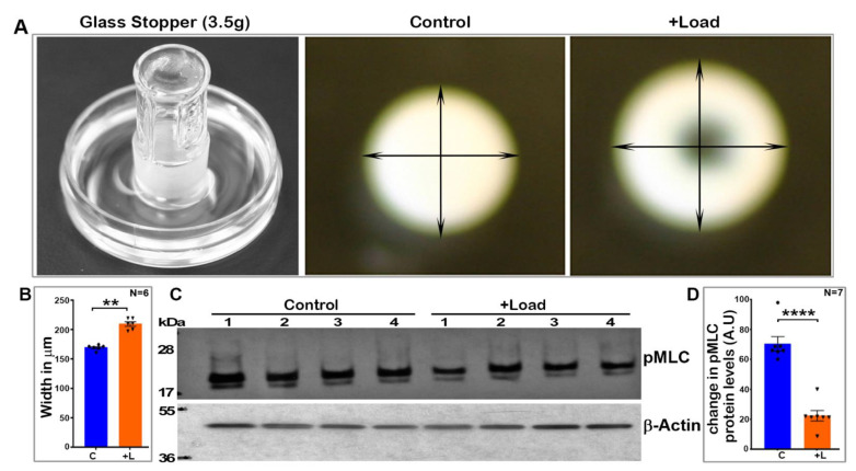
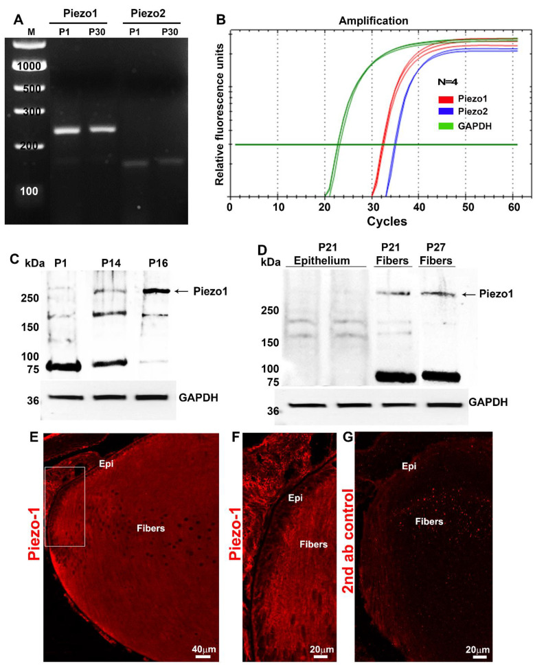
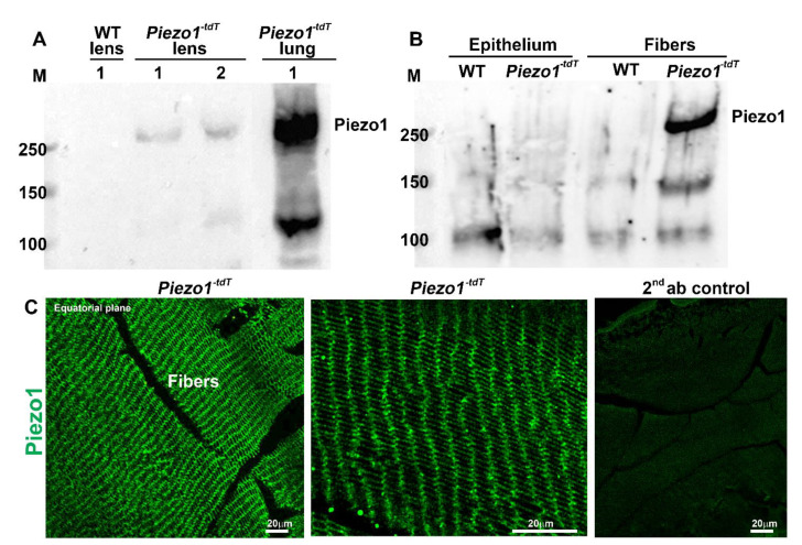
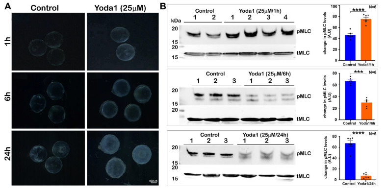
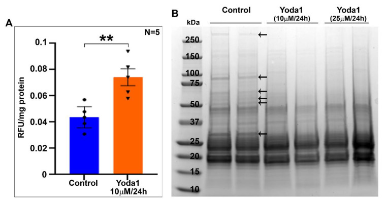
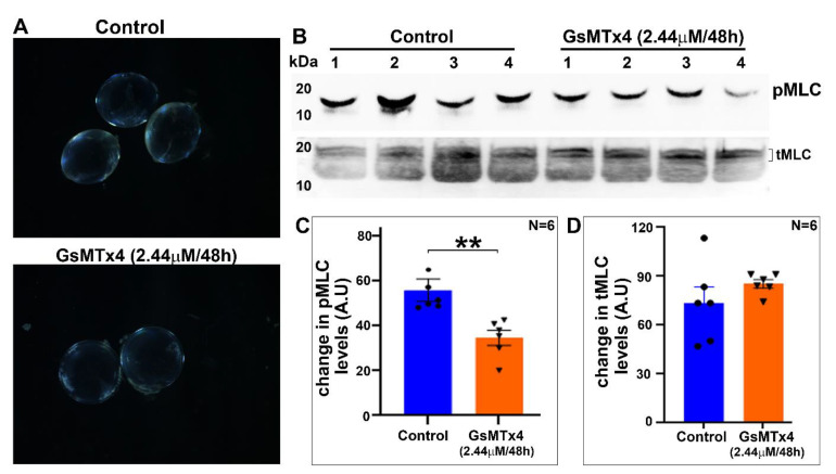
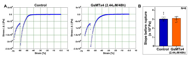
The cytoarchitecture and tensile characteristics of ocular lenses play a crucial role in maintaining their transparency and deformability, respectively, which are properties required for the light focusing function of ocular lens. Calcium-dependent myosin-II-regulated contractile characteristics and mechanosensitive ion channel activities are presumed to influence lens shape change and clarity. Here, we investigated the effects of load-induced force and the activity of Piezo channels on mouse lens myosin II activity. Expression of the Piezo1 channel was evident in the mouse lens based on immunoblot and immufluorescence analyses and with the use of a Piezo1-tdT transgenic mouse model. Under ex vivo conditions, change in lens shape induced by the load decreased myosin light chain (MLC) phosphorylation. While the activation of Piezo1 by Yoda1 for one hour led to an increase in the levels of phosphorylated MLC, Yoda1 treatment for an extended period led to opacification in association with increased calpain activity and degradation of membrane proteins in ex vivo mouse lenses. In contrast, inhibition of Piezo1 by GsMTx4 decreased MLC phosphorylation but did not affect the lens tensile properties. This exploratory study reveals a role for the mechanical load and Piezo1 channel activity in the regulation of myosin II activity in lens, which could be relevant to lens shape change during accommodation.

Keywords: calpain; cataract; lens; mechanotransduction; myosin II; piezo channel; stiffness.


Conflict of interest statement

The authors declare no conflict of interest.

Figures

Figure 1
Figure 1
Mechanical-loading-induced lens deformation decreases intracellular myosin II activity. (A). To evaluate whether lens shape change induced by external mechanical loading influences myosin II activity, a glass stopper weighing ~3.5 g was placed on freshly enucleated lenses (derived from 4-week-old mice) for 60 s before fixation of the lenses with TCA as described in the Methods section. As shown in panel A (representative images), lenses subjected to mechanical loading exhibited an obvious deformation with a significant increase (by ~24%, n = 6) in width but no rupture, compared to control lenses (B,C). Analysis of phosphorylated MLC by immunoblotting revealed a significant decrease (>65%, n = 7) in the levels of phospho-MLC in the load-induced deformed lenses (lanes 1 to 4) relative to their respective controls (lanes 1 to 4) (D). Phospho-MLC levels were normalized to β-actin. The levels of β-actin were found to be the same between the deformed and control lenses (C). C: Control; L: Load-applied lenses. Values presented as Mean ± SEM. ** p < 0.01; **** p < 0.0001. A.U.: Arbitrary Units.
Figure 2
Figure 2
Load-induced lens deformation using glass coverslips decreases myosin II activity. For this, either 5 (total weight = 1.07 g) or 10 (total weight = 2.14 g) glass coverslips were stacked on 2 different sets of mouse lenses (from 4-week-old mice) for 60 s. Following lens compression, half of the total lenses from each set were immediately treated with TCA, while the other half were treated with TCA sixty seconds after the glass coverslips were removed, to determine the levels of phospho-MLC and total MLC. The lens-compression-induced use of glass coverslips for 60 s (A,B, 5 coverslips; C,D, 10 coverslips) was associated with an obvious decrease (ranging from 35–45%) in the levels of phospho-MLC compared to the control lenses (n = 5). There was a slight but significant increase (~25%) in the levels of phospho-MLC upon removal of the glass coverslips during the 60 s resilience period in the lens group loaded with 5 coverslips (B). Overall, however, there was still a marked decrease in the levels of phospho-MLC (by >20 and 60%, for 5- and 10-coverslip-loaded lenses, respectively) in compressed lenses allowed to ‘recover’ for 60 s after the removal of glass coverslips relative to the control lenses. Total MLC levels were not different between the compressed and uncompressed (control) lenses (A,C). Cnt: Control; L: Lenses loaded with glass coverslips for 60 s; L + R: Lenses loaded with coverslips for 60 s and allowed to ‘recover’ for 60 s prior to analysis. Values are presented as Mean ± SEM. * p < 0.5; ** p < 0.01; *** p < 0.001; ****p < 0.0001. A.U.: Arbitrary Unites.
Figure 3
Figure 3
Expression and distribution of Piezo channels in mouse lenses. (A). RT-PCR-based confirmation of Piezo1 and Piezo2 expression in P1 and P30 mouse lenses. (B). qRT-PCR analysis revealed a relatively much higher level of Piezo1 expression in the lens (P30) compared to Piezo2. (C). Total lysates (800× g supernatants; 75 µg protein) derived from the P1, P14, and P16 mouse lenses analyzed using a Piezo1 polyclonal antibody exhibited immunopositive bands with an expected molecular mass of >250 kDa and >150 kDa. There was also a prominent immunopositive band at >75 kDa in the P1 and P14 lenses, the levels of which appeared to be decreased in the P16 lenses. (D). Piezo1 immunopositive bands of >250 and >75 kDa were present predominantly in the lens fiber samples (P21 and P27) compared to the lens epithelium (P21). (E,F). Immunofluorescence analysis of Piezo1 in the P1 mouse lens (the sagittal plane of the cryosection) revealed that the protein distributes predominantly to lens fibers relative to the epithelium (boxed area in panel (E) was magnified and shown in panel (F)). (G) Shows background immunofluorescence with secondary antibody alone. GAPDH: Loading control; Epi: Epithelium; Bars: Image magnification.
Figure 4
Figure 4
The piezo1-tdT mouse confirms the expression and distribution of Piezo1 in lens fibers. (A). The Piezo1-tdT mouse model expressing a fusion protein of Piezo1 and tdTomato (Piezo1-tdT) was used to determine the distribution pattern of Piezo1 in the mouse lens. Similar to what was found in the wild-type lenses (Figure 3), Piezo1-tdT exhibiting the expected molecular mass of >250 kDa was detected only in P30 lens homogenates derived from the Piezo1-tdT mice, but not in the wild-type lens. The positive control (lung tissue lysate from the Piezo1-tdT mice) also showed a robust expression of Piezo1-tdT. Lanes 1 and 2 represent two different loads of the total protein (75 and 150 µg, respectively). (B) The Piezo1-tdT fusion protein was detected predominantly in fiber cell lysates compared to lens epithelial lysates. (C). Immunofluorescence analysis revealed the Piezo1-tdT fusion protein distributing to lens fibers with localization to both the short and long arms of the hexagonal lens fibers (Left and middle panels are with low and high magnification, respectively). The right panel shows a second antibody (Alexa Flour 488) background control fluorescence staining in the Piezo1-tdT mouse lens section. Bars: Image magnification.
Figure 5
Figure 5
Piezo1 activation in ex vivo lenses by Yoda1: Effects on lens clarity and myosin II activity. (A). To determine the effects of Piezo1 activation on lens transparency, lenses derived from P30 mice and maintained under culture conditions were treated with 25 µM Yoda1, an agonist of Piezo1, and changes in lens transparency were monitored for 24 h. Yoda1-treated lenses exhibited a slight haziness starting at the 1 h interval that increased progressively with time, with lenses exhibiting significant increases in swelling and a slight nuclear opacity after 24 h compared to the control lenses. (B). In lenses treated with Yoda1 (25 µM) for 1 h, the levels of phospho-MLC were significantly elevated compared to the untreated control lenses. On the other hand, in the 6 and 24 h Yoda1-treated lenses, there was a significant and progressive decrease in the levels of phospho-MLC (pMLC) compared to the control lenses. The levels of total MLC (tMLC), however, were found to be similar between the Yoda1-treated and control lenses throughout the course of drug treatment. Lanes 1 to 3 represent 3 experimental replicates. Bars denote image magnification. *** p < 0.001; **** p < 0.0001. A.U.: Arbitrary Units.
Figure 6
Figure 6
Piezo channel agonist Yoda1 treatment induces calpain activity and membrane protein proteolysis in lenses. (A). Calpain activity, which is calcium-dependent, was evaluated in mouse lenses treated (ex vivo) with 10 µM Yoda1 for 24 h. A significant increase (by ~69%, n = 5) in calpain activity was detected in the treated lenses compared to the control lenses. Values are represented as Mean ± SEM. ** p < 0.01. RFU: Relative fluorescence units. (B). Having found increased calpain activity and haziness in Yoda1-treated lenses, we then examined the integrity of lens membrane protein fractions derived from the Yoda1-treated (10 and 25 µM for 24 h) and control lenses by SDS-PAGE analysis. As shown in the figure, the Coomassie-blue-stained SDS-PAGE profile of the membrane-enriched fraction from Yoda1-treated lenses showed a markedly reduced staining of several protein bands (indicated with arrows) compared to the control lenses, indicating increased proteolysis, perhaps due to increased calcium-dependent calpain activity associated with Piezo1 activation (A). Data are shown for two representative lenses per group.
Figure 7
Figure 7
Decreased myosin II activity in Piezo1-inhibitor-treated lenses. (A) Lenses (from P30 mice) treated ex vivo for 48 h with 2.44 µM GsMTx4, a specific inhibitor of Piezo1, remained largely transparent, with no significant change in wet weight (4.33 ± 0.156 mg/lens, n = 5) compared to the control (4.14 ± 0.036 mg/lens, n = 5) lenses. (B,C) Relative to the control lenses, the GsMTx4-treated (48 h) lenses showed a significant (by ~38%) decrease in their levels of phospho-MLC. (D) The levels of total MLC in GsMTx4-treated lenses were not different relative to the control lenses (based on densitometric evaluation of the top doublet bands of tMLC shown in panel B). Values represent Mean ± SEM. ** p < 0.01. A.U.: Arbitrary Units.
Figure 8
Figure 8
The mouse lens maintains normal tensile properties under Piezo1 channel inhibition. (A) A representative microstrain analyzer generated stress/strain tracings of mouse lenses from the control and GsMTx4-treated groups. (B) Changes in Young’s modulus calculated from the lens prerupture stress/strain slopes were not different between the GsMTx4-treated and control lenses. Values are denoted as Mean ± SEM.

References

    1. Liu Y.C., Wilkins M., Kim T., Malyugin B., Mehta J.S. Cataracts. Lancet. 2017;390:600–612. doi: 10.1016/S0140-6736(17)30544-5. - DOI - PubMed
    1. Fricke T.R., Tahhan N., Resnikoff S., Papas E., Burnett A., Ho S.M., Naduvilath T., Naidoo K.S. Global Prevalence of Presbyopia and Vision Impairment from Uncorrected Presbyopia: Systematic Review, Meta-analysis, and Modelling. Ophthalmology. 2018;125:1492–1499. doi: 10.1016/j.ophtha.2018.04.013. - DOI - PubMed
    1. Fisher R.F. The force of contraction of the human ciliary muscle during accommodation. J. Physiol. 1977;270:51–74. doi: 10.1113/jphysiol.1977.sp011938. - DOI - PMC - PubMed
    1. Knaus K.R., Hipsley A., Blemker S.S. The action of ciliary muscle contraction on accommodation of the lens explored with a 3D model. Biomech. Model. Mechanobiol. 2021;20:879–894. doi: 10.1007/s10237-021-01417-9. - DOI - PubMed
    1. Nakazawa Y., Petrova R.S., Sugiyama Y., Nagai N., Tamura H., Donaldson P.J. Regulation of the Membrane Trafficking of the Mechanosensitive Ion Channels TRPV1 and TRPV4 by Zonular Tension, Osmotic Stress and Activators in the Mouse Lens. Int. J. Mol. Sci. 2021;22:12658. doi: 10.3390/ijms222312658. - DOI - PMC - PubMed

LinkOut - more resources