. 2022 Apr 27;23(9):4813.
doi: 10.3390/ijms23094813.

Nanoencapsulation of Gla-Rich Protein (GRP) as a Novel Approach to Target Inflammation

Affiliations

Nanoencapsulation of Gla-Rich Protein (GRP) as a Novel Approach to Target Inflammation

Carla S B Viegas et al. Int J Mol Sci. .

Abstract

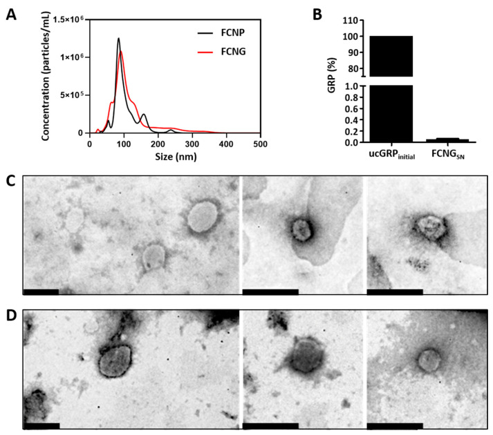
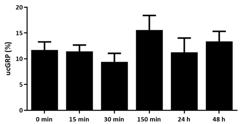
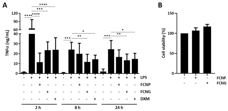
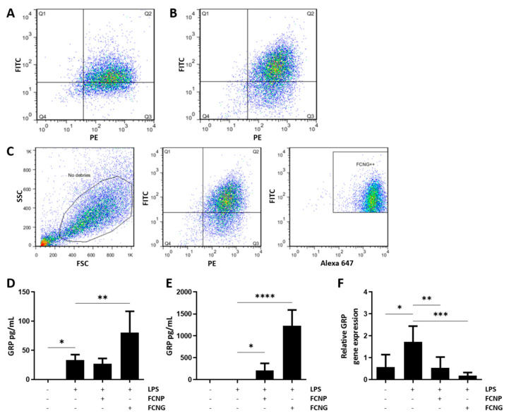
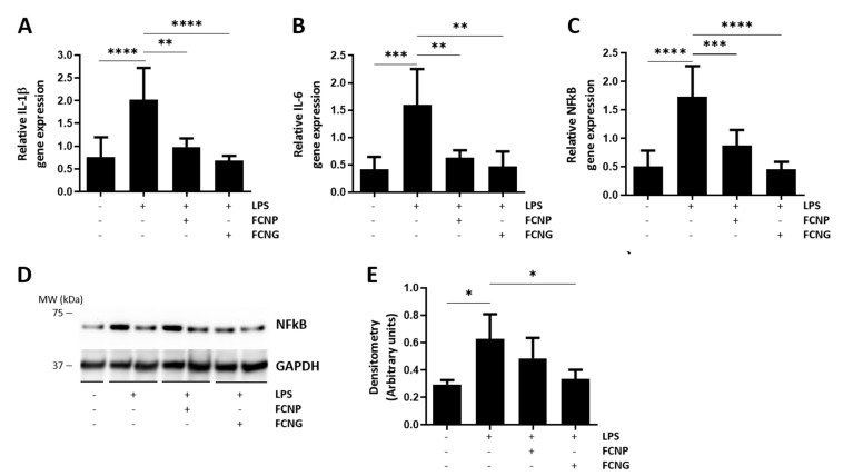
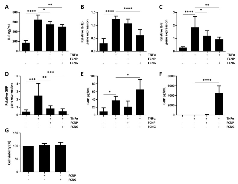
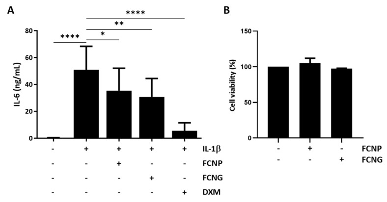
Chronic inflammation is a major driver of chronic inflammatory diseases (CIDs), with a tremendous impact worldwide. Besides its function as a pathological calcification inhibitor, vitamin K-dependent protein Gla-rich protein (GRP) was shown to act as an anti-inflammatory agent independently of its gamma-carboxylation status. Although GRP's therapeutic potential has been highlighted, its low solubility at physiological pH still constitutes a major challenge for its biomedical application. In this work, we produced fluorescein-labeled chitosan-tripolyphosphate nanoparticles containing non-carboxylated GRP (ucGRP) (FCNG) via ionotropic gelation, increasing its bioavailability, stability, and anti-inflammatory potential. The results indicate the nanosized nature of FCNG with PDI and a zeta potential suitable for biomedical applications. FCNG's anti-inflammatory activity was studied in macrophage-differentiated THP1 cells, and in primary vascular smooth muscle cells and chondrocytes, inflamed with LPS, TNFα and IL-1β, respectively. In all these in vitro human cell systems, FCNG treatments resulted in increased intra and extracellular GRP levels, and decreased pro-inflammatory responses of target cells, by decreasing pro-inflammatory cytokines and inflammation mediators. These results suggest the retained anti-inflammatory bioactivity of ucGRP in FCNG, strengthening the potential use of ucGRP as an anti-inflammatory agent with a wide spectrum of application, and opening up perspectives for its therapeutic application in CIDs.

Keywords: Gla-rich protein (GRP); chronic inflammatory diseases (CIDs); inflammation; nanoparticles; vitamin K-dependent protein (VKDP).


Conflict of interest statement

Dina C. Simes and Carla Viegas are cofounders of GenoGla Diagnostics. The authors declare that there is no conflict of interests regarding the publication of this paper. The tools and methods described in this manuscript are included in a PCT patent application PCT/PT2009000046, which is owned by University of Algarve and the Centre of Marine Sciences (CCMAR), and the exclusive rights are licensed to GenoGla Diagnostics. The funders had no role in the design of the study; in the collection, analyses, or interpretation of data; in the writing of the manuscript, or in the decision to publish the results.

Figures

Figure 1
Figure 1
FCNP and FCNG size distribution, ucGRP incorporation and morphology. (A) Representative analysis of FCNP and FCNG by nanotracking analysis (NTA), showing the concentration of particles as a function of size. (B) Percentage of ucGRP initially used for FCNG synthesis (ucGRPinitial), and in the supernatants after separation of pelleted nanoparticles (FCNGSN), representing non-incorporated ucGRP, determined by ELISA. Data are representative of six independent experiments. (C,D) Ultrastructural characterization of FCNP (C) and FCNG (D) by transmission electron microscopy (TEM). Scale bar of 200 nm.
Figure 2
Figure 2
Levels of ucGRP released from FCNG during a 48 h study. ucGRP in cell culture media (RPMI) was measured by ELISA, at different time points from 0 min to 48 h. The data are representative of three independent experiments, and the differences are statistically non-significant.
Figure 3
Figure 3
Anti-inflammatory effect and toxicity of FCNP and FCNG in LPS-stimulated THP-1 macrophages (THP-1-MoM). (A) The evaluation of the inflammatory marker TNFα was performed by ELISA in the cell culture media of THP-1 MoM treated for 2 h, 8 h or 24 h with (11.7 ± 4.5) × 109 particles/mL of FCNP or FCNG, and then stimulated with LPS (100 ng/mL) for a further 24 h. Dexametasone (DXM) (2 μM) was used as a positive anti-inflammatory control and non-stimulated cells were used as controls for LPS stimulation. (B) Viability of THP-1-MoM exposed to (11.7 ± 4.5) × 109 particles/mL of FCNP or FCNG for 48 h. Data are representative of three independent experiments, and presented as mean ± SD. Two-way ANOVA and multiple comparisons were achieved with the Dunnett’s test and are presented relative to the LPS-stimulated cells. Statistical significance was defined as p ≤ 0.05 (*), p ≤ 0.01 (**), p ≤ 0.001 (***) and p ≤ 0.0001 (****).
Figure 4
Figure 4
Binding/uptake of FCNP and FCNG by THP1-MoM cells. (AC) Flow cytometry analysis of FCNP and FCNG in THP1-MoM cells. (A,B) Dot plots of THP-1 MoM cells exposed to (11.7 ± 4.5) × 109 particles/mL of FCNP (A) and FCNG (B) for 2 h. The Q2 quadrant represents cells that have, simultaneously, fluorescein-labeled nanoparticles and the THP-1 MoM marker labeled with PE (double positive for FITC and PE). Q2 is 67.1% for FCNP (A) and 73.7% for FCNG (B). (C) Gating strategy used for the analysis of FCNG in THP1-MoM cells. The first plot shows the debris exclusion in the side scatter (SSC) vs. the forward scatter (FSC). In the second plot, the double positive population for fluorescein-labeled nanoparticles and the THP-1 MoM marker was gated (73.7% are double positive for FITC and PE). Finally, the last plot shows the selection of the population of interest, FCNG++, which represents THP-1 MoM cells with fluorescein-labeled nanoparticles containing GRP labeled with Alexa 647 (73.3% are triple positive for FITC, PE and Alexa 647). (D,E) Quantification of GRP present in THP1-MoM cell protein extracts (D) and in the cell culture media (E) by ELISA, after pre-treatments with (11.7 ± 4.5) × 109 particles/mL of FCNP and FCNG for 24 h, followed by stimulation with LPS (100 ng/mL) for an additional 24 h. Non-stimulated cells were used as controls for LPS stimulation. (F) Relative GRP gene expression determined by quantitative polymerase chain reaction (qPCR) of experiments described in (D,E). Data in (DF) are representative of three independent experiments and presented as mean ± SD. Two-way ANOVA and multiple comparisons were achieved with the Dunnett’s test, and presented relative to the LPS-stimulated cells. Statistical significance was defined as p ≤ 0.05 (*), p ≤ 0.01 (**), p ≤ 0.001 (***) and p ≤ 0.0001 (****).
Figure 5
Figure 5
The anti-inflammatory activity of FCNG in THP1-MoM cells is mediated by downregulation of pro-inflammatory cytokines. (AC) Gene expression analysis of IL-1β (A), IL-6 (B) and NFkB (C) by qPCR of THP1-MoM cells pre-treated with (11.7 ± 4.5) × 109 particles/mL of FCNP and FCNG for 24 h, followed by stimulation with LPS (100 ng/mL) for an additional 24 h. Non-stimulated cells were used as controls for LPS stimulation. (D,E) Total protein extracts of THP1-MoM cells treated as described in (AC) were analyzed by Western blot to detect NFkB. The positions of relevant molecular mass markers (kDa) are indicated on the right side and GAPDH was used as the loading control. (E) Quantification of NFkB levels was performed by densitometry using ImageJ software, and is presented relatively to the GAPDH loading control as arbitrary units. Data are representative of three independent experiments and presented as mean ± SD. Two-way ANOVA and multiple comparisons were performed with the Dunnett’s test, and are presented relative to the LPS-stimulated cells. Statistical significance was defined as p ≤ 0.05 (*), p ≤ 0.01 (**), p ≤ 0.001 (***) and p ≤ 0.0001 (****).
Figure 6
Figure 6
Anti-inflammatory activity of FCNG in primary human VSMCs. VSMCs were pre-treated with (11.7 ± 4.5) E + 9 particles/mL of FCNP and FCNG for 24 h, followed by stimulation with TNFα (20 ng/mL) for an additional 24 h, then analyzed for levels of IL-6 present in the cell culture media by ELISA (A) and levels of gene expression of IL-1β (B), IL-8 (C) and GRP (D) by qPCR. (E,F) Quantification of GRP present in VSMCs protein extracts (E) and in the cell culture media (F) by ELISA. Non-stimulated cells were used as controls for TNFα stimulation. (G) Viability of VSMCs exposed to (11.7 ± 4.5) × 109 particles/mL of FCNP or FCNG for 48 h. Data are representative of three independent experiments and presented as mean ± SD. Two-way ANOVA and multiple comparisons were performed with the Dunnett’s test, and are presented relative to the TNFα-stimulated cells. Statistical significance was defined as p ≤ 0.05 (*), p ≤ 0.01 (**), p ≤ 0.001 (***) and p ≤ 0.0001 (****).
Figure 7
Figure 7
Anti-inflammatory activity of FCNG in primary human articular chondrocytes. (A) Chondrocytes were pre-treated with (11.7 ± 4.5) × 109 particles/mL of FCNP and FCNG for 24 h, or dexametasone (DXM, 2 µM), followed by stimulation with IL-1β (10 ng/mL) for an additional 24 h, and analyzed for levels of IL-6 present in the cell culture media by ELISA. Non-stimulated cells were used as controls of IL-1β stimulation. (B) Viability of articular chondrocytes exposed to (11.7 ± 4.5) × 109 particles/mL of FCNP or FCNG for 48 h. Data are representative of three independent experiments and are presented as mean ± SD. Two-way ANOVA and multiple comparisons were performed with the Dunnett’s test, and are presented relative to the IL-1β-stimulated cells. Statistical significance was defined as p ≤ 0.05 (*), p ≤ 0.01 (**), and p ≤ 0.0001 (****).

References

    1. Pahwa R., Goyal A., Bansal P., Jialal I. Chronic Inflammation. StatPearls Publishing; Treasure Island, FL, USA: 2021.
    1. Furman D., Campisi J., Verdin E., Carrera-Bastos P., Targ S., Franceschi C., Ferrucci L., Gilroy D.W., Fasano A., Miller G.W., et al. Chronic inflammation in the etiology of disease across the life span. Nat. Med. 2019;25:1822–1832. doi: 10.1038/s41591-019-0675-0. - DOI - PMC - PubMed
    1. Chen L., Deng H., Cui H., Fang J., Zuo Z., Deng J., Li Y., Wang X., Zhao L. Inflammatory responses and inflammation-associated diseases in organs. Oncotarget. 2018;9:7204–7218. doi: 10.18632/oncotarget.23208. - DOI - PMC - PubMed
    1. Wongrakpanich S., Wongrakpanich A., Melhado K., Rangaswami J.A. Comprehensive Review of Non-Steroidal Anti-Inflammatory Drug Use in The Elderly. Aging Dis. 2018;9:143–150. doi: 10.14336/AD.2017.0306. - DOI - PMC - PubMed
    1. Viegas C.S.B., Costa R.M., Santos L., Videira P.A., Silva Z., Araújo N., Macedo A.L., Matos A.P., Vermeer C., Simes D.C. Gla-rich protein function as an anti-inflammatory agent in monocytes/macrophages: Implications for calcification-related chronic inflammatory diseases. PLoS ONE. 2017;12:e0177829. doi: 10.1371/journal.pone.0177829. - DOI - PMC - PubMed

LinkOut - more resources