. 2022 Apr 27;23(9):4833.
doi: 10.3390/ijms23094833.

Single-Cell RNA Sequencing Analysis for Oncogenic Mechanisms Underlying Oral Squamous Cell Carcinoma Carcinogenesis with Candida albicans Infection

Affiliations

Single-Cell RNA Sequencing Analysis for Oncogenic Mechanisms Underlying Oral Squamous Cell Carcinoma Carcinogenesis with Candida albicans Infection

Yi-Ping Hsieh et al. Int J Mol Sci. .

Abstract

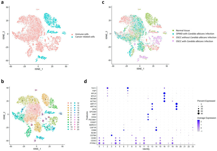
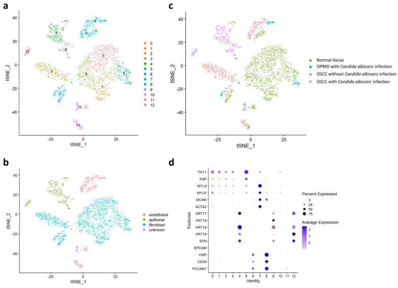
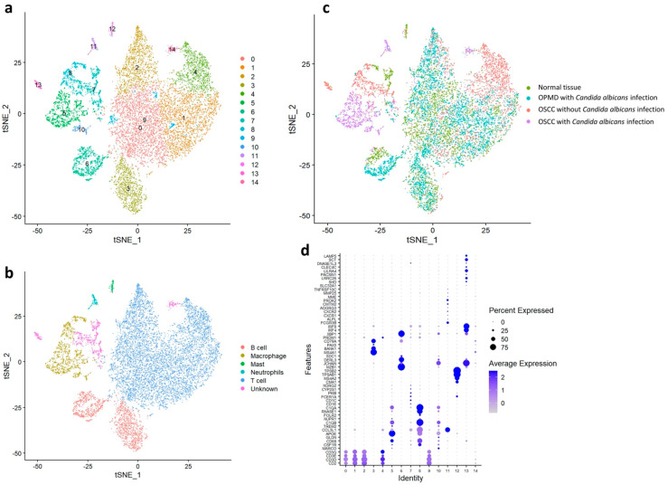
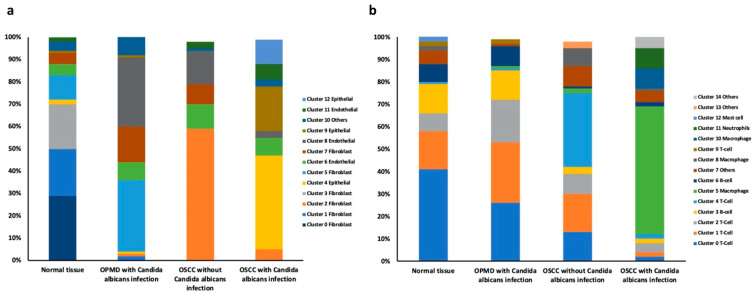
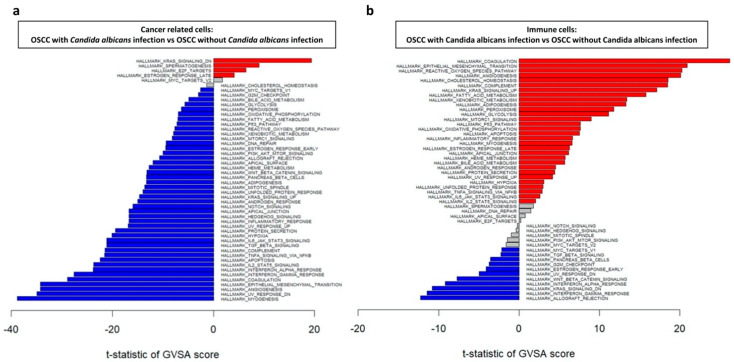
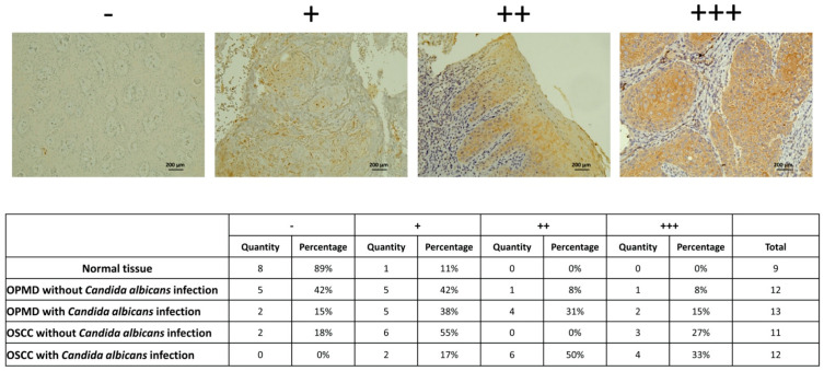
Oral squamous cell carcinoma (OSCC) carcinogenesis involves heterogeneous tumor cells, and the tumor microenvironment (TME) is highly complex with many different cell types. Cancer cell-TME interactions are crucial in OSCC progression. Candida albicans (C. albicans)-frequently pre-sent in the oral potentially malignant disorder (OPMD) lesions and OSCC tissues-promotes malignant transformation. The aim of the study is to verify the mechanisms underlying OSCC car-cinogenesis with C. albicans infection and identify the biomarker for the early detection of OSCC and as the treatment target. The single-cell RNA sequencing analysis (scRNA-seq) was performed to explore the cell subtypes in normal oral mucosa, OPMD, and OSCC tissues. The cell composi-tion changes and oncogenic mechanisms underlying OSCC carcinogenesis with C. albicans infec-tion were investigated. Gene Set Variation Analysis (GSVA) was used to survey the mechanisms underlying OSCC carcinogenesis with and without C. albicans infection. The results revealed spe-cific cell clusters contributing to OSCC carcinogenesis with and without C. albicans infection. The major mechanisms involved in OSCC carcinogenesis without C. albicans infection are the IL2/STAT5, TNFα/NFκB, and TGFβ signaling pathways, whereas those involved in OSCC carcinogenesis with C. albicans infection are the KRAS signaling pathway and E2F target down-stream genes. Finally, stratifin (SFN) was validated to be a specific biomarker of OSCC with C. albicans infection. Thus, the detailed mechanism underlying OSCC carcinogenesis with C. albicans infection was determined and identified the treatment biomarker with potential precision medicine applications.

Keywords: Candida albicans; oral potentially malignant disorder; oral squamous cell carcinoma; single-cell RNA sequencing analysis; tumor heterogeneity; tumor microenvironment.


Conflict of interest statement

The authors declare no conflict of interest.

Figures

Figure 1
Figure 1
Visual distribution of dimensionality reduction in all cell populations. (a) Cell distribution of all samples generated through t-distributed stochastic neighbor embedding. The cells are divided into cancer-related cells and immune cells. (b) Classification of cells into 24 subtypes by using the Louvain algorithm. (c) Separation of the four major groups: normal tissue, OPMD lesion with Candida albicans infection, OSCC tissue with C. albicans infection, and OSCC tissue without C. albicans infection. (d) Classification of cell types in each cluster by using specific biomarkers.
Figure 2
Figure 2
Cancer-related cell distribution in the normal tissue, OPMD lesion with Candida albicans infection, OSCC tissue with C. albicans infection, and OSCC tissue without C. albicans infection. (a) Classification of cancer−related cells into 13 subtypes by using the Louvain algorithm. (b) Classification of cancer-related cells into endothelial, epithelial, fibroblast, and unknown cells. (c) Separation of the four major groups. (d) Classification of cell types in each cluster by using specific biomarkers.
Figure 3
Figure 3
Immune cell distribution in the normal tissue, OPMD lesion with Candida albicans infection, OSCC tissue with C. albicans infection, and OSCC tissue without C. albicans infection. (a) Classification of immune cells into 15 subtypes by using the Louvain algorithm. (b) Classification of immune cells into B cells, macrophages, mast cells, neutrophils, T cells, and unknown cells. (c) Separation of the four major groups. (d) Classification of cell types in each cluster by using specific biomarkers.
Figure 4
Figure 4
Comparison of the compositions of cell subtypes within the normal tissue, OPMD lesion with Candida albicans infection, OSCC tissue with C. albicans infection, and OSCC tissue without C. albicans infection. (a) Cancer-related cell ratios and (b) Immune cell ratios of these clusters within the four groups.
Figure 5
Figure 5
GSVA of regulatory pathways in (a) cancer-related cells and (b) immune cells in OSCC tissue with and without Candida albicans infection. Red and blue bars indicate that gene expression was significantly increased in OSCC tissue with C. albicans infection compared with that without C. albicans infection and OSCC tissue without C. albicans infection compared with that with C. albicans infection, respectively (both p < 0.05).
Figure 6
Figure 6
SFN significantly increased in OPMD lesions and OSCC tissues with Candida albicans infection. IHC staining data shows increased SFN expression in tissues with C. albicans infection, especially OSCC tissue. SFN staining intensity was interpreted by two researchers. −means negative stain; + means weak stain; ++ means moderate stain; +++ means strong stain.

References

    1. Sagaert X., Vanstapel A., Verbeek S. Tumor Heterogeneity in Colorectal Cancer: What Do We Know So Far? Pathobiology. 2018;85:72–84. doi: 10.1159/000486721. - DOI - PubMed
    1. Marusyk A., Polyak K. Tumor heterogeneity: Causes and consequences. Biochim. Biophys. Acta. 2010;1805:105–117. doi: 10.1016/j.bbcan.2009.11.002. - DOI - PMC - PubMed
    1. El-Sayes N., Vito A., Mossman K. Tumor Heterogeneity: A Great Barrier in the Age of Cancer Immunotherapy. Cancers. 2021;13:806. doi: 10.3390/cancers13040806. - DOI - PMC - PubMed
    1. Dagogo-Jack I., Shaw A.T. Tumour heterogeneity and resistance to cancer therapies. Nat. Rev. Clin. Oncol. 2018;15:81–94. doi: 10.1038/nrclinonc.2017.166. - DOI - PubMed
    1. Anderson N.M., Simon M.C. The tumor microenvironment. Curr. Biol. 2020;30:R921–R925. doi: 10.1016/j.cub.2020.06.081. - DOI - PMC - PubMed