Review
. 2022 Apr 27;23(9):4855.
doi: 10.3390/ijms23094855.

Exosomal miRNAs as a Promising Source of Biomarkers in Colorectal Cancer Progression

Affiliations
Review

Exosomal miRNAs as a Promising Source of Biomarkers in Colorectal Cancer Progression

Tahani Bakhsh et al. Int J Mol Sci. .

Abstract

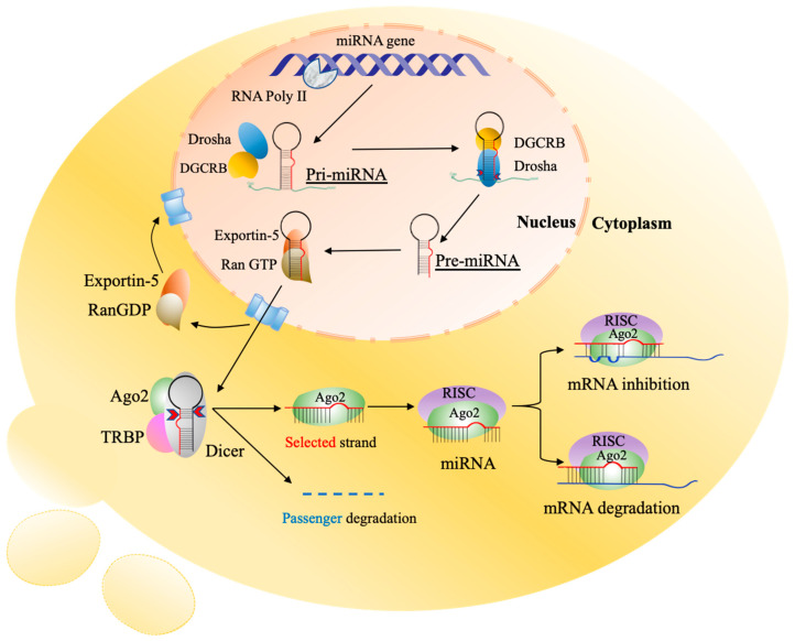
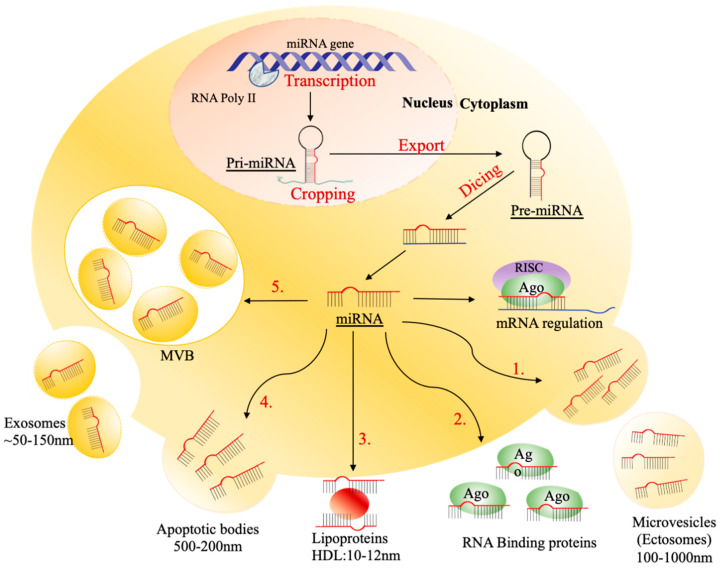
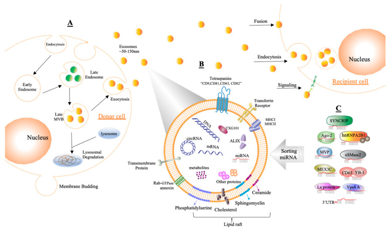
Colorectal cancer (CRC) is the third most common type of cancer worldwide amongst males and females. CRC treatment is multidisciplinary, often including surgery, chemotherapy, and radiotherapy. Early diagnosis of CRC can lead to treatment initiation at an earlier stage. Blood biomarkers are currently used to detect CRC, but because of their low sensitivity and specificity, they are considered inadequate diagnostic tools and are used mainly for following up patients for recurrence. It is necessary to detect novel, noninvasive, specific, and sensitive biomarkers for the screening and diagnosis of CRC at earlier stages. The tumor microenvironment (TME) has an essential role in tumorigenesis; for example, extracellular vesicles (EVs) such as exosomes can play a crucial role in communication between cancer cells and different components of TME, thereby inducing tumor progression. The importance of miRNAs that are sorted into exosomes has recently attracted scientists' attention. Some unique sequences of miRNAs are favorably packaged into exosomes, and it has been illustrated that particular miRNAs can be directed into exosomes by special mechanisms that occur inside the cells. This review illustrates and discusses the sorted and transported exosomal miRNAs in the CRC microenvironment and their impact on CRC progression as well as their potential use as biomarkers.

Keywords: biomarkers; colorectal cancer; exosomes; extracellular vesicles; miRNAs; tumor microenvironment.


Conflict of interest statement

The authors declare that there is no conflict of interest regarding the publication of this paper.

Figures

Figure 1
Figure 1
Biogenesis of miRNAs. The initiation process is started in the nucleus by RNA polymerase II which transcribes protein-coding genes into pri-miRNAs with a large cap and polyadenylation. The pri-miRNAs are processed by a complex of Drosha and RNA-binding protein DGCR8 to produce the stem-looped structures of 59–89 nucleotides (nt) known as pre-miRNAs. Then, pre-miRNAs are transported into the cytoplasm by exportin-5/Ran-GTP, and further processing occurs to generate mature miRNAs where Dicer cleaves stem-looped structures into double-strand miRNAs. One functional miRNA strand (red) is loaded with Ago2 into the RISC, and that complex, miRISC, binds to the complementary sequences of target mRNA in the 3′UTR. The result of these interactions is degradation and suppression of translation of the specific mRNA. pri-miRNAs, primary miRNAs; pre-miRNAs, processor miRNA; Dicer, RNase III nuclease; RISC, RNA-induced silencing complex; Ago2, and Argonaute 2. The image is adapted from Strubberg and Madison [18].
Figure 2
Figure 2
The mechanism of transporting circulating miRNAs. The process is initiated by transcription of the miRNA gene, cropping pri-miRNA, exporting pre-miRNA, and dicing to form a mature miRNA. The mature miRNA either regulates mRNA or is passaged into extracellular circulation. There are five different ways of passaging circulating miRNAs: (1) collecting in 500–200 nm of apoptotic bodies; (2) binding to Ago2 protein; (3) enveloping into 100–1000 nm of microvesicles (MVs), including ectosomes, (4) binding to 10–12 high-density lipoproteins (HDLs); (5) packaging into ~50–150 nm of exosomes.
Figure 3
Figure 3
Demonstrating the composition and generation of exosomes. (A) Exosomes are formed by endosomal multivesicular bodies (MVBs) budding inside the cell. Some of the MVBs that develop are delivered to the membrane of the cells after becoming late MVBs. MVBs can be dissolved during fusing with the lysosome or can release exosomes into the extracellular area by fusion with the plasma membrane through the exocytosis process, and the size range of exosomes is around ~50–150 nm. Acceptor cells receive exosomes through fusion, endocytosis, and/or signaling processes to insert their content. (B) Exosomes are enclosed by a phospholipid bilayer that contains a variety of components on its surface, such as tetraspanins (CD9, CD81, CD63, CD82), transferrin receptors, transmembrane proteins, molecular histocompatibility complex (MHCI, MHCII), Rab-GTPase annexin, and lipid rafts, while inward components contain biological species such as RNA (circRNA, mRNA, miRNA), proteins, DNA, and metabolites. In addition, tumor susceptibility gene 101 (TSG101) and apoptosis-linked gene 2-interacting protein X (ALIX) can be used as markers for exosomes. (C) Sorting miRNAs into exosomes can be regulated via different binding processes such as those of synaptotagmin-binding cytoplasmic RNA-interacting protein (SYNCRIP), sumoylated hnRNPA2B, Argonaute protein (Ago2), neutral sphingomyelinase 2 (nSMase2), major vault protein (MVP), CD63 with Y-box protein I (YBP1), Mex-3 RNA-binding family member C (MEX3C), protein 4A (Vps4A), lupus La protein (La protein), or the 3′ end of miRNA (3′UTR).

References

    1. Ding L., Lan Z., Xiong X., Ao H., Feng Y., Gu H., Yu M., Cui Q. The Dual Role of MicroRNAs in Colorectal Cancer Progression. Int. J. Mol. Sci. 2018;19:2791. doi: 10.3390/ijms19092791. - DOI - PMC - PubMed
    1. Augestad K., Bakaki P., Rose J., Crawshaw B., Lindsetmo R., Dørum L., Koroukian S., Delaney C. Metastatic spread pattern after curative colorectal cancer surgery. A retrospective, longitudinal analysis. Cancer Epidemiol. 2015;39:734–744. doi: 10.1016/j.canep.2015.07.009. - DOI - PubMed
    1. Toiyama Y., Okugawa Y., Fleshman J., Boland C.R., Goel A. MicroRNAs as potential liquid biopsy biomarkers in colorectal cancer: A systematic review. Biochim. Biophys. Acta Rev. Cancer. 2018;1870:274–282. doi: 10.1016/j.bbcan.2018.05.006. - DOI - PMC - PubMed
    1. Swiderska M., Choromańska B., Dąbrowska E., Konarzewska-Duchnowska E., Choromańska K., Szczurko G., Myśliwiec P., Dadan J., Ladny J.R., Zwierz K. The diagnostics of colorectal cancer. Contemp. Oncol. 2014;18:1–6. doi: 10.5114/wo.2013.39995. - DOI - PMC - PubMed
    1. Tan S., Xia L., Yi P., Han Y., Tang L., Pan Q., Tian Y., Rao S., Oyang L., Liang J. Exosomal miRNAs in tumor microenvironment. J. Exp. Clin. Cancer Res. 2020;39:67. doi: 10.1186/s13046-020-01570-6. - DOI - PMC - PubMed