. 2022 Apr 27;23(9):4856.
doi: 10.3390/ijms23094856.

Inactivation of E. coli, S. aureus, and Bacteriophages in Biofilms by Humidified Air Plasma

Affiliations

Inactivation of E. coli, S. aureus, and Bacteriophages in Biofilms by Humidified Air Plasma

Xinni Liu et al. Int J Mol Sci. .

Abstract

In this study, humidified air dielectric barrier discharge (DBD) plasma was used to inactivate Escherichia coli (E. coli), Staphylococcus aureus (S. aureus), and bacteriophages in biofilms containing DNA, NaCl, carbohydrates, and proteins. The humidified DBD plasma was very effective in the inactivation of microbes in the (≤1.0 μm) biofilms. The number of surviving E. coli, S. aureus, and bacteriophages in the biofilms was strongly dependent on the constituent and thickness of the biofilms and was greatly reduced when the plasma treatment time increased from 5 s to 150 s. Our analysis shows that the UV irradiation was not responsible for the inactivation of microbes in biofilms. The short-lived RONS generated in the humidified air DBD plasma were not directly involved in the inactivation process; however, they recombined or reacted with other species to generate the long-lived RONS. Long-lived RONS diffused into the biofilms to generate very active species, such as ONOOH and OH. This study indicates that the geminated NO2 and OH pair formed due to the homolysis of ONOOH can cause the synergistic oxidation of various organic molecules in the aqueous solution. Proteins in the biofilm were highly resistant to the inactivation of microbes in biofilms, which is presumably due to the existence of the unstable functional groups in the proteins. The unsaturated fatty acids, cysteine-rich proteins, and sulfur-methyl thioether groups in the proteins were easily oxidized by the geminated NO2 and OH pair.

Keywords: E. coli; bacteriophage; biofilms; dielectric barrier discharge; disinfection; humidified air plasma; plasma inactivation; proteins.


Conflict of interest statement

The authors declare no conflict of interest.

Figures
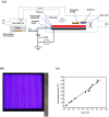
Figure 1
Figure 1
Humidified air surface dielectric barrier discharge (DBD) system for the inactivation of Escherichia coli (E. coli), Staphylococcus aureus (S. aureus), and bacteriophages in biofilm. (a) Schematic of the humidified air surface DBD plasma system. The plasma inactivation system consists of a tank filled with distilled water, a discharge chamber, surface DBD device, and a power supply. The surface DBD device consists of a layer of aluminum tape (0.13 mm thick) serving as an H.V. electrode, an insulating alumina plate (1.0 mm × 175 mm × 175 mm), and a patterned ground electrode (30 μm thick) covered by a layer of Kapton polyimide tape. An AC power source was capable of generating the 1.0–10.0 kV peak-to-peak voltage (VPP) with a frequency of 10 kHz. One capacitor of 2.2 μF in series to ground was used to obtain a Lissajous figure of this charge. The air was humidified when passing over the water surface, and the humidity level was 60–70% at a room temperature of 20 °C. The steel discs with dry biofilms were placed into the humidified air DBD chamber for plasma inactivation. Chemical probes were used to capture the RONS in the gas phase; (b) The photo of the surface DBD plasma obtained at VPP = 8.25 kV. The surface DBD plasma was generated over the alumina plate with its surface area of 155 mm × 155 mm; (c) The discharge power as a function of VPP. Measurements for the charges across this capacitor and the applied voltage across the DBD device resulted in the Lissajous figure, which was used to calculate the discharge power.
Figure 2
Figure 2
The number of surviving E. coli (a), S. aureus (b), and bacteriophages (c) in the protein biofilms as a function of the plasma treatment time. The plasma inactivation was performed at VPP = 8.25 kV and a power of 460 W; 10 μL of the aqueous solutions containing microbes and proteins was spread onto each steel disc (1.0 cm2) and placed for 10 min under air flow in a biosafety cabinet to allow drying. The thickness of the protein biofilms varied from 0.05 μm to 1.0 μm. The steel discs with dry biofilms were placed into the humidified air DBD chamber for plasma inactivation. The control samples were not covered with protein films. The samples with the treatment time of 0 s were the untreated control. Means ± standard deviations of experiments carried out at least in triplicate are shown.
Figure 2
Figure 2
The number of surviving E. coli (a), S. aureus (b), and bacteriophages (c) in the protein biofilms as a function of the plasma treatment time. The plasma inactivation was performed at VPP = 8.25 kV and a power of 460 W; 10 μL of the aqueous solutions containing microbes and proteins was spread onto each steel disc (1.0 cm2) and placed for 10 min under air flow in a biosafety cabinet to allow drying. The thickness of the protein biofilms varied from 0.05 μm to 1.0 μm. The steel discs with dry biofilms were placed into the humidified air DBD chamber for plasma inactivation. The control samples were not covered with protein films. The samples with the treatment time of 0 s were the untreated control. Means ± standard deviations of experiments carried out at least in triplicate are shown.
Figure 3
Figure 3
The number of surviving E. coli in the biofilms containing proteins, carbohydrates, DNA, salt or their mixture as a function of the plasma treatment time. The plasma inactivation was performed at VPP = 8.25 kV and a power of 460 W; 10 μL of the aqueous solutions containing E. coli and constituents of biofilms was spread onto each steel disc (1.0 cm2) and placed for 10 min under air flow in a biosafety cabinet to allow drying. After drying, the thicknesses of proteins, carbohydrates, DNA, salt, and their mixed biofilms were 1.15 μm, 0.68 μm, 0.04 μm, 0.3 μm, and 1.87 μm, respectively. The mixed biofilms contained 2% DNA, 19% NaCl, 29% carbohydrates, and 50% proteins. The steel discs with dry biofilms were placed into the humidified air DBD chamber for plasma inactivation. Means ± standard deviations of experiments carried out at least in triplicate are shown.
Figure 4
Figure 4
The number of surviving bacteriophages in the biofilms containing proteins, carbohydrates, DNA, salt or their mixture as a function of the plasma treatment time. The plasma inactivation was performed at VPP = 8.25 kV and a power of 460 W; 10 μL of the aqueous solutions containing bacteriophage and constituents of biofilms was spread onto each steel disc (1.0 cm2) and placed for 10 min under air flow in a biosafety cabinet to allow drying. After drying, the thicknesses of proteins, carbohydrates, DNA, salt, and their mixed biofilms were 1.15 μm, 0.68 μm, 0.04 μm, 0.3 μm, and 1.87 μm, respectively. The mixed biofilms contained 2% DNA, 19% NaCl, 29% carbohydrates, and 50% proteins. The steel discs with dry biofilms were placed into the humidified air DBD chamber for plasma inactivation. Means ± standard deviations of experiments carried out at least in triplicate are shown.
Figure 5
Figure 5
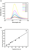
Measurements of RONS in the aqueous solution based on terephthalic acid (TA). Terephthalic acid (TA) was oxidized into 2-hydroxyterephthalic acid (HTA) by RONS in aqueous solution. When the TA/HTA solution was irradiated by UV light (λ = 310 nm), HTA molecules emitted light at λ = 425 nm. (a) The fluorescence spectra of the HTA solutions obtained by changing the plasma treatment time. The initial concentrations of TA and NaOH in the aqueous solution were 4 nM and 10 nM, respectively. (b) The surface density of HTA obtained in the treated solutions as a function of treatment time. To quantify the concentration of TA molecules oxidized into HTA, a calibration curve was obtained by using the standard HTA solution.
Figure 6
Figure 6
The fluorescence spectra of the 2,7-dichlorofluorescein (DCF) solutions obtained by changing the treatment time; 2,7-dichlorodihydrofluorescein diacetate (H2DCFDA) represents a non-fluorescent form of the dye. After the deacetylation in the NaOH solution, the non-fluorescent form (2,7-dichlorofluorescein, DCFH) was oxidized into the highly fluorescent form 2,7-dichlorofluorescein (DCF) by the RONS in the aqueous solution. The process of deacetylation was shown by hydrolysis with NaOH by mixing 0.5 mL of stock H2DCFDA solution with 2 mL of 0.1 M NaOH solution. The fluorescence was obtained at an excitation wavelength of 495 nm.
Figure 7
Figure 7
Pathways for the oxidation of terephthalic acid (TA) by ONOOH. The homolysis of ONOOH into OH and NO2 radicals leads to the oxidation of TA into 2-hydroxyterephthalic acid or 2-nitrogrephthalic acid.
Figure 8
Figure 8
Pathways for the oxidation of 2,7-dichlorodihydrofluorescein (DCFH) by ONOOH. The homolysis of ONOOH into OH and NO2 radicals leads to the oxidation of DCFH into 2,7-dichlorofluorescein.
Figure 9
Figure 9
Mechanism for the inactivation of microbes in the biofilms by humidified air plasma. The humidified air plasma generates the long-lived RONS in the gas phase, such as Y(H2O)n, O3, NO, NO2, N2O5, N2O4, H2O2, and HNO2. These long-lived RONS diffuse into the biofilms and react with each other to generate very active species, such as OH and ONOOH, leading to the oxidation and inactivation of microbes in the biofilms.

References

    1. Ki S.H., Baik K.Y., Choi E.H. Effects of humidity on room disinfection by dielectric barrier discharge plasma. J. Phys. D Appl. Phys. 2019;42:425204. doi: 10.1088/1361-6463/ab3066. - DOI
    1. Kogelheide F., Voigt F., Hillebrand B., Moeller R., Fiebrandt M. The role of humidity and UV-C emission in the inactivation of B. subtilis spores during atmospheric-pressure dielectric barrier discharge treatment. J. Phys. D Appl. Phys. 2020;29:295201. doi: 10.1088/1361-6463/ab77cc. - DOI
    1. Elena V.S., Aleksandra Y.L., Roman A.L., Elena V.V., Mariam A.A., Svetlana A.E., Aleksey V.S. Bidirectional mass transfer-based generation of plasma-activated water mist with antibacterial properties. Plasma Process. Polym. 2020;17:e2000058.
    1. Moldgy A., Nayak G., Aboubakr H.A., Goyal S.M., Bruggeman P.J. Inactivation of virus and bacteria using cold atmospheric pressure air plasmas and the role of reactive nitrogen species. J. Phys. D Appl. Phys. 2020;43:434004. doi: 10.1088/1361-6463/aba066. - DOI
    1. Los A., Ziuzina D., Boehm D., Cullen P.J., Bourke P. A comparison of inactivation efficacy and mechanisms of gas plasma and plasma activated water against Aspergillus flavus spores and biofilms. Appl. Environ. Microbiol. 2020;9:e02619-19. - PMC - PubMed

Substances