. 2022 Apr 27;23(9):4868.
doi: 10.3390/ijms23094868.

Mesenchymal Stem Cells and Extracellular Vesicles Derived from Canine Adipose Tissue Ameliorates Inflammation, Skin Barrier Function and Pruritus by Reducing JAK/STAT Signaling in Atopic Dermatitis

Affiliations

Mesenchymal Stem Cells and Extracellular Vesicles Derived from Canine Adipose Tissue Ameliorates Inflammation, Skin Barrier Function and Pruritus by Reducing JAK/STAT Signaling in Atopic Dermatitis

Sung Youl Kim et al. Int J Mol Sci. .

Abstract

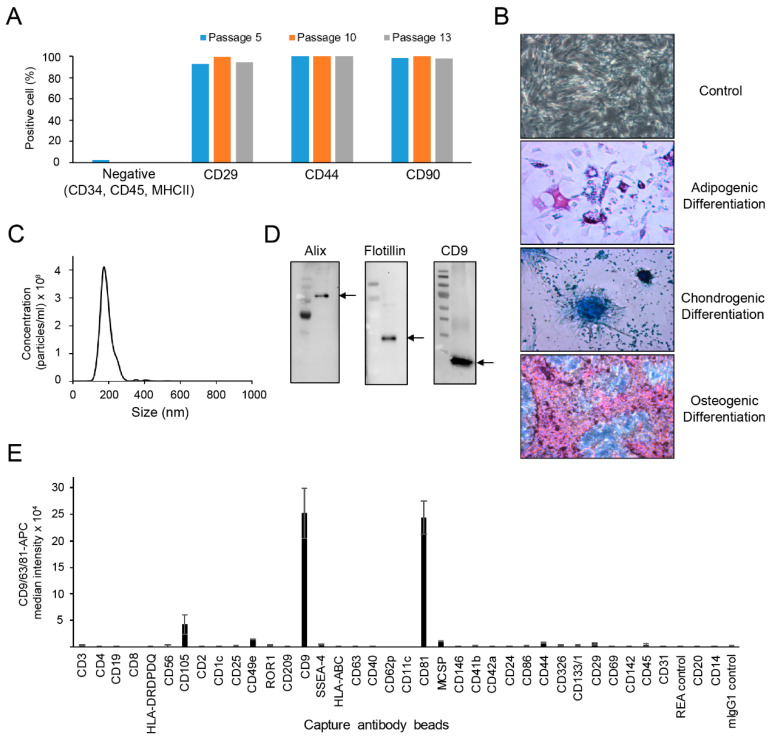
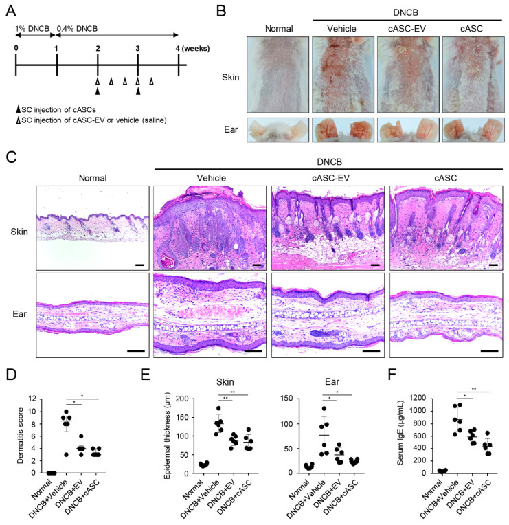
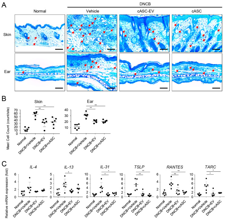
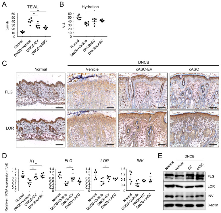
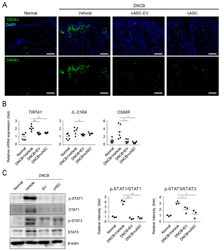
Canine atopic dermatitis (AD) is a common chronic inflammatory skin disorder resulting from imbalance between T lymphocytes. Current canine AD treatments use immunomodulatory drugs, but some of the dogs have limitations that do not respond to standard treatment, or relapse after a period of time. Thus, the purpose of this study was to evaluate the immunomodulatory effect of mesenchymal stem cells derived from canine adipose tissue (cASCs) and cASCs-derived extracellular vesicles (cASC-EVs) on AD. First, we isolated and characterized cASCs and cASCs-EVs to use for the improvement of canine atopic dermatitis. Here, we investigated the effect of cASCs or cASC-EVs on DNCB-induced AD in mice, before using for canine AD. Interestingly, we found that cASCs and cASC-EVs improved AD-like dermatitis, and markedly decreased levels of serum IgE, (49.6%, p = 0.002 and 32.1%, p = 0.016 respectively) epidermal inflammatory cytokines and chemokines, such as IL-4 (32%, p = 0.197 and 44%, p = 0.094 respectively), IL-13 (47.4%, p = 0.163, and 50.0%, p = 0.039 respectively), IL-31 (64.3%, p = 0.030 and 76.2%, p = 0.016 respectively), RANTES (66.7%, p = 0.002 and 55.6%, p = 0.007) and TARC (64%, p = 0.016 and 86%, p = 0.010 respectively). In addition, cASCs or cASC-EVs promoted skin barrier repair by restoring transepidermal water loss, enhancing stratum corneum hydration and upregulating the expression levels of epidermal differentiation proteins. Moreover, cASCs or cASC-EVs reduced IL-31/TRPA1-mediated pruritus and activation of JAK/STAT signaling pathway. Taken together, these results suggest the potential of cASCs or cASC-EVs for the treatment of chronic inflammation and damaged skin barrier in AD or canine AD.

Keywords: atopic dermatitis; canine; extracellular vesicle; inflammation; mesenchymal stem cell; pruritus; skin barrier function.


Conflict of interest statement

S.Y.K. and T.H.Y. are employees of GNG CELL Co., Ltd., R&D Center. T.-W.C. is CEO and stockholder of JIN BioCell Co., Ltd. Sang-Soo Lee who is funder and CEO (founder) of GNG CELL Co., Ltd., had no role in data collection, design, analyses, interpretation of data, manuscript preparation, or publication. All rights is reserved by GNG CELL, although this paper was written by supporting technique and idea through a joint research contract (GNG CELL Co./Kyungpook National University/JIN BioCell Co., Ltd.). No potential conflict of interest was reported by all other authors.

Figures

Figure 1
Figure 1
Characterization of canine adipose tissue-derived mesenchymal stem cells (cASC) and cASC-EV. (A) Bar graph obtained from flow cytometry data show the expression of MSC-positive markers such as CD29, CD44 and CD90 in cASCs populations. (B) Representative data showing the differentiation phenotypes of cASC. (C) Representative histogram of particle concentration and size distribution of cASC-EV measured by nanoparticle tracking analysis (NTA). (D) The presence of EV markers such as Alix, Flotilin and CD9 in cASC-EV as determined by Western blots. (E) Surface signature of cASC-EV quantified by MACSPlex EV kit in conjunction with flow cytometry. Data indicate APC median signal intensities of ASC-EV incubated with the 39 capture beads and stained with a mixture of CD9-, CD63-, and CD81-APC antibodies. Background was corrected by subtraction of median fluorescence APC intensity.
Figure 2
Figure 2
cASC and cASC-EV improve DNCB-induced atopic dermatitis-like skin lesion in Balb/c mice. (A) Schematic diagram for development of atopic dermatitis model by topical application of DNCB in Balb/c mice. After mice were sensitized with DNCB for 7 days, DNCB was further topically applied to the shaved dorsal skin and ear for 3 weeks. cASC was applied twice and cASC-EV was applied 5 times for 2 weeks (n = 6/group). (B) Representative dorsal skin and ear photographs of each treatment group showing comparison of AD-like skin lesions. (C) Tissue sections from the back skin and ear stained with hematoxylin and eosin (H&E). Scale bar, 100 μm. (D) The severity of dermatitis evaluated by a 3-point scoring index of atopic dermatitis. Dermatitis score was graded as 0 (absent), 1 (mild), 2 (moderate), or 3 (severe) based on the sum of the scores of clinical signs such as excoriation/erosion, scaling/dryness, edema, and erythema/hemorrhage. (E) Epidermal thickness of back skin and ear (n = 6/group). (F) The concentration of the serum total IgE levels measured by an ELISA. Dermatitis score represent median [IQR] (min–max) and the others represent mean ± SD. Significant value was * p < 0.05, ** p < 0.01 vs. DNCB + Vehicle.
Figure 3
Figure 3
cASC and cASC-EV reduce the level of multiple inflammatory cytokines in atopic dermatitis-like skin lesions of Balb/c mice. (A) Tissue sections from the back skin and ears stained with Toluidine blue (TB). Red arrow indicates mast cell infiltrated into skin or ear tissue. Scale bar, 100 μm. (B) The number of mast cells counted from five random section areas (n = 6/group). (C) The expression of IL-4, IL-13, IL31, TSLP, RANTES and TARC genes on skin tissues as determined using real-time PCR (n = 6/group). The expression of TARC gene represent median [IQR] (min–max) and the others represent mean ± SD. Significant value was * p < 0.05, ** p < 0.01 vs. DNCB + Vehicle.
Figure 4
Figure 4
cASC and cASC-EV improve epidermal barrier function in atopic dermatitis-like skin lesion of Balb/c mice. (A) TEWL and (B) hydration of the AD-like skin lesions (n = 6/group). (C) Expression of filaggrin and loricrin as evaluated by immunohistochemical stain. Scale bar, 100 μm. (D) Transcripts of keratin 1, filaggrin, loricrin, and involucrin were quantified using real-time PCR (n = 6/group). (E) Immunoblots with anti-filaggrin, anti-loricrin, anti-involucrin, and anti-β-actin antibodies using lysates from skin. All data represent mean ± SD. Significant value was * p < 0.05, ** p < 0.01 vs. DNCB + Vehicle.
Figure 5
Figure 5
cASC and cASC-EV inhibit pruritogen responses through downregulation of phosphorylation of STATs in atopic dermatitis-like skin lesion of Balb/c mice. (A) Tissue sections from back skins stained with TRPA1 using Immunofluorescence. Scal bar = 100 μm. (B) The expression of TRPA1, OSMR and IL-31RA genes in skin tissues; each group represented using real-time PCR (n = 6/group). (C) Immunoblots with anti-p-STAT1, anti-STAT1, anti-p-STAT3, anti-STAT3 or anti-β-actin antibodies using lysates from skin. Western blots were analyzed quantitatively. All data represent mean ± SD. Significant value was * p < 0.05, ** p < 0.01 vs. DNCB + Vehicle.

Similar articles

Cited by

References

    1. Anjos-Afonso F., Siapati E.K., Bonnet D. In vivo contribution of murine mesenchymal stem cells into multiple cell-types under minimal damage conditions. J. Cell Sci. 2004;117:5655–5664. doi: 10.1242/jcs.01488. - DOI - PubMed
    1. Murphy M.B., Moncivais K., Caplan A.I. Mesenchymal stem cells: Environmentally responsive therapeutics for regenerative medicine. Exp. Mol. Med. 2013;45:e54. doi: 10.1038/emm.2013.94. - DOI - PMC - PubMed
    1. Kapur S.K., Katz A.J. Review of the adipose derived stem cell secretome. Biochimie. 2013;95:2222–2228. doi: 10.1016/j.biochi.2013.06.001. - DOI - PubMed
    1. Kang J.W., Kang K.-S., Koo H.C., Park J.R., Choi E.W., Park Y.H. Soluble factors–mediated immunomodulatory effects of canine adipose tissue–derived mesenchymal stem cells. Stem Cells Dev. 2008;17:681–694. doi: 10.1089/scd.2007.0153. - DOI - PubMed
    1. Carrade D.D., Borjesson D.L. Immunomodulation by mesenchymal stem cells in veterinary species. Comp. Med. 2013;63:207–217. - PMC - PubMed

MeSH terms