Review
. 2022 Apr 28;23(9):4883.
doi: 10.3390/ijms23094883.

Retinitis Pigmentosa: Progress in Molecular Pathology and Biotherapeutical Strategies

Affiliations
Review

Retinitis Pigmentosa: Progress in Molecular Pathology and Biotherapeutical Strategies

Wanqin Liu et al. Int J Mol Sci. .

Abstract

Retinitis pigmentosa (RP) is genetically heterogeneous retinopathy caused by photoreceptor cell death and retinal pigment epithelial atrophy that eventually results in blindness in bilateral eyes. Various photoreceptor cell death types and pathological phenotypic changes that have been disclosed in RP demand in-depth research of its pathogenic mechanism that may account for inter-patient heterogeneous responses to mainstream drug treatment. As the primary method for studying the genetic characteristics of RP, molecular biology has been widely used in disease diagnosis and clinical trials. Current technology iterations, such as gene therapy, stem cell therapy, and optogenetics, are advancing towards precise diagnosis and clinical applications. Specifically, technologies, such as effective delivery vectors, CRISPR/Cas9 technology, and iPSC-based cell transplantation, hasten the pace of personalized precision medicine in RP. The combination of conventional therapy and state-of-the-art medication is promising in revolutionizing RP treatment strategies. This article provides an overview of the latest research on the pathogenesis, diagnosis, and treatment of retinitis pigmentosa, aiming for a convenient reference of what has been achieved so far.

Keywords: cell death; gene therapy; induced pluripotent stem cells; optogenetics; retinal remodeling; retinitis pigmentosa (RP).


Conflict of interest statement

The authors declare that the research was conducted in the absence of any commercial or financial relationships that could be construed as a potential conflict of interest.

Figures

Figure 1
Figure 1
Retinal laminae and photoreceptor cell structure. (a) The retina consists of ten layers, of which photoreceptor cells (rod and cone) and retinal pigment epithelium (RPE) are the main target cells for the treatment of RP and other inherited retinal dystrophies. (b) The final morphological structure of photoreceptor cell development includes an inner segment, outer segment, and a synaptic terminal. Connecting Cilium transports components, such as proteins, from the inner segment to the outer segment to the sensory discs stacked in the outer segment in order to mediate the onset of light signal transduction.
Figure 2
Figure 2
Three common types of cell death in RP. Damage signals from intracellular macromolecules cause necrosis, apoptosis, and autophagy, respectively.
Figure 3
Figure 3
Part of the cell death mechanism. Schematic diagrams are represented as: (a) Activation of multiple protein complexes by caspase-8 induces caspase-dependent apoptosis; (b) AIF-mediated mitochondrial pathway induces caspase-independent apoptosis; (c) Necroptosis performed by RIPK1 and/or RIPK3; (d) Pyroptosis caused by the immune response activation of caspase family members; (e) Ferroptosis caused by the excessive oxidation of membrane lipids; (f) Atg family-mediated autophagy-dependent cell death.
Figure 4
Figure 4
Biochemical reactions, such as protein aggregation, oxidative stress, the immune response, and metabolic dysfunction that occur during retinal degeneration cause retinal cell death. When gene mutations trigger macromolecular aggregation, one leads to endoplasmic reticulum stress, activating the unfolded protein response (UPR), but when UPR activation is not sufficient to relieve stress, cell death is induced by activating pro-apoptotic pathways (e.g., Caspases activation, Ca2+ release, and mitochondrial signaling); second, the oxidative system and antioxidant system imbalance and cyclically aggravate retinal oxidative stress. Third, it triggers the mechanism’s immune defense, when the active markers are immune cells and immune factors. The result of these unsustainable reactions ultimately points to the death of photoreceptor cells in various ways. Another pathway of cell death is autophagy, while excessive autophagy triggers apoptosis and necrosis. In addition, the accumulation of these reactions to a certain extent causes metabolic dysfunction, which is also closely associated with autophagy.
Figure 5
Figure 5
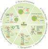
Six current treatment strategies for retinitis pigmentosa (RP). (a) Neuroprotective agents mainly include neurotrophic factors, anti-apoptotic agents, and antioxidants; they are usually used in the early stages of the disease and can also serve as the adjunctive treatment in other stages; (b) Gene therapy takes effect via the virus-mediated injection of a therapeutic gene tool into the retina in vitro to replace the disease-causing gene; (c) Introducing photosensitive optic proteins into the degenerated retina for ectopic expression in damaged cell membranes to restore the cone function and conferring photosensitive ability to residual retinal cells, such as bipolar cells or ganglion cells; (d) Injecting neural stem cells cultured in vitro into the retinal injury site induces differentiation into the injured cell type and replacement of injured cells, with the remaining retinal neurons forming synaptic connections; (e) Retinal prosthesis implantation at the site of retinal damage; the implantation sites of the artificial retina vary from subretinal, epiretinal membranes to intra-scleral; (f) injecting residual non-photoreceptor cells with genes that express ion channel proteins, and attaching “photoswitches”—chemical molecules that change shape when exposed to light—to the ion channel proteins.
Figure 6
Figure 6
(a) In vitro mRNAs delivered into the cytoplasm via special materials are directly translated by ribosomes into various proteins that exert their corresponding effects. (b) Antisense oligonucleotides (ASO) as chemically modified short RNA or DNA molecules that bind target mRNAs and can lead to RNase H-induced cleavage (bottom) or inhibit translation (top). (c,d) RNAi therapies involving small interfering RNAs (SiRNAs) or similar molecules (microRNAs) that are 21–23 nucleotides long to degrade mRNA and prevent its translation into proteins.

References

    1. Dias M.F., Joo K., Kemp J.A., Fialho S.L., da Silva Cunha A., Jr., Woo S.J., Kwon Y.J. Molecular genetics and emerging therapies for retinitis pigmentosa: Basic research and clinical perspectives. Prog. Retin. Eye Res. 2018;63:107–131. doi: 10.1016/j.preteyeres.2017.10.004. - DOI - PubMed
    1. Tsang S.H., Sharma T. Retinitis Pigmentosa (Non-syndromic) Adv. Exp. Med. Biol. 2018;1085:125–130. doi: 10.1007/978-3-319-95046-4_25. - DOI - PubMed
    1. Michalakis S., Koch S., Sothilingam V., Garcia Garrido M., Tanimoto N., Schulze E., Becirovic E., Koch F., Seide C., Beck S.C., et al. Gene therapy restores vision and delays degeneration in the CNGB1(-/-) mouse model of retinitis pigmentosa. Adv. Exp. Med. Biol. 2014;801:733–739. doi: 10.1007/978-1-4614-3209-8_92. - DOI - PubMed
    1. Jordan S.A., Farrar G.J., Kenna P., Humphries M.M., Sheils D.M., Kumar-Singh R., Sharp E.M., Soriano N., Ayuso C., Benitez J., et al. Localization of an autosomal dominant retinitis pigmentosa gene to chromosome 7q. Nat. Genet. 1993;4:54–58. doi: 10.1038/ng0593-54. - DOI - PubMed
    1. Banerjee P., Kleyn P.W., Knowles J.A., Lewis C.A., Ross B.M., Parano E., Kovats S.G., Lee J.J., Penchaszadeh G.K., Ott J., et al. TULP1 mutation in two extended Dominican kindreds with autosomal recessive retinitis pigmentosa. Nat. Genet. 1998;18:177–179. doi: 10.1038/ng0298-177. - DOI - PubMed

MeSH terms