. 2022 Apr 28;23(9):4905.
doi: 10.3390/ijms23094905.

Modulation of Mismatch Repair and the SOCS1/p53 Axis by microRNA-155 in the Colon of Patients with Primary Sclerosing Cholangitis

Affiliations

Modulation of Mismatch Repair and the SOCS1/p53 Axis by microRNA-155 in the Colon of Patients with Primary Sclerosing Cholangitis

Monika Adamowicz et al. Int J Mol Sci. .

Abstract

Deficient mismatch repair (MMR) proteins may lead to DNA damage and microsatellite instability. Primary sclerosing cholangitis (PSC) is a risk factor for colitis-associated colon cancer. MiR-155 is suggested to act as a key regulating node, linking inflammation and tumorigenesis. However, its involvement in the chronic colitis of PSC-UC patients has not been examined. We investigated the involvement of miR-155 in the dysregulation of MMR genes and colitis in PSC patients. Colon tissue biopsies were obtained from patients with PSC, PSC with concomitant ulcerative colitis (PSC-UC), uncomplicated UC, and healthy controls (n = 10 per group). In the ascending colon of PSC and PSC-UC patients, upregulated miR-155 promoted high microsatellite instability and induced signal transducer and activator of transcription 3 (STAT-3) expression via the inhibition of suppressors of cytokine signalling 1 (SOCS1). In contrast, the absence of miR-155 overexpression in the sigmoid colon of PSC-UC patients activated the Il-6/S1PR1 signalling pathway and imbalanced the IL17/FOXP3 ratio, which reinforces chronic colitis. Functional studies on human intestinal epithelial cells (HT-29 and NCM460D) confirmed the role of miR-155 over-expression in the inhibition of MMR genes and the modulation of p53. Moreover, those cells produced more TNFα upon a lipopolysaccharide challenge, which led to the suppression of miR-155. Additionally, exposure to bile acids induced upregulation of miR-155 in Caco-2 cell lines. Thus, under different conditions, miR-155 is involved in either neoplastic transformation in the ascending colon or chronic colitis in the sigmoid colon of patients with PSC. New insight into local modulation of microRNAs, that may alter the course of the disease, could be used for further research on potential therapeutic applications.

Keywords: biomarker; colorectal cancer; inflammation; miR-155; microsatellite instability; primary sclerosing cholangitis.


Conflict of interest statement

The authors declare no conflict of interest. The funders had no role in the design of the study; in the collection, analyses, or interpretation of data; in the writing of the manuscript, or in the decision to publish the results.

Figures

Figure 1
Figure 1
Expression of miR-155, suppressor of cytokine signalling 1 (SOCS1), and signal transducer and activator of transcription 3 (STAT3) mRNAs in human colon tissue. A scatter dot plot showing the relative expression levels of miR-155 (a,d), SOCS1 (b,e), (STAT3) (c,f) in both the ascending (ac) and sigmoid colons (df) of controls, primary sclerosing cholangitis patients (PSC), PSC and concomitant ulcerative colitis patients (PSC-UC), and ulcerative colitis (UC). Levels of gene expression were normalised to an endogenous reference, miR-191 for miRNA or 18S RNA for other genes, and presented as a fold-change relative to healthy controls. Results are representative of 10 independent experiments per group. Dots illustrate each patient, and data are presented as mean plus interquartile range (IQR) * p-value < 0.05, ** p-value < 0.01, *** p-value < 0.001.
Figure 2
Figure 2
Relative expression of miR-155, SOCS1, and the concentration of p-STAT3 protein in PBMCs. Peripheral blood mononuclear cells were obtained from PSC and PSC-UC patients, and healthy subjects. Significant upregulation of miR-155 in the PSC and PSC-UC groups was accompanied by the inhibition of SOCS1 and enhancement of p-STAT3. Bars indicate the mean ± SEM * p-value < 0.05, ** p-value < 0.01.
Figure 3
Figure 3
The expression patterns of mismatch repair (MMR) components in colonic tissue. Levels of gene expression were normalised to 18S RNA and presented as a fold-change relative to healthy controls. Bars indicate the mean ± SEM * p-value < 0.05, ** p-value < 0.01.
Figure 4
Figure 4
Distinct intestinal IL-17/FOXP3 ratio and the expression of IL-6 and S1PR1 mRNAs in the colonic tissue of PSC patients with and without UC. * Scatter dot plot showing IL-17 and FOXP3 proportions (a,d), and the relative expression levels of IL-6 (b,e) and S1PR1 (c,f) in the ascending (ac) and sigmoid colons (df) of controls, PSC, PSC with UC, and UC patients. Levels of gene expression presented as fold-change relative to healthy controls were normalised to the endogenous reference 18S RNA. Results are representative of 10 independent experiments per group. Dots illustrate each patient and data are presented as means plus interquartile ranges (IQRs) * p-value < 0.05, ** p-value < 0.01, *** p-value < 0.001.
Figure 5
Figure 5
The effect of miRNA inhibition on miR-155 and S1PR1 expression (a). Dose-dependent effect of GCDCA on miR-155 expression (b). The basal miR-155 expression in colonic cell lines (c). miR-155 and TNF α mRNA levels in Caco-2 after lipopolysaccharide (LPS) exposure (d). The expression of miR-155, TNFα, SOCS1 and S1PR1 in HT29 and NCM460D cells following 24 h treatment with LPS (e). Modulation of MMR (MLH1, MSH2, MSH6) and p53 gene expression levels after miR-155 mimic transfection in colonic cell lines (f). At least three independent experiments were conducted. The dotted lines represent control values. Levels of gene expression were normalised to the endogenous reference miR-191/miR-16 for miRNA, or 18S RNA for other genes. Bars indicate the mean ± SEM * p-value < 0.05, ** p-value < 0.01, *** p-value < 0.001.
Figure 6
Figure 6
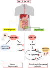
Schematic figure showing the contribution of miR-155 in inflammation and neoplastic transformation in the colon of patients with primary sclerosing cholangitis.

Similar articles

Cited by

References

    1. Hirschfield G.M., Karlsen T.H., Lindor K.D., Adams D.H. Primary sclerosing cholangitis. Lancet. 2013;382:1587–1599. doi: 10.1016/S0140-6736(13)60096-3. - DOI - PubMed
    1. Torres J., Pineton de Chambrun G., Itzkowitz S., Sachar D.B., Colombel J.F. Review article: Colorectal neoplasia in patients with primary sclerosing cholangitis and inflammatory bowel disease. Aliment. Pharm. Ther. 2011;34:497–508. doi: 10.1111/j.1365-2036.2011.04753.x. - DOI - PubMed
    1. Soetikno R.M., Lin O.S., Heidenreich P.A., Young H.S., Blackstone M.O. Increased risk of colorectal neoplasia in patients with primary sclerosing cholangitis and ulcerative colitis: A meta-analysis. Gastrointest. Endosc. 2002;56:48–54. doi: 10.1067/mge.2002.125367. - DOI - PubMed
    1. Broomé U., Lindberg G., Löfberg R. Primary sclerosing cholangitis in ulcerative colitis—A risk factor for the development of dysplasia and DNA aneuploidy? Gastroenterology. 1992;102:1877–1880. doi: 10.1016/0016-5085(92)90308-L. - DOI - PubMed
    1. Valeri N., Gasparini P., Fabbri M., Braconi C., Veronese A., Lovat F., Adair B., Vannini I., Fanini F., Bottoni A., et al. Modulation of mismatch repair and genomic stability by miR-155. Proc. Natl. Acad. Sci. USA. 2010;107:6982–6987. doi: 10.1073/pnas.1002472107. - DOI - PMC - PubMed

MeSH terms

Substances