. 2022 Apr 28;23(9):4926.
doi: 10.3390/ijms23094926.

At Embryo Implantation Site IL-35 Secreted by Trophoblast, Polarizing T Cells towards IL-35+ IL-10+ IL-4+ Th2-Type Cells, Could Favour Fetal Allograft Tolerance and Pregnancy Success

Affiliations

At Embryo Implantation Site IL-35 Secreted by Trophoblast, Polarizing T Cells towards IL-35+ IL-10+ IL-4+ Th2-Type Cells, Could Favour Fetal Allograft Tolerance and Pregnancy Success

Letizia Lombardelli et al. Int J Mol Sci. .

Abstract

We investigated the role of rhIL-35, at low concentrations compatible with those produced by human trophoblast cells (less than 1 ng/mL), on human T helper (Th) cell functions and the presence of decidual IL-35-producing Th cells in human pregnancy. We found that human trophoblast cells produced IL-35 but not IL-4 or IL-10. RhIL-35, at concentrations produced by human trophoblasts, polarized T cells towards IL-35+, IL-10+, IL-4+ Th2-type cells and to Foxp3+ EBI3+ p35+ T reg cells producing IL-35 but not IL-10 and IL-4. Moreover, rhIL-35 at low concentrations did not suppress the proliferation of Th cells but stimulated IL-4 and IL-10 production by established Th clones. In particular, Th1-type clones acquired the capacity to produce IL-4. In addition, purified human trophoblast cell supernatants containing IL-35 upregulated IL-4 and IL-10 production by Th clones. Finally, IL-35+, IL-10+, IL-4+ Th2-type cells, which were found to be induced by low concentrations of IL-35 compatible with those produced by human trophoblasts, are exclusively present in the decidua of a successful pregnancy and at the embryo implantation site, suggesting their stringent dependence on trophoblast cells. Thus, the proximity of Th cells to IL-35-producing trophoblasts could be the determining factor for the differentiation of IL-35+, IL-10+, IL-4+ Th2-type cells that are crucial for human pregnancy success.

Keywords: IL-10; IL-35; IL-4; Th2 cells; Treg cells; ectopic pregnancy; implantation; p35; pregnancy; trophoblast.


Conflict of interest statement

The authors declare no commercial or financial conflict of interest.

Figures

Figure 1
Figure 1
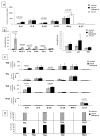
Effect of rhIL-35 on the cytokine production and on the cytokine mRNA expression of SK-specific T-cell lines and on the cytokine profile of macrophages. PBMCs from 10 different donors were stimulated with SK in the absence or presence of rhIL-35 at 300 pg/mL. (A) The levels of IL-4, IL-13, IL-10, IL-22, IL-17A, IL-17F, IL-35, and IL-5 in response to PMA plus anti-CD3 mAb in the T-cell lines specific for SK generated in the presence and absence of rh IL-35 (300 pg/mL) were measured with a multiplex bead-based assay. (B) T-cell lines specific for SK generated in the presence or absence of rhIL-35 (300 pg/mL) in bulk PBMC cultures were also analyzed with RT-PCR for the ability to express mRNA for IFN-γ, IL-4, IL-10, IL-17A, p35, and EBI3 in response to PMA plus anti-CD3 mAb. (C) Purified macrophages from PBMC obtained from five healthy donors stimulated for 5 days with the antigen SK in the absence or in the presence of rh IL-35 (300 pg/mL). MIP-3α, IL-1β, IL-23, IL-6, IL-12, IL-27, IL-10, and TNF-α were measured in the supernatants of macrophages with a multiplex bead-based assay.
Figure 2
Figure 2
rhIL-35 favors the development of IL-35 + IL-10 + IL4 + Th2-type cells in SK-specific CD4+ T-cell lines. T-cell blasts from SK-specific T-cell lines generated in the absence or in the presence of rhIL-35 (300 pg/mL) derived from the peripheral blood of two different donors were cloned under limiting dilution conditions. The SK-specific T-cell clones derived from the SK-specific T-cell lines generated with rhIL-35 and without rhIL-35 were assessed for their cytokine profiles in response to stimulation with PMA plus anti-CD3 mAb. The percentage of CD4+ T-cell clones, Th2-type T-cell clones, and Th0-type T-cell clones producing IL-35, IL-10, and IL-4 was determined.
Figure 3
Figure 3
rhIL-35 favors the development of Foxp3 Treg cells producing IL-35 in SK-specific CD4+ T-cell lines. T-cell blasts from SK-specific T-cell lines generated in the absence or in the presence of rhIL-35 (300 pg/mL) derived from the peripheral blood of two different donors were cloned under limiting dilution conditions. (A) The derived CD4+ T-cell clones were analyzed with RT-PCR for their ability to express mRNA for Foxp3, EBI3, and p35 and for the ability to produce IL-35, IL-4, and IL-10 in response to PMA plus anti-CD3 mAb. (B) The Treg-type clones expressing Foxp3 derived from the SK-specific T-cell lines generated with rhIL-35 and without rhIL-35 were assessed for IL-35, IL-10, and IL-4 production in response to PMA plus anti-CD3 mAb stimulation.
Figure 4
Figure 4
Direct effects of rh IL-35 on the cytokine profile of established CD4+ T-cell clones. (A) To provide evidence of the direct effect of rhIL-35 (300 pg/mL) on the cytokine production by T cells, IL-4, IL-5, IL-13, IL-17A, IFN-γ, and IL-10 production were measured with multiplex assays in the supernatant of 15 established T-cell clones stimulated by an immobilized anti-CD3 mAb. (B) To provide evidence of the direct effect of rh IL-35 (300 pg/mL) on the mRNA expression of IFN-γ, IL-4, IL-10, IL-17A, GATA3, T-bet, p35, EBI3, and RORC, RT-PCR was performed on five additional established T-cell clones stimulated by an immobilized anti-CD3 mAb. (C) To provide evidence of the direct effect of rh IL-35 (300 pg/mL) on different CD4+T cell subpopulations, five additional CD4+ Th1 T-cell clones, five additional CD4+ Th2 T-cell clones, five additional CD4+ Th17 T-cell clones, and five additional CD4+ Th0 2 T-cell clones were stimulated with an insolubilized anti-CD3 monoclonal antibody in the absence or presence of rhIL-35 (300 pg/mL). The levels of IL-4, IL-5, IL-13, IL-10, IL-17A, and IFN-γ were measured in the T-cell clone supernatants by multiplex bead-based assays. (D) To provide evidence of the direct effect of rhIL-35 (300 pg/mL) on different CD4+ T cell subpopulations, six additional CD4+ Th1, six additional CD4+ Th2, six additional CD4+ Th17, and six additional CD4+ Th0 T-cell clones were stimulated with an insolubilized anti-CD3 monoclonal antibody in the absence or presence of rhIL-35 (300 pg/mL). The percentages of T-cell clones producing IFN-γ alone (Th1-type T-cell clones); producing IL-4, IL-5, IL-13, and IL-10 (Th2-type T-cell clones); producing IL-17A (Th17-type T-cell clones); and producing IFN-γ, IL-4, IL-5, IL-13, IL-10, and IL-17A (Th0-type T-cell clones) were determined to evaluate the possible cytokine profile modification induced by rhIL-35 on established CD4+ T-cell clones.
Figure 5
Figure 5
Trophoblast cells, through the spontaneous production of IL-35, stimulate IL-4 and IL-10 production by CD4+ T helper cells. (A) IL-35 was measured with a bead-based assay in the supernatant of 16 specimens of isolated human first-trimester cytotrophoblast cells and in the supernatant of 3 specimens from the trophoblast cell line HTR8. (B) Fourteen CD4+ T-cell clones derived from the peripheral blood of healthy subjects stimulated with immobilized anti-CD3 mAb were cultured for 36 h in the absence and in the presence of IL-35 derived from four isolated human first-trimester cytotrophoblast cell supernatants at two different final concentrations, 25 pg/mL and 50 pg/mL.
Figure 6
Figure 6
RhIL-35, at low concentrations compatible with those produced by human primary trophoblasts, does not inhibit the CD4+ T-cell clone proliferation, but at high concentrations it inhibits T-cell clone proliferation. Nine additional CD4+ T-cell clones generated from the peripheral blood of healthy subjects were stimulated with immobilized anti-CD3 mAb in the absence and in the presence of different concentrations of rhIL-35 (range 100 pg/mL- 100 ng/mL), and the proliferative activity of these T-cell clones was evaluated by 3H thymidine incorporation (c.p.m.).
Figure 7
Figure 7
Th2 CD4+ T cells producing IL-35, IL-10, and IL-4 are exclusively present at the embryo implantation site and in the decidua of normal human pregnancy. (A) mRNA expression for p35 and EBI3 was evaluated in three decidua specimens of first-trimester normal pregnancy. (B) Seventy CD4+ T-cell clones were generated from each of these tissues: at the embryo implantation site and far from the implantation site of ectopic tubal pregnancies, from the decidua of the same women with ectopic pregnancy, where the implantation site was not present in the deciduae, but in the fallopian tube and from the deciduae of women with normal pregnancy with an embryo implantation site. The percentages of CD4+ T-cell clones, Th2-type T-cell clones, and Th0-type T-cell clones producing IL-35, IL-10, and IL-4 were determined in the different tissues.

References

    1. Chen H., Wang W., Xie H., Xu X., Wu J., Jiang Z., Zhang M., Zhou L., Zheng S. A pathogenic role of IL-17 at the early stage of corneal allograft rejection. Transpl. Immunol. 2009;21:155–161. doi: 10.1016/j.trim.2009.03.006. - DOI - PubMed
    1. Yuan X., Paez-Cortez J., Schmitt-Knosalla I., D’Addio F., Mfarrej B., Donnarumma M., Habicht A., Clarkson M.R., Iacomini J., Glimcher L.H., et al. A novel role of CD4 Th17 cells in mediating cardiac allograft rejection and vasculopathy. J. Exp. Med. 2008;205:3133–3144. doi: 10.1084/jem.20081937. - DOI - PMC - PubMed
    1. Burns W.R., Wang Y., Tang P.C., Ranjbaran H., Iakimov A., Kim J., Cuffy M., Bai Y., Pober J.S., Tellides G. Recruitment of CXCR3+ and CCR5+ T cells and production of interferon-gamma-inducible chemokines in rejecting human arteries. Am. J. Transplant. 2005;5:1226–1236. doi: 10.1111/j.1600-6143.2005.00892.x. - DOI - PubMed
    1. Waaga A.M., Gasser M., Kist-van Holthe J.E., Najafian N., Müller A., Vella J.P., Womer K.L., Chandraker A., Khoury S.J., Sayegh M.H. Regulatory functions of self-restricted MHC class II allopeptide-specific Th2 clones in vivo. J. Clin. Investig. 2001;107:909–916. doi: 10.1172/JCI11427. - DOI - PMC - PubMed
    1. Strom T.B., Roy-Chaudury R., Manfro R., Zheng X.X., Nickerson P.W., Wood K., Bushell A. The Th1/Th2 paradigm and allograft response. Curr. Opin. Immunol. 1996;8:688–693. doi: 10.1016/S0952-7915(96)80087-2. - DOI - PubMed