. 2022 Apr 30;23(9):4983.
doi: 10.3390/ijms23094983.

Transcriptomic Profiling of Peripheral Edge of Lesions to Elucidate the Pathogenesis of Psoriasis Vulgaris

Affiliations

Transcriptomic Profiling of Peripheral Edge of Lesions to Elucidate the Pathogenesis of Psoriasis Vulgaris

Suphagan Boonpethkaew et al. Int J Mol Sci. .

Abstract

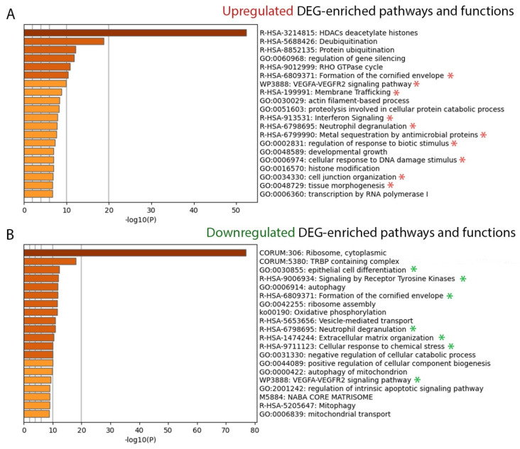
Elucidating transcriptome in the peripheral edge of the lesional (PE) skin could provide a better understanding of the molecules or signalings that intensify inflammation in the PE skin. Full-thickness biopsies of PE skin and uninvolved (UN) skin were obtained from psoriasis patients for RNA-seq. Several potential differentially expressed genes (DEGs) in the PE skin compared to those in the UN skin were identified. These DEGs enhanced functions such as angiogenesis, growth of epithelial tissue, chemotaxis and homing of cells, growth of connective tissues, and degranulation of myeloid cells beneath the PE skin. Moreover, the canonical pathways of IL-17A, IL-6, and IL-22 signaling were enriched by the DEGs. Finally, we proposed that inflammation in the PE skin might be driven by the IL-36/TLR9 axis or IL-6/Th17 axis and potentiated by IL-36α, IL-36γ, IL-17C, IL-8, S100A7, S100A8, S100A9, S100A15, SERPINB4, and hBD-2. Along with IL-36α, IL-17C, and IκBζ, ROCK2 could be an equally important factor in the pathogenesis of psoriasis, which may involve self-sustaining circuits between innate and adaptive immune responses via regulation of IL-36α and IL-36γ expression. Our finding provides new insight into signaling pathways in PE skin, which could lead to the discovery of new psoriasis targets.

Keywords: RNA sequencing; chronic skin diseases; cytokines; gene; inflammation.


Conflict of interest statement

The authors have no conflict of interest to declare.

Figures

Figure 1
Figure 1
The differentially expressed genes (DEGs). A total of l202 genes was identified as the DEGs in the peripheral edge of the lesional (PE) skin vs. uninvolved (UN) skin. Of these, 653 (54%) were upregulated and 549 (46%) were downregulated. Among these DEGs, 135 DEGs are overlapped with previously reported psoriasis-associated genes or proteins. Red presents upregulation in the PE skin and green represents downregulation in the PE skin. Proteins of the 86 upregulated DEGs might be potential molecules involved in psoriasis pathogenesis in the PE skin. The DEGs are listed based on their molecule type.
Figure 2
Figure 2
DEG-enriched pathway and function analysis (A) The top 20 upregulated DEG-enriched pathways and functions. (B) The top 20 downregulated DEG-enriched pathways and functions. A −log10 (10) value is the p-value in log base 10 to create the height of each bar chart. The associated pathways and functions are annotated with *. CORUM, comprehensive resource of mammalian protein complexes; DEG, differentially expressed gene; GO, gene ontology; ko, Kyoto Encyclopedia of Genes and Genomes (KEGG) pathway; M, canonical pathways; R-HSA, reactome gene sets; WP, WikiPathways.
Figure 3
Figure 3
Potential upregulated differentially expressed genes (DEGs). The circles represent the fold change of expressed genes; the larger the circle, the more upregulated the expression of DEGs. A2M, alpha 2 macroglobulin; A2ML1, alpha 2 macroglobulin-like 1; ADAM17, ADAM metallopeptidase domain 17; C10orf99, chromosome 10 open reading frame 99; CXCL8, C-X-C motif chemokine ligand 8; DEFB4A/DEFB4B, defensin beta 4A; DSC2, desmocollin2; DSG3, desmoglein 3; EDNRB, endothelin receptor type B; FLG, filaggrin; GFRA1, GDNF family receptor alpha 1; GJB2, gap junction protein beta 2; IL36A, interleukin 36 alpha; ITGB1, integrin subunit beta 1; KLK7, kallikrein related peptidase 7; KRT16P1, keratin 16 pseudogene 1; LAMA2, laminin subunit alpha 2; LCE3A, late cornified envelope 3A; LCN2, lipocalin 2; mDC, myeloid dendritic cells; NAMPT, nicotinamide phosphoribosyltransferase; pDC, plasmacytoid dendritic cell; PLAT, plasminogen activator, tissue type; PLEC, plectin; PLSCR1, phospholipid scramblase 1; PMN, polymorphonuclear leukocytes; S100A7A, S100 calcium binding protein A7A; SERPINB4, serpin family B member 4; Th 0, T helper 0 cell; TNFRSF1A, TNF receptor superfamily member 1A; VAV3, vav guanine nucleotide exchange factor 3; XDH, xanthine dehydrogenase.
Figure 4
Figure 4
DEG-enriched canonical pathways. (A,C) The height of the bar represents the level of significance. The predicted activation pathways are shown by orange bars and the predicted inhibition pathways are shown by blue bars. The more intense the color, the stronger the activity prediction. The orange line connecting square points in each bar indicates the ratio of the dataset DEGs that are members of each known pathway divided by the total molecules’ pathway. (B,D) Percentage of DEGs overlapping in each pathway. Percentage of the non-overlapped pathway’s molecules is shown by the white segment of the bar. The total number of molecules in each canonical pathway are shown on the top of the bar. CCR3, C-C Motif Chemokine Receptor 3; CNTF, ciliary neurotrophic factor; CXCL8, C-X-C Motif Chemokine Ligand 8; DEG, differentially expressed gene; eIF2, eukaryotic initiation factor 2; fMLP, formyl-methionyl-leucyl-phenylalanine; GM-CSF, granulocyte-macrophage colony-stimulating factor; HER-2, human epidermal growth factor receptor; IL, interleukin; JAK, Janus Kinase; LXR/RXR, liver X receptor/retinoid X receptor; NER, Nucleotide excision repair.
Figure 5
Figure 5
Potential regulators and their relationship to the target dataset’s DEGs. In each radiant, the molecule at the center is the predicted potential regulator and the outer circular molecules are the dataset’s DEGs. DEGs, differentially expressed genes; PE skin, peripheral edge of the lesional skin.
Figure 6
Figure 6
The two most useful regulator effect networks. (A) The highest consistency score network. (B) The second-highest consistency score network. Each network shows how predicted upstream regulators (top tier) control their dataset’s DEGs (middle tier) and the downstream functions of the dataset’s DEGs (bottom tier). DEGs, differentially expressed genes; PE skin, peripheral edge of the lesional skin.
Figure 7
Figure 7
Quantitative multiplex Real-Time PCR analysis of DEGs and upstream regulators. The mRNA level of each gene was calculated relative to GAPDH, the housekeeping gene. (AJ) The relative RNA expression of selected DEGs. (KM) The relative mRNA expression levels of selected upstream regulators. (N) The fold change of significantly altered genes. All graphs were designated by geometric mean and standard deviation. The significance was determined using *; * p < 0.05, ** p < 0.01 and *** p < 0.001. n ≥ 10 for each gene. CXCL8, C-X-C motif chemokine ligand 8; DEFB103A/B, defensin beta 103A/B; DEGs, differentially expressed genes; IL17C, interleukin 17C; NFKBIZ, nuclear factor kappa B inhibitor zeta; PE skin, peripheral edge of lesional skin; PLSCR1, phospholipid scramblase 1; ROCK2, rho-associated kinase 2; S100A7, S100 calcium binding protein A7; SERPINB4, serpin family B member 4; UN skin, uninvolved skin.
Figure 8
Figure 8
The proposed pathogenesis network at the peripheral edge (PE) of the lesional skin. Injured keratinocytes secrete interleukin (IL)-8, IL-36α, and IL-36γ. IL-36 activates plasmacytoid dendritic cells (pDCs) to secrete interferon-alpha (IFN)-α in a Toll-like receptor (TLR)-9-dependent manner (IL-36/TLR-9 axis). IL-36α and IL-36γ activate myeloid dendritic cells (mDCs) to mature and secrete IL-6, which drives the differentiation of T helper (h) cells (IL-6/Th17 axis). ROCK2 (rho-associated kinase 2) regulates CD4+ T cells to secrete IL-17. Note that inflammation in the PE skin is potentiated by IL-36α, IL-36γ, IL-17C, IL-8, S100A7, S100A8, S100A9, S100A15, SERPINB4, and hBD-2 and that a self-sustaining circuit between the innate and adaptive immune response in the PE skin is mediated through IL-36α and IL-36γ.
Figure 9
Figure 9
Peripheral edge of the lesional skin. The biopsy landmark was identified by the well-demarcated edge of the plaque. The piece of biopsy included equal sides of normal phenotypic skin and lesional skin.

Similar articles

Cited by

References

    1. Griffiths C.E.M., Armstrong A.W., Gudjonsson J.E., Barker J.N.W.N. Psoriasis. Lancet. 2021;397:1301–1315. doi: 10.1016/S0140-6736(20)32549-6. - DOI - PubMed
    1. Dastoli S., Nisticò S.P., Morrone P., Patruno C., Leo A., Citraro R., Gallelli L., Russo E., De Sarro G., Bennardo L. Colchicine in Managing Skin Conditions: A Systematic Review. Pharmaceutics. 2022;14:294. doi: 10.3390/pharmaceutics14020294. - DOI - PMC - PubMed
    1. Amoruso G., Nisticò S., Iannone L., Russo E., Rago G., Patruno C., Bennardo L. Ixekizumab May Improve Renal Function in Psoriasis. Healthcare. 2021;9:543. doi: 10.3390/healthcare9050543. - DOI - PMC - PubMed
    1. Harden J.L., Krueger J.G., Bowcock A.M. The immunogenetics of Psoriasis: A comprehensive review. J. Autoimmun. 2015;64:66–73. doi: 10.1016/j.jaut.2015.07.008. - DOI - PMC - PubMed
    1. Di Meglio P., Villanova F., Nestle F.O. Psoriasis. Cold Spring Harb. Perspect. Med. 2014;4:a015354. doi: 10.1101/cshperspect.a015354. - DOI - PMC - PubMed