. 2022 Apr 30;23(9):5012.
doi: 10.3390/ijms23095012.

Urine-Derived Stem Cell-Secreted Klotho Plays a Crucial Role in the HK-2 Fibrosis Model by Inhibiting the TGF-β Signaling Pathway

Affiliations

Urine-Derived Stem Cell-Secreted Klotho Plays a Crucial Role in the HK-2 Fibrosis Model by Inhibiting the TGF-β Signaling Pathway

Sang-Heon Kim et al. Int J Mol Sci. .

Abstract

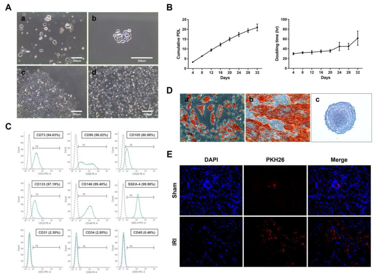
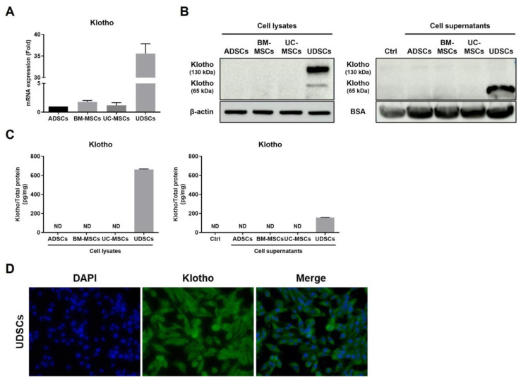
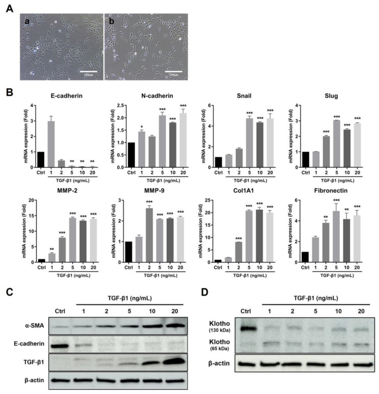
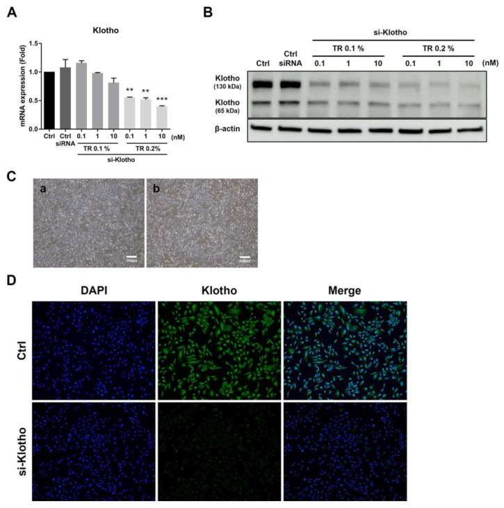
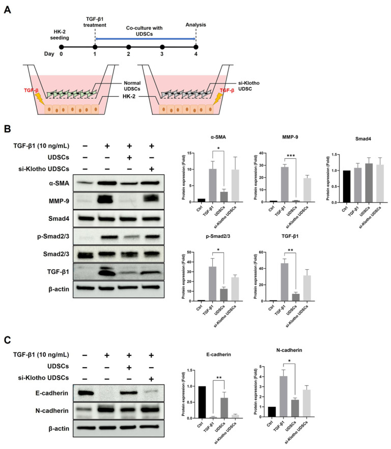
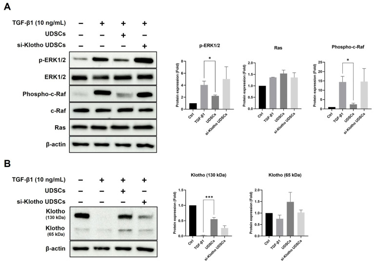
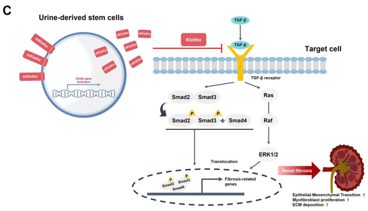
Renal fibrosis is an irreversible and progressive process that causes severe dysfunction in chronic kidney disease (CKD). The progression of CKD stages is highly associated with a gradual reduction in serum Klotho levels. We focused on Klotho protein as a key therapeutic factor against CKD. Urine-derived stem cells (UDSCs) have been identified as a novel stem cell source for kidney regeneration and CKD treatment because of their kidney tissue-specific origin. However, the relationship between UDSCs and Klotho in the kidneys is not yet known. In this study, we discovered that UDSCs were stem cells that expressed Klotho protein more strongly than other mesenchymal stem cells (MSCs). UDSCs also suppressed fibrosis by inhibiting transforming growth factor (TGF)-β in HK-2 human renal proximal tubule cells in an in vitro model. Klotho siRNA silencing reduced the TGF-inhibiting ability of UDSCs. Here, we suggest an alternative cell source that can overcome the limitations of MSCs through the synergetic effect of the origin specificity of UDSCs and the anti-fibrotic effect of Klotho.

Keywords: chronic kidney disease; klotho; mesenchymal stem cells; renal fibrosis; urine-derived stem cells.


Conflict of interest statement

All authors declare that no support, financial or otherwise, has been received from any organization that may have an interest in the submitted work and there are no other relationships or activities that could appear to have influenced the submitted work. EHLBIO Co., LTD. had no role in the design of the study; in the collection, analyses, or interpretation of data; in the writing of the manuscript, or in the decision to publish the results.

Figures

Figure 1
Figure 1
Characterization and homing effect of UDSCs. (A) Isolation and culture of UDSC. (a) Non-adherent cells in s fresh urine sample (Day 0). (b) Cell attachment (Day 3). (c) Colony formation of rice grain-shaped cells (Day 10). (d) Morphological changes into spindle-shaped cells (Passage 1). (B) Representative growth curve of the UDSCs. (a) The cumulative population doubling level (CPDL) and (b) doubling time (DT) were analyzed at each passage. (C) The representative surface marker expression was detected by flow cytometry at passage 5. (D) Multilineage differentiation of UDSCs. (a) Adipogenic differentiation; Oil Red O staining for lipid droplets (Day 21). (b) Osteogenic differentiation; Alizarin Red S staining for calcium deposits (Day 21). (c) Chondrogenic differentiation; Alcian Blue staining for glycosaminoglycan accumulation (Day 21). (E) PKH26-labeled UDSCs (red) were intravenously administered to wild-type (Sham) and ischemia-reperfusion injury (IRI) mice.
Figure 2
Figure 2
Klotho expression patterns of UDSCs. (A) The Klotho gene expression level was analyzed by real-time PCR using cell lysates. (B) The Klotho protein expression level was detected by Western blotting using cell lysates and supernatants. (C) The Klotho protein concentration was quantified by ELISA using cell lysates and supernatants. The results are shown as the mean ± SEM. (D) The Klotho expression was visualized by fluorescence staining (green) in UDSCs.
Figure 3
Figure 3
TGF-β1-induced HK-2 fibrosis model. (A) HK-2 cells were treated with 10 ng/mL TGF-β1 for 72 h. The morphological changes were observed using a microscope. (a) Normal HK-2 cells. (b) TGF-β1 treated HK-2 cells. (B) Fibrosis-related genes were analyzed by real-time PCR using HK-2 cell lysate. (C,D) Fibrosis-related proteins and Klotho were detected by Western blotting using HK-2 cell lysate. The results are shown as the mean ± SEM. * p < 0.05, ** p < 0.01, *** p < 0.001 vs. Control.
Figure 4
Figure 4
Klotho siRNA transfection efficiency in UDSCs. (A,B) UDSCs were treated with various concentrations of transfection reagent (TR) and Klotho siRNA. The transfection efficiency was evaluated using real-time PCR and Western blotting. Universal negative siRNA control was used as a transfection control. (C) UDSCs were treated with 0.2% transfection reagent and 10 nM Klotho siRNA for 48 h. The morphological changes were observed using a microscope. (a) Normal UDSCs, and (b) si-Klotho UDSCs. (D) Klotho expression was visualized by fluorescence staining (green) in normal and si-Klotho UDSCs. The results are shown as the mean ± SEM. ** p < 0.01, *** p < 0.001 vs. Control.
Figure 5
Figure 5
UDSCs suppress fibrosis in HK-2 cells via the TGF-β/Smad signaling pathway. (A) The experimental design of an indirect co-culture assay. (B) In total, 10 ng/mL TGF- β1-treated HK-2 were co-cultured with normal UDSCs or si-Klotho UDSCs for 72 h. The TGF-β/Smad signaling pathway was analyzed by Western blotting using HK-2 cell lysates. (C) EMT-related proteins were analyzed by Western blotting using HK-2 cell lysates. The results are shown as the mean ± SEM. * p < 0.05, ** p < 0.01, *** p < 0.001 vs. Control.
Figure 6
Figure 6
UDSCs suppress fibrosis in HK-2 cells via the TGF-β/ERK signaling pathway. (A) In total, 10 ng/mL TGF- β1-treated HK-2 were co-cultured with normal UDSCs or si-Klotho UDSCs for 72 h. The TGF-β/ERK signaling pathway was analyzed by Western blotting using HK-2 cell lysates. (B) Klotho expressions were analyzed by Western blotting using HK-2 cell lysates. The results are shown as the mean ± SEM. * p < 0.05, *** p < 0.001 vs. Control. (C) Schematic diagram of the mechanism of UDSC-derived Klotho.
Figure 6
Figure 6
UDSCs suppress fibrosis in HK-2 cells via the TGF-β/ERK signaling pathway. (A) In total, 10 ng/mL TGF- β1-treated HK-2 were co-cultured with normal UDSCs or si-Klotho UDSCs for 72 h. The TGF-β/ERK signaling pathway was analyzed by Western blotting using HK-2 cell lysates. (B) Klotho expressions were analyzed by Western blotting using HK-2 cell lysates. The results are shown as the mean ± SEM. * p < 0.05, *** p < 0.001 vs. Control. (C) Schematic diagram of the mechanism of UDSC-derived Klotho.

Similar articles

Cited by

References

    1. Zelmer J.L. The economic burden of end-stage renal disease in Canada. Kidney Int. 2007;72:1122–1129. doi: 10.1038/sj.ki.5002459. - DOI - PubMed
    1. Chow F.Y., Briganti E.M., Kerr P.G., Chadban S.J., Zimmet P.Z., Atkins R.C. Health-related quality of life in Australian adults with renal insufficiency: A population-based study. Am. J. Kidney Dis. 2003;41:596–604. doi: 10.1053/ajkd.2003.50121. - DOI - PubMed
    1. Kusek J.W., Greene P., Wang S.R., Beck G., West D., Jamerson K., Agodoa L.Y., Faulkner M., Level B. Cross-sectional study of health-related quality of life in African Americans with chronic renal insufficiency: The African American Study of Kidney Disease and Hypertension Trial. Am. J. Kidney Dis. 2002;39:513–524. doi: 10.1053/ajkd.2002.31401. - DOI - PubMed
    1. Pagels A.A., Soderkvist B.K., Medin C., Hylander B., Heiwe S. Health-related quality of life in different stages of chronic kidney disease and at initiation of dialysis treatment. Health Qual. Life Outcomes. 2012;10:71. doi: 10.1186/1477-7525-10-71. - DOI - PMC - PubMed
    1. Mujais S.K., Story K., Brouillette J., Takano T., Soroka S., Franek C., Mendelssohn D., Finkelstein F.O. Health-related quality of life in CKD Patients: Correlates and evolution over time. Clin. J. Am. Soc. Nephrol. 2009;4:1293–1301. doi: 10.2215/CJN.05541008. - DOI - PMC - PubMed