. 2022 Apr 30;23(9):5013.
doi: 10.3390/ijms23095013.

SIRT2 Is Critical for Sheep Oocyte Maturation through Regulating Function of Surrounding Granulosa Cells

Affiliations

SIRT2 Is Critical for Sheep Oocyte Maturation through Regulating Function of Surrounding Granulosa Cells

Xiaohuan Fang et al. Int J Mol Sci. .

Abstract

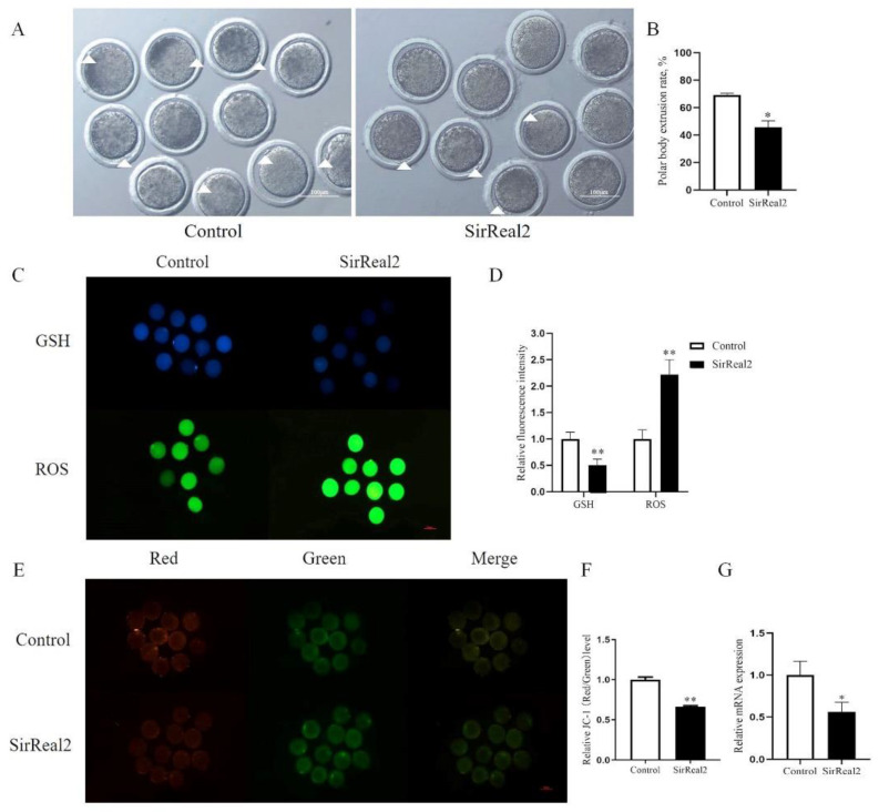
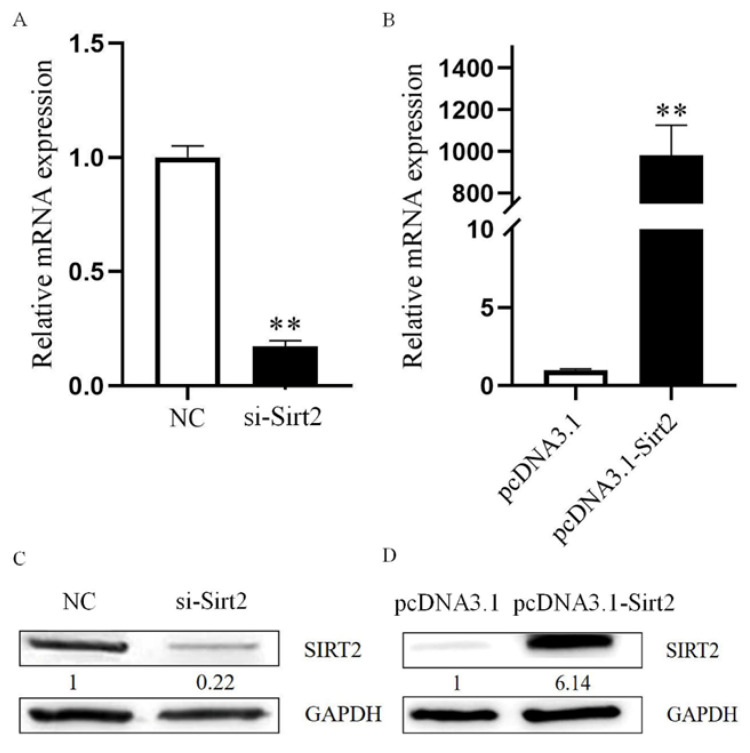
Oocyte in vitro maturation is crucial for in vitro embryo production technology, which provides oocytes resources for in vitro fertilization and somatic cell nuclear transfer. Previous studies proved that SIRT2, a member of the sirtuin family, plays a role in oocyte meiosis, but its role in sheep oocyte maturation and its regulating mechanism remains unknown. Firstly, we confirmed the role of Sirt2 in sheep oocytes maturation by supplementation of SIRT2 inhibitor and activator. To further explore the specific mechanism, we performed knockdown of Sirt2 in granulosa cells and then cocultured them with oocytes. Moreover, we determined the effects of Sirt2 on granulosa cell oxidative apoptosis, cell migration, and diffusion, and examined its effects on granulosa cell mitochondrial function, mitophagy, and steroid hormone levels. The results showed that supplementation of SIRT2 inhibitor decreased the oocytes maturation rate (69.28% ± 1.28 vs. 45.74% ± 4.74, p < 0.05), while resveratrol, a SIRT2 activator, increased its maturation rate (67.44% ± 1.68 vs. 78.52 ± 1.28, p < 0.05). Knockdown of Sirt2 in sheep granulosa cells also reduced the oocytes maturation rate (47.98% ± 1.43 vs. 33.60% ± 1.77, p < 0.05), and led to decreased cell migration and expansion ability, oxidative apoptosis, abnormal mitochondrial gene expression, decreased mitochondrial membrane potential and ATP level, and increased mitophagy level. Overexpression of Sirt2 improved mitochondrial membrane potential and ATP level and improved mitochondrial function. Furthermore, we found that Sirt2 knockdown in granulosa cells promotes the secretion of P4 through regulating p-ERK1/2. In conclusion the present study showed that SIRT2 is critical for sheep oocyte maturation through regulating the function of ovarian granulosa cells, especially affecting its mitochondrial function.

Keywords: SIRT2; mitochondria; mitophagy; oocytes in vitro maturation; sheep granulosa cells.


Conflict of interest statement

The authors declare no conflict of interest.

Figures

Figure 1
Figure 1
Inhibition of SIRT2 in oocytes affects maturation rate, GSH, ROS, and mitochondrial membrane potential. (A) Representative images of oocyte maturation after Sirt2 inhibition. The white triangle represents the extrusion of the polar body. Bar = 100 μm. (B) The maturation rate after inhibition of SIRT2 in oocytes. (C) Representative images of GSH and ROS after SIRT2 inhibition in oocytes. Bar = 100 μm. (D) The relative levels of GSH and ROS in oocytes after SIRT2 was inhibited. (E) Red and green fluorescent images of JC-1 staining after inhibiting SIRT2 in oocytes. Bar = 100 μm. (F) The relative level of JC-1 after inhibiting SIRT2 in oocytes. (G) Sirt2 mRNA level after Sirt2 inhibition. The graph shows the mean ± SEM of the results obtained in three independent experiments. *, significant difference (p < 0.05). **, significant difference (p < 0.01).
Figure 2
Figure 2
Resveratrol affects maturation rate, GSH, ROS, and mitochondrial membrane potential in oocytes. (A) Representative images of oocyte maturation after resveratrol treatment. The white triangle represents the extrusion of the polar body. Bar = 100 μm. (B) The maturation rate after treatment with different concentrations of resveratrol in oocytes. (C) Representative images of GSH and ROS after treatment with different concentrations of resveratrol in oocytes. Bar = 100 μm. (D) The relative levels of GSH and ROS in oocytes after treatment with different concentrations of resveratrol. (E) Red and green fluorescent images of JC-1 staining after treatment with different concentrations of resveratrol in oocytes. Bar = 100 μm. (F) The relative level of JC-1 after treatment with different concentrations of resveratrol in oocytes. (G) Sirt2 mRNA level after 1μM resveratrol treatment. The graph shows the mean ± SEM of the results obtained in three independent experiments. *, significant difference (p < 0.05).
Figure 3
Figure 3
Determination of Sirt2 knockdown and overexpression efficiency in present study. (A) Detection of mRNA level of Sirt2 by qRT-PCR after siRNA knockdown. (B) Detection of mRNA level of Sirt2 by qRT-PCR after overexpression. (C) Detection of protein level of SIRT2 by Western blot after siRNA knockdown. (D) Detection of protein level of SIRT2 by Western blot after overexpression. **, significant difference (p < 0.01). The results were obtained in three independent experiments.
Figure 4
Figure 4
Sirt2 of granulosa cells is critical for sheep oocyte maturation and granulosa cell migration. (A) Representative images of oocyte maturation after Sirt2 knockdown in granulosa cells. The white triangle represents the extrusion of the polar body. Bar = 100 μm. (B) Oocyte maturation rate after Sirt2 knockdown in granulosa cells. (C) qRT-PCR detected the expression of oxidative and apoptotic genes in granulosa cells after Sirt2 knockdown. (D) The levels of E2, P4, and T in SIRT2-inhibited granulosa cells detected by ELISA. (E) Images showing scratch widths at 0, 6, 12, and 24 h after Sirt2 knockdown. Bar = 100 μm. (F) The granulosa cells migration distance compared between Sirt2 knockdown and control groups after 0, 6, 12, and 24 h. (G) The mRNA expression levels of granulosa cell expansion genes including has2, ptx3, ptgs2, and tnfaip6. The graph shows the mean ± SEM of the results obtained in three independent experiments. *, significant difference (p < 0.05). **, significant difference (p < 0.01).
Figure 5
Figure 5
Sirt2 knockdown in granulosa cells affects mitochondrial function. (A) Images of red and green fluorescence in the NC and Sirt2 knockdown groups after JC-1 treatment. Bar = 100 μm. (B) Relative levels of mitochondrial membrane potential after Sirt2 knockdown. (C) ATP relative levels after Sirt2 knockdown. (D) Western blot detected the protein level of mitochondrial protein TOMM20 after Sirt2 knockdown. (E) The expression of granulosa cell mitochondrial genes was detected by qRT-PCR after Sirt2 knockdown. The graph shows the mean ± SEM of the results obtained in three independent experiments. *, significant difference (p < 0.05). **, significant difference (p < 0.01).
Figure 6
Figure 6
Overexpression of Sirt2 in granulosa cells affects mitochondrial function. (A) Mitochondrial membrane potential (red and green fluorescence) in the control group and Sirt2 overexpression group after JC-1 treatment. Bar = 100 μm. (B) Intensity of mitochondrial membrane potential after Sirt2 overexpression. (C) ATP relative levels after overexpression of Sirt2. (D) Western blot detected the protein level of mitochondrial protein TOMM20 after Sirt2 overexpression. (E) The expression of granulosa cell mitochondrial genes (mfn1, opa1, mff, fis1, drp1, and tomm20) was detected by qRT-PCR after overexpression of Sirt2. The graph shows the mean ± SEM of the results obtained in three independent experiments. *, significant difference (p < 0.05). **, significant difference (p < 0.01).
Figure 7
Figure 7
Sirt2 knockdown in granulosa cells affects mitophagy. (A) Immunofluorescence staining images of mitophagy proteins PINK1, Parkin, and LC3 after Sirt2 knockdown. Bar = 100 μm. (B) The relative expression level of mitophagy proteins by immunofluorescence staining after Sirt2 knockdown. (C) The mRNA relative expression level of mitophagy genes (pink1, parkin, and lc3) after Sirt2 knockdown. (D) The protein expression level of Parkin after Sirt2 knockdown. The graph shows the mean ± SEM of the results obtained in three independent experiments. *, significant difference (p < 0.05). **, significant difference (p < 0.01).
Figure 8
Figure 8
Overexpression of Sirt2 in granulosa cells had no effect on mitophagy. (A) Immunofluorescence staining images of mitophagy proteins PINK1, Parkin, and LC3 after Sirt2 overexpression. Bar = 100 μm. (B) The relative expression level of mitophagy proteins by immunofluorescence staining after Sirt2 overexpression. (C) The mRNA relative expression level of mitophagy genes (pink1, parkin, and lc3) after Sirt2 overexpression was detected by qRT-PCR. (D) The protein level of mitophagy protein Parkin after Sirt2 overexpression. The graph shows the mean ± SEM of the results obtained in three independent experiments.
Figure 9
Figure 9
Sirt2 knockdown in granulosa cells affects steroid hormone secretion through ERK1/2. (A) Western blot representative images of p-ERK1/2 and ERK1/2 in different treatment groups. (B) ERK1/2 protein expression levels in different treatment groups. (C) p-ERK1/2 protein expression levels in different treatment groups. (D) The levels of E2, P4, and T secreted by granulosa cells in different treatment groups were detected by ELISA. The graph shows the mean ± SEM of the results obtained in three independent experiments. *, significant difference (p < 0.05).
Figure 10
Figure 10
Mechanism of SIRT2 influencing sheep oocyte maturation through regulating surrounding granulosa cells. SIRT2 in granulosa cells is required for sheep oocyte maturation by affecting mitochondrial membrane potential and ATP level, mitophagy, and increasing the P4 secretion through regulating p-ERK1/2.

Similar articles

Cited by

References

    1. Eppig J.J. Reproduction: Oocytes Call, Granulosa Cells Connect. Curr. Biol. 2018;28:R354–R356. doi: 10.1016/j.cub.2018.03.005. - DOI - PubMed
    1. Sanchez-Lazo L., Brisard D., Elis S., Maillard V., Uzbekov R., Labas V., Desmarchais A., Papillier P., Monget P., Uzbekova S. Fatty acid synthesis and oxidation in cumulus cells support oocyte maturation in bovine. Mol. Endocrinol. 2014;28:1502–1521. doi: 10.1210/me.2014-1049. - DOI - PMC - PubMed
    1. Li H.J., Sutton-McDowall M.L., Wang X., Sugimura S., Thompson J.G., Gilchrist R.B. Extending prematuration with cAMP modulators enhances the cumulus contribution to oocyte antioxidant defence and oocyte quality via gap junctions. Hum. Reprod. 2016;31:810–821. doi: 10.1093/humrep/dew020. - DOI - PubMed
    1. Sugiyama M., Sumiya M., Shirasuna K., Kuwayama T., Iwata H. Addition of granulosa cell mass to the culture medium of oocytes derived from early antral follicles increases oocyte growth, ATP content, and acetylation of H4K12. Zygote. 2016;24:848–856. doi: 10.1017/S0967199416000198. - DOI - PubMed
    1. Richani D., Dunning K.R., Thompson J.G., Gilchrist R.B. Metabolic co-dependence of the oocyte and cumulus cells: Essential role in determining oocyte developmental competence. Hum. Reprod. Update. 2021;27:27–47. doi: 10.1093/humupd/dmaa043. - DOI - PubMed

LinkOut - more resources