. 2022 May 3;23(9):5068.
doi: 10.3390/ijms23095068.

Overexpression of EgrIAA20 from Eucalyptus grandis, a Non-Canonical Aux/ IAA Gene, Specifically Decouples Lignification of the Different Cell-Types in Arabidopsis Secondary Xylem

Affiliations

Overexpression of EgrIAA20 from Eucalyptus grandis, a Non-Canonical Aux/ IAA Gene, Specifically Decouples Lignification of the Different Cell-Types in Arabidopsis Secondary Xylem

Hong Yu et al. Int J Mol Sci. .

Abstract

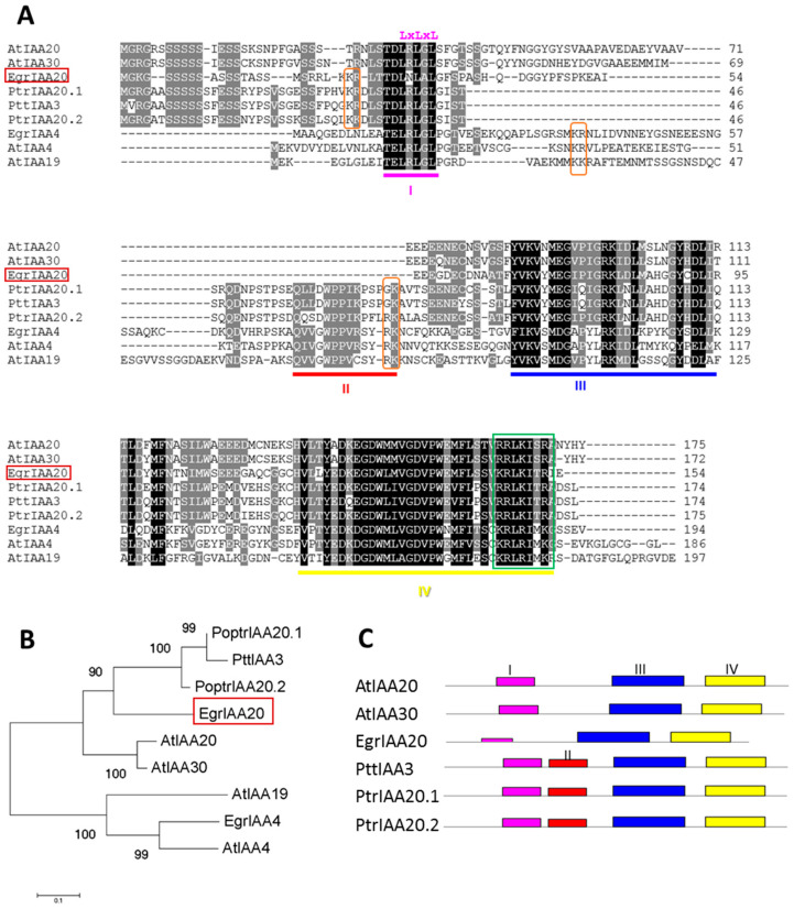
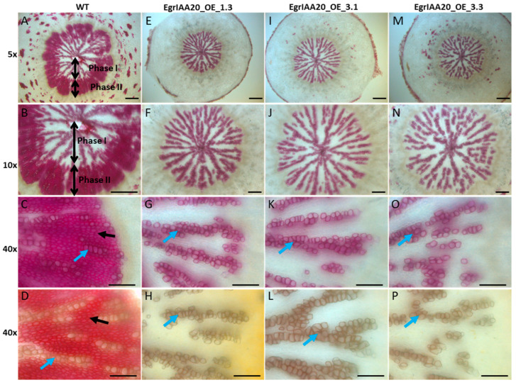
Wood (secondary xylem) formation is regulated by auxin, which plays a pivotal role as an integrator of developmental and environmental cues. However, our current knowledge of auxin-signaling during wood formation is incomplete. Our previous genome-wide analysis of Aux/IAAs in Eucalyptus grandis showed the presence of the non-canonical paralog member EgrIAA20 that is preferentially expressed in cambium. We analyzed its cellular localization using a GFP fusion protein and its transcriptional activity using transactivation assays, and demonstrated its nuclear localization and strong auxin response repressor activity. In addition, we functionally tested the role of EgrIAA20 by constitutive overexpression in Arabidopsis to investigate for phenotypic changes in secondary xylem formation. Transgenic Arabidopsis plants overexpressing EgrIAA20 were smaller and displayed impaired development of secondary fibers, but not of other wood cell types. The inhibition in fiber development specifically affected their cell wall lignification. We performed yeast-two-hybrid assays to identify EgrIAA20 protein partners during wood formation in Eucalyptus, and identified EgrIAA9A, whose ortholog PtoIAA9 in poplar is also known to be involved in wood formation. Altogether, we showed that EgrIAA20 is an important auxin signaling component specifically involved in controlling the lignification of wood fibers.

Keywords: Arabidopsis; Eucalyptus; IAA20; auxin; cambium differentiation; non-canonical Aux/IAA; secondary fiber; secondary xylem; syringyl lignin; wood.


Conflict of interest statement

The authors declare no conflict of interest. The funders had no role in the design of the study; in the collection, analyses, or interpretation of data; in the writing of the manuscript; or in the decision to publish the results.

Figures

Figure 1
Figure 1
EgrIAA20 transcript (Eucgr.K00561) shows preferential accumulation in the cambium of Eucalyptus. (A) Ratio of relative mRNA abundance of all Eucalyptus Aux/IAA members in vascular tissues including: juvenile vascular cambium (Cabium-7-year-old tree), mature vascular cambium (Cambium-25-year-old tree), phloem (7-year-old tree), and xylem (7-year-old tree). (B) Real-time PCR expression levels of EgrIAA20 in various Eucalyptus tissues and organs. Each relative mRNA abundance was normalized to a control sample (in vitro Eucalyptus plantlets). Error bars indicate mean expression values ±SE from three independent experiments. (C) Pictographic view of EgrIAA20 expression across a diverse range of RNAseq expression datasets by exImage (https://eucgenie.org/exImage, accessed on 25 April 2022).
Figure 2
Figure 2
EgrIAA20 defines a non-canonical Aux/IAA protein which lacks highly conserved Aux/IAA characteristic Domain II. (A) Sequence Analysis of EgrIAA20. Sequence comparison of EgrIAA20, putative orthologs from other plants, and paralog of Aux/IAA protein family. Conserved residues are shaded in black; gray shading indicates similar residues in at least 5 out of 9 of the sequences. Four conserved domains I, II, III, IV are underlined. Conserved basic residues that putatively function as NLS are indicated by orange frames (Type I) and green frame (Type II). The core repression EAR motif in Domain I is indicated by “LxLxL” on the top of the alignment. (B) The phylogenetic relationship of EgrIAA20 with its related putative orthologs from Arabidopsis and poplar defines a distinct clade with Arabidopsis AtIAA20 and AtIAA30, and previously characterized wood-related aspen member PttIAA3. (C) Shema of proteins structures for EgrIAA20 and its closely related putative orthologs from poplar and Arabidopsis. The domains of Aux/IAA proteins were predicted by Pfam (http://pfam.xfam.org, accessed on 23 April 2020) and are indicated by different colors; Domain II (red box) was absent for EgrIAA20, AtIAA20, and AtIAA30, but present in PtrIAA20.1, PtrIAA20.2, and PttIAA3.
Figure 3
Figure 3
Subcellular localization and repressor activity of auxin response of EgrIAA20 protein on a synthetic DR5 promotor. (A) Subcellular localization of EgrIAA20-GFP fusion protein in BY-2 tobacco protoplasts. The merged images of green fluorescence (left panel) and the corresponding bright-field image (middle panels) are shown in the right panels. Canonical Aux/IAA member EgrIAA4-GFP serves as a positive control. Scale bar = 10 µm. (B) Transcriptional repression of auxin response by EgrIAA20 protein on a synthetic DR5 promotor. Effector and reporter constructs were co-expressed in tobacco protoplasts in the presence or absence of a synthetic auxin (50 µM 2,4-D). A mock effector construct (empty vector) was used as negative control, and EgrIAA4 construct was used as positive control. Three independent experiments were performed, and similar results were obtained. In each experiment, protoplast transformations were performed in independent biological triplicates. The figure indicates the data from one experiment. Error bars represent SE of mean fluorescence. Significant statistical differences (Student’s t-test, n > 400, p < 0.01) from the control are marked with **. The schemes of the reporter and effector constructs are illustrated on the top of the figure.
Figure 4
Figure 4
EgrIAA20 overexpressing lines displayed reduced rosette development with helical twisting leaves and reduced root development. (A) EgrIAA20 mRNA accumulation in three phenotypically representative independent transgenic lines. The EgrIAA20 overexpressing lines (IAA20_OE_3.1 and IAA20_OE_3.3) presented reduced rosette development with helices twisting and backward rolling leaf blades and reduced leaf size (B), significantly reduced diameter of rosette (C), significantly reduced leaves number (D), significantly reduced primary root length (E), and greatly reduced lateral roots number (F). Error bars represent standard error. Asterisks indicate values found to be significantly different from the wild-type control (Student’s t-test, n > 10); ** indicates p < 0.01, and * indicates p < 0.05.
Figure 5
Figure 5
Phenotypes of EgrIAA20 overexpressing lines. Comparison of plant architecture of aerial parts at 64 days old (A) and 71 days old (B); EgrIAA20 overexpressing lines displayed a bushier plants architecture compared to the normal plant, with floppy inflorescence stems, in contrast to wild-type control up-right growth inflorescence stems. (C) Growth curve of the Arabidopsis primary inflorescence stem. The elongation was reduced in EgrIAA20 overexpressing lines with significantly reduced inflorescence diameter (D); error bars represent standard error, n > 12; stem diameter was measured at the base (<1 cm to the rosette level) when the first silique fully developed. Asterisks indicate values found to be significantly different (student’s t-test, n > 12) from the wild-type control. ** indicates p < 0.01. (E) The stamens of EgrIAA20 overexpressing lines were dramatically shorter than the wild-type control.
Figure 6
Figure 6
Over-expressing EgrIAA20 in Arabidopsis regulated vascular patterning in cotyledons through the interaction with AtARF5. (A) EgrIAA20 overexpression impaired vascular patterning in cotyledons. The vascular patterning was assessed by the number of secondary vein loops originating from the mid-vein. Class I presents four complete vascular loops; class II presents partially incomplete venation with at least two loops; class III presents entirely incompletely venation with no entire venation loop. In wild type seedlings, all the cotyledon venation patterns belonged to classes I and II, and 45% of them exhibited a more complex pattern (class I with four complete loops). In contrast, cotyledon venation patterns of all the transgenic plants belonged to classes II and III; none of them showed the complete vascular patterning with four loops (class I). Values in bracket indicate the percentage contribution of each class. The scale bars represent 0.5 mm. (B) EgrIAA20 interacts with Arabidopsis ARF5/MP (AtARF5) and its potential ortholog in Eucalyptus EgrARF5 in yeast-2-hybrid assay. The EgrIAA20 and AtARF/EgrARF5 proteins were fused with GAL4 DNA-binding domain (BD) and a GAL4 activation domain (AD), respectively. Yeast of co-transformed EgrIAA20-BD and AtARF5-AD or EgrARF5-AD grew on quadruple dropout medium lacking leucine, tryptophan, histidine, and adenine (THLA), and then scratched again on a TLHA plate; AD-T7 were used as negative controls.
Figure 7
Figure 7
EgrIAA20 overexpression specifically repressed secondary fibers but not primary fibers nor secondary vessels in Arabidopsis inflorescence stems. Left panel (AD) shows cross section of inflorescence stems at the basal part of wild-type control, <1 cm to the rosette level, 62-day-old plants; 5×, 20×, and 40× objective image for the first raw, second and third raw, respectively, using Phloroglucinol-HCl staining, which stains the lignin polymers in the SCW of xylem cells into red-purple; the last raw was 40× objective observation image stained with Maüle method, which stains the fiber cells into bright red color due to the syringyl unit (S unit) of lignin, indicated by yellow and black arrows, and stains the vessels in to brown due to the G unit of lignin, indicated by blue arrow. The corresponding cross sections from three independent EgrIAA20 overexpressing lines are shown in the middle and right panels: EgrIAA20_OE_1.3 (strong line, middle left panel (EH)), EgrIAA20_OE_3.1 (strong line, middle right panel (IL)), EgrIAA20_OE_3.3 (weak line, right panel, (MP)). Green arrows indicate primary xylem cells in fascicular bundles; blue arrows indicate secondary vessel cells (vessel II); yellow arrows indicate primary fiber cells (fiber I); black arrows indicate secondary fiber cells (Fiber II). * indicates staining gaps dispersed in the secondary growth region. The first to third raw used Phloroglucinol-HCl staining; the bottom raw used Maüle staining methods. The secondary growth regions are indicated by double-arrow lines in the images stained by Maüle. For 5× objective observation images, scale bar = 200 µm, and for 20× and 40× objective observation images, scale bar = 50 µm.
Figure 8
Figure 8
EgrIAA20 overexpression specifically inhibits the lignification of fibers in secondary but not primary fibers nor any tracheary elements in Arabidopsis hypocotyl. Left panel (AD) shows cross section of hypocotyl of wild-type control, 5×, 10×, and 40× objective image for the first raw, second and third raw, respectively, using Phloroglucinol-HCl staining, which stains the lignin polymers in the SCW of xylem cells into red-purple; the last raw was 40× objective observation image stained with Maüle method, which stains the fiber cells into bright red color, indicated by black arrow, and stains the vessels in to brown, indicated by blue arrow. The corresponding hypocotyl cross sections from three independent EgrIAA20 overexpressing lines are shown in the middle and right panels: EgrIAA20_OE_1.3 (strong line, middle left panel, (EH)), EgrIAA20_OE_3.1 (strong line, middle right panel, (IL)), EgrIAA20_OE_3.3 (weak line, right panel, (MP)). Phase I growth region and phase II growth region are indicated by double-arrow lines in wild type cross section. Blue arrows indicate secondary vessel cells (vessel II); black arrows indicate secondary fiber cells (Fiber II). The first to third raw used Phloroglucinol-HCl staining, the bottom raw used Maüle staining methods. For 5× and 10× images, scale bar = 200 µm, and for 40× images, scale bar = 50 µm.
Figure 9
Figure 9
Yeast-two-hybrid analysis of protein interactions between EgrIAA20 and xylem expressing EgrARFs and EgrIAA proteins. The EgrIAA20 and EgrARF/EgrIAA proteins were fused with Gal4 DNA-binding domain (BD) and a GAL4 activation domain (AD), respectively. The interaction between BD-Lam and AD-T was used as negative control, while the interaction between BD-p53 and AD-T was used as positive control. Yeast cells were inoculated on selective medium in a 10-fold gradient dilution. SD/-Leu-Trp: double dropout medium lacking leucine and tryptophan; SD/-Leu-Trp-His-Ade: quadruple dropout medium lacking leucine, tryptophan, histidine, and adenine.
None

Similar articles

Cited by

References

    1. Boudet A.M., Kajita S., Grima-Pettenati J., Goffner D. Lignins and lignocellulosics: A better control of synthesis for new and improved uses. Trends Plant Sci. 2003;8:576–581. doi: 10.1016/j.tplants.2003.10.001. - DOI - PubMed
    1. Ye Z.H., Zhong R. Molecular control of wood formation in trees. J. Exp. Bot. 2015;66:4119–4131. doi: 10.1093/jxb/erv081. - DOI - PubMed
    1. Barros J., Serk H., Granlund I., Pesquet E. The cell biology of lignification in higher plants. Ann. Bot. London. 2015;115:1053–1074. doi: 10.1093/aob/mcv046. - DOI - PMC - PubMed
    1. Boerjan W., Ralph J., Baucher M. Lignin biosynthesis. Annu. Rev. Plant Biol. 2003;54:519–546. doi: 10.1146/annurev.arplant.54.031902.134938. - DOI - PubMed
    1. Pesquet E., Wagner A., Grabber J.H. Cell culture systems: Invaluable tools to investigate lignin formation and cell wall properties. Curr. Opin. Biotech. 2019;56:215–222. doi: 10.1016/j.copbio.2019.02.001. - DOI - PubMed

MeSH terms

Substances

LinkOut - more resources