Review
. 2022 May 6;23(9):5214.
doi: 10.3390/ijms23095214.

Immune and Inflammatory Networks in Myocardial Infarction: Current Research and Its Potential Implications for the Clinic

Affiliations
Review

Immune and Inflammatory Networks in Myocardial Infarction: Current Research and Its Potential Implications for the Clinic

Atsushi Anzai et al. Int J Mol Sci. .

Abstract

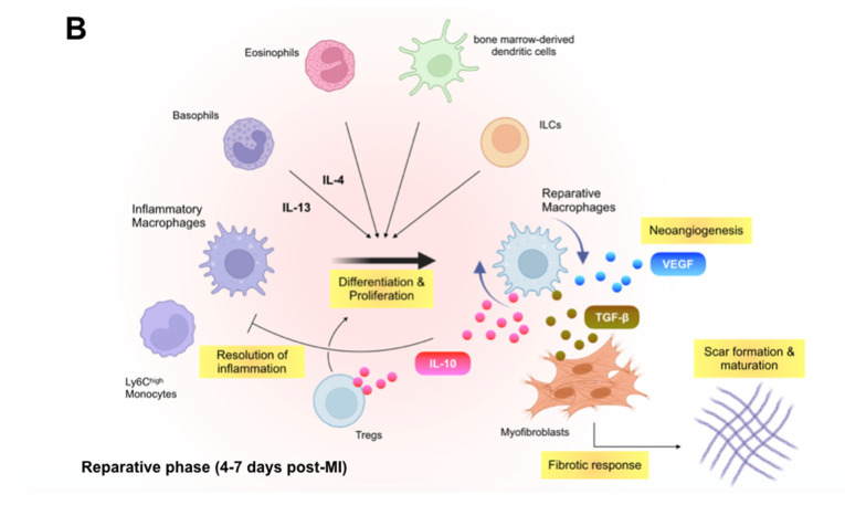
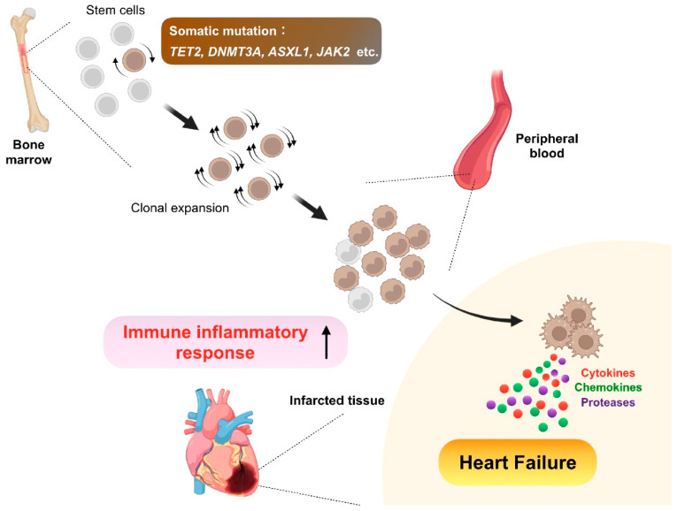
Despite recent scientific and technological advances, myocardial infarction (MI) still represents a major global health problem, leading to high morbidity and mortality worldwide. During the post-MI wound healing process, dysregulated immune inflammatory pathways and failure to resolve inflammation are associated with maladaptive left ventricular remodeling, progressive heart failure, and eventually poor outcomes. Given the roles of immune cells in the host response against tissue injury, understanding the involved cellular subsets, sources, and functions is essential for discovering novel therapeutic strategies that preserve the protective immune system and promote optimal healing. This review discusses the cellular effectors and molecular signals across multi-organ systems, which regulate the inflammatory and reparative responses after MI. Additionally, we summarize the recent clinical and preclinical data that propel conceptual revolutions in cardiovascular immunotherapy.

Keywords: adaptive immunity; chemokines; clinical trial; clonal hematopoiesis; cytokines; growth factors; heart failure; hematopoiesis; immune cells; immunotherapy; inflammation; innate immunity; myocardial infarction.


Conflict of interest statement

The authors have no conflict of interest to declare.

Figures

Figure 1
Figure 1
Immune inflammatory response within the infarcted heart. (A): Shortly after the onset of myocardial infarction (MI), endogenous damage-associated molecular patterns (DAMPs) released from necrotic cardiomyocytes and other injured cells stimulate surviving resident cells and recruited immune cells to produce inflammatory cytokines, chemokines, and proteases (e.g., interleukin [IL]-1β, C-C motif chemokine ligand 2 [CCL2], and matrix metalloproteinases), which initiate innate immune response. Inflammatory macrophages scavenge dying cardiomyocyte-derived DNA fragments to activate interferon regulatory factor 3 (IRF3)-dependent type I interferon (IFN) signals that enhance infarct inflammation. Cardiac fibroblasts produce and release granulocyte-macrophage colony-stimulating factor (GM-CSF) in response to the engagement of their pattern recognition receptors (PRRs), which not only endows monocytes and macrophages with a pro-inflammatory signature but also stimulates bone marrow hematopoiesis. B cell-derived CCL7 further enhance monocyte accumulation in the infarcted heart. (B): In the later phase of ischemic injury, inflammatory macrophages convert their features to the anti-inflammatory phenotype. The transition is supported partly by regulatory T cells (Tregs), basophils, eosinophils, dendritic cells, and innate lymphoid cells (ILCs) and defines the reparative response in the wound healing process. Reparative macrophages as well as Tregs produce anti-inflammatory cytokines (e.g., IL-10) that resolve inflammation. The reparative macrophages likewise produce transforming growth factor (TGF)-β and vascular endothelial growth factor (VEGF), thereby promoting angiogenesis, fibrosis, and scar formation.
Figure 1
Figure 1
Immune inflammatory response within the infarcted heart. (A): Shortly after the onset of myocardial infarction (MI), endogenous damage-associated molecular patterns (DAMPs) released from necrotic cardiomyocytes and other injured cells stimulate surviving resident cells and recruited immune cells to produce inflammatory cytokines, chemokines, and proteases (e.g., interleukin [IL]-1β, C-C motif chemokine ligand 2 [CCL2], and matrix metalloproteinases), which initiate innate immune response. Inflammatory macrophages scavenge dying cardiomyocyte-derived DNA fragments to activate interferon regulatory factor 3 (IRF3)-dependent type I interferon (IFN) signals that enhance infarct inflammation. Cardiac fibroblasts produce and release granulocyte-macrophage colony-stimulating factor (GM-CSF) in response to the engagement of their pattern recognition receptors (PRRs), which not only endows monocytes and macrophages with a pro-inflammatory signature but also stimulates bone marrow hematopoiesis. B cell-derived CCL7 further enhance monocyte accumulation in the infarcted heart. (B): In the later phase of ischemic injury, inflammatory macrophages convert their features to the anti-inflammatory phenotype. The transition is supported partly by regulatory T cells (Tregs), basophils, eosinophils, dendritic cells, and innate lymphoid cells (ILCs) and defines the reparative response in the wound healing process. Reparative macrophages as well as Tregs produce anti-inflammatory cytokines (e.g., IL-10) that resolve inflammation. The reparative macrophages likewise produce transforming growth factor (TGF)-β and vascular endothelial growth factor (VEGF), thereby promoting angiogenesis, fibrosis, and scar formation.
Figure 2
Figure 2
Remote organs contributing to immune and inflammatory networks after myocardial infarction (MI). Bone marrow generates billions of immune cells and supplies them to the injured heart, thereby essentially contributing to the immune inflammatory response after MI. Infarct-derived inflammatory cytokines (e.g., interleukin [IL]-1β), damage-associated molecular patterns (e.g., S100A8), and growth factors (e.g., granulocyte-macrophage colony-stimulating factor [GM-CSF]) as well as increased sympathetic nervous signals stimulate bone marrow hematopoietic stem cell proliferation and immune cell production. Spleen also has stem cells and produces mainly myeloid cells in a process known as extramedullary hematopoiesis. Splenic reservoir monocytes exit the spleen and accumulate in injured tissue depending on angiotensin II signaling during the first day of MI. The miR21/HIF-1α axis in splenic marginal zone B cells is essential for C-C motif chemokine ligand 7 production, thereby promoting inflammatory monocyte mobilization to the infarcted heart. B cells also reside in pericardial adipose tissue where GM-CSF-producing B cells potentially enhance infarct inflammation, whereas IL-10-producing CD5+ B cells promote the resolution of MI-induced inflammation. Innate lymphoid cells (ILCs) also expand in the pericardial adipose tissue after MI and exhibit beneficial function. Pericardial fluid contains a discrete subset of macrophages expressing GATA binding protein 6, which may be protective against diastolic heart failure following MI. Mediastinal lymph nodes are heart-draining lymph nodes where regulatory T cells (Tregs) expand and proliferate with antigen presentation to regulate repair of the ischemic myocardium.
Figure 3
Figure 3
Clonal hematopoiesis. As we age, somatic mutations accumulate in bone marrow hematopoietic stem cells, some of which confer a competitive advantage or fitness, leading to clonal expansion of the mutated cells. Consequently, the bone marrow expels clonal descendants of the mutant stem cells into peripheral circulation. In this premalignant condition called clonal hematopoiesis, genes frequently mutated are Tet methylcytosine dioxygenase 2 (TET2), DNA methyltransferase 3 alpha (DNMT3A), addition of sex combs like 1 (ASXL1), and Janus kinase 2 (JAK2), and the somatic mutations confer both increased proliferation capacity and inflammatory signature to the mutated cells, thereby leading to adverse post-infarction heart failure with enhanced inflammation.

References

    1. Jernberg T., Hasvold P., Henriksson M., Hjelm H., Thuresson M., Janzon M. Cardiovascular risk in post-myocardial infarction patients: Nationwide real world data demonstrate the importance of a long-term perspective. Eur. Heart J. 2015;36:1163–1170. doi: 10.1093/eurheartj/ehu505. - DOI - PubMed
    1. Tsao C.W., Aday A.W., Almarzooq Z.I., Alonso A., Beaton A.Z., Bittencourt M.S., Boehme A.K., Buxton A.E., Carson A.P., Commodore-Mensah Y., et al. Heart Disease and Stroke Statistics-2022 Update: A Report from the American Heart Association. Circulation. 2022;145:e153–e639. - PubMed
    1. Frazier C.G., Alexander K.P., Newby L.K., Anderson S., Iverson E., Packer M., Cohn J., Goldstein S., Douglas P.S. Associations of gender and etiology with outcomes in heart failure with systolic dysfunction: A pooled analysis of 5 randomized control trials. J. Am. Coll. Cardiol. 2007;49:1450–1458. doi: 10.1016/j.jacc.2006.11.041. - DOI - PubMed
    1. Shoji S., Shiraishi Y., Kohsaka S., Fukuda K., Yoshikawa T. Long-Term Outcomes According to Etiology May Alter under Different Circumstances. JACC Heart Fail. 2020;8:83–84. doi: 10.1016/j.jchf.2019.08.021. - DOI - PubMed
    1. Shore S., Grau-Sepulveda M.V., Bhatt D.L., Heidenreich P.A., Eapen Z.J., Hernandez A.F., Yancy C.W., Fonarow G.C. Characteristics, Treatments, and Outcomes of Hospitalized Heart Failure Patients Stratified by Etiologies of Cardiomyopathy. JACC Heart Fail. 2015;3:906–916. doi: 10.1016/j.jchf.2015.06.012. - DOI - PubMed