Review
. 2022 May 8;23(9):5252.
doi: 10.3390/ijms23095252.

Recent Progress on the Localization of PLK1 to the Kinetochore and Its Role in Mitosis

Affiliations
Review

Recent Progress on the Localization of PLK1 to the Kinetochore and Its Role in Mitosis

Taekyung Kim. Int J Mol Sci. .

Abstract

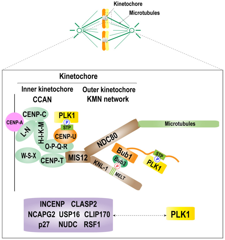
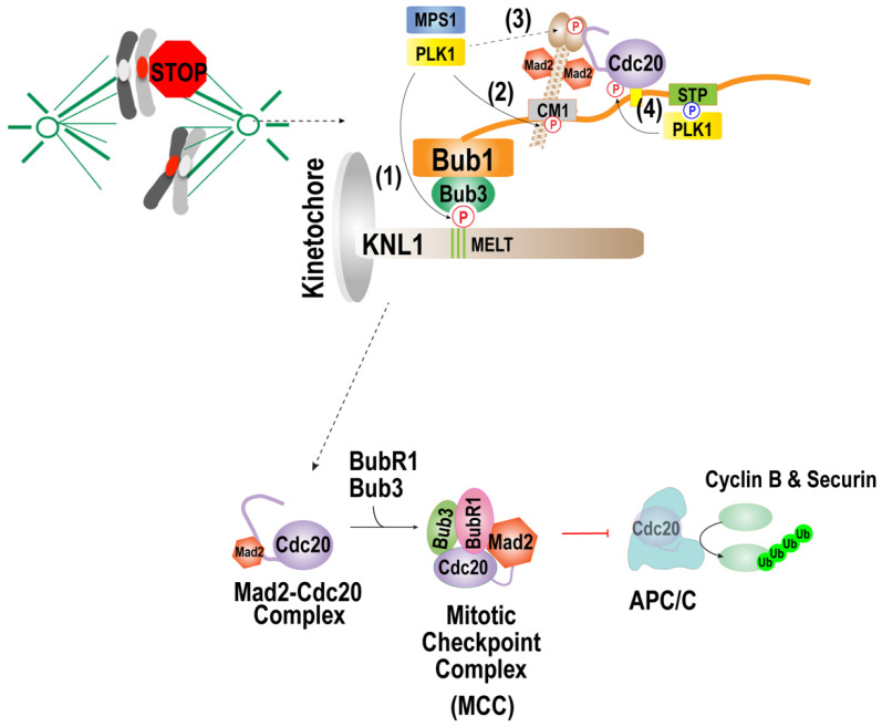
The accurate distribution of the replicated genome during cell division is essential for cell survival and healthy organismal development. Errors in this process have catastrophic consequences, such as birth defects and aneuploidy, a hallmark of cancer cells. PLK1 is one of the master kinases in mitosis and has multiple functions, including mitotic entry, chromosome segregation, spindle assembly checkpoint, and cytokinesis. To dissect the role of PLK1 in mitosis, it is important to understand how PLK1 localizes in the specific region in cells. PLK1 localizes at the kinetochore and is essential in spindle assembly checkpoint and chromosome segregation. However, how PLK1 localizes at the kinetochore remains elusive. Here, we review the recent literature on the kinetochore recruitment mechanisms of PLK1 and its roles in spindle assembly checkpoint and attachment between kinetochores and spindle microtubules. Together, this review provides an overview of how the local distribution of PLK1 could regulate major pathways in mitosis.

Keywords: PLK1 kinase; PP1; PP2A; cell cycle; chromosome segregation; kinetochore; spindle assembly checkpoint.


Conflict of interest statement

The author declares no conflict of interest.

Figures

Figure 1
Figure 1
Domain organization of PLK1. A schematic diagram of human PLK1 protein and its domain organization. PLK1 contains the serine/threonine kinase domain (KD: orange), the polo-box domain (PBD: green), and the linker between two domains. The PBD consists of polo-box 1 and 2 (PB1 and PB2: yellow). PLK1 activation requires the phosphorylation of conserved Thr residues within the T-Loop.
Figure 2
Figure 2
Molecular mechanism of PLK1 localization at the kinetochore. A schematic diagram of the kinetochore structure and the mechanism of kinetochore recruitment of PLK1. The kinetochore consists of an inner and an outer kinetochore. The inner kinetochore is composed of a complex of CCAN, CENP-C, CENP-HIK-LN, CENP-T, and CENP-QU. The outer kinetochore is composed of KNL1 complex, Mis12 complex, and the NDC80 complex (KMN network). PLK1 is recruited to the kinetochore by interacting with CENP-U and Bub1, which are located in the inner kinetochore and outer kinetochore, respectively. There are other known PLK1 receptors, including the kinetochore components Bub1, BubR1, and CENP-U (also called PBIP1), and other proteins, including nuclear distribution protein C (NUDC), CLIP-170, dynactin subunit p27, INCENP, CLASP2, Survivin, NCAPG2, USP16, and RSF1. The relationship between PLK1 and these components during mitosis requires further characterization.
Figure 3
Figure 3
Function of the PLK1 in spindle assembly checkpoint activation. Reprinted/adapted with permission from Ref. [133], 2021, Taekyung Kim. When kinetochores are not attached to the microtubules, PLK1 activates the checkpoint by phosphorylating (1) MELT motifs of KNL1 and (2) the CM1 motif of Bub1, (3) C-terminal region of Mad1, and (4) Cdc20. The PLK1 can substitute Mps1 kinase in phosphorylating MELT motifs of KNL1, which promotes kinetochore localization of the Bub1/Bub3 complex and the CM1 motif of bub1, mediating the interaction with Mad1. PLK-1 in C. elegans was shown to phosphorylate the C-terminal region of Mad1, which promotes the interaction with Mad1. PLK1 that interacts with Bub1 activates the MCC formation by phosphorylating Cdc20.

Similar articles

Cited by

References

    1. Santaguida S., Amon A. Short- and long-term effects of chromosome mis-segregation and aneuploidy. Nat. Rev. Mol. Cell Biol. 2015;16:473. doi: 10.1038/nrm4025. - DOI - PubMed
    1. Funk L.C., Zasadil L.M., Weaver B.A. Living in CIN: Mitotic Infidelity and Its Consequences for Tumor Promotion and Suppression. Dev. Cell. 2016;39:638–652. doi: 10.1016/j.devcel.2016.10.023. - DOI - PMC - PubMed
    1. Lara-Gonzalez P., Pines J., Desai A. Spindle assembly checkpoint activation and silencing at kinetochores. Semin. Cell Dev. Biol. 2021;117:86–98. doi: 10.1016/j.semcdb.2021.06.009. - DOI - PMC - PubMed
    1. Musacchio A., Desai A. A Molecular View of Kinetochore Assembly and Function. Biology. 2017;6:5. doi: 10.3390/biology6010005. - DOI - PMC - PubMed
    1. Saurin A.T. Kinase and Phosphatase Cross-Talk at the Kinetochore. Front. Cell Dev. Biol. 2018;6:62. doi: 10.3389/fcell.2018.00062. - DOI - PMC - PubMed

Substances

LinkOut - more resources