. 2022 May 9;23(9):5265.
doi: 10.3390/ijms23095265.

Hedgehog Signaling Pathway Orchestrates Human Lung Branching Morphogenesis

Affiliations

Hedgehog Signaling Pathway Orchestrates Human Lung Branching Morphogenesis

Randa Belgacemi et al. Int J Mol Sci. .

Abstract

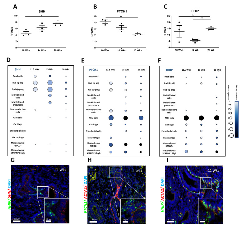
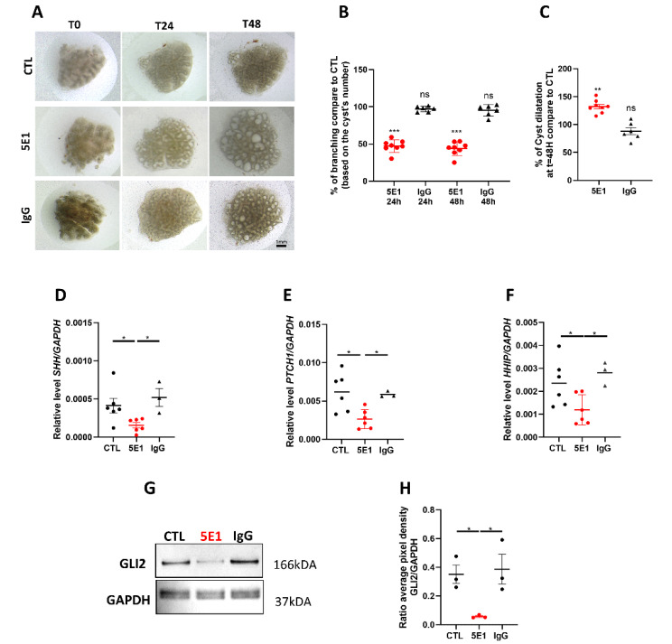
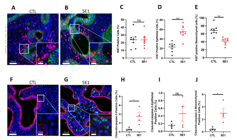
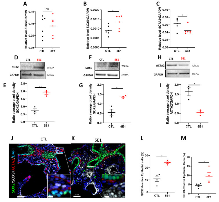
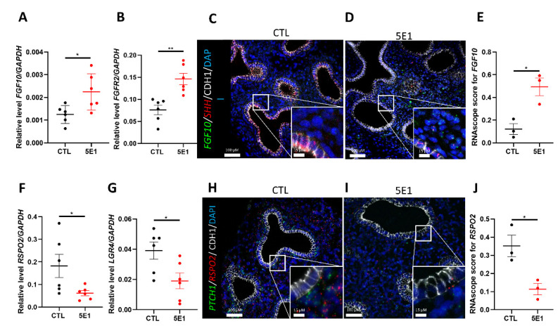
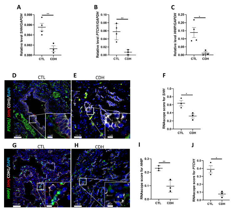
The Hedgehog (HH) signaling pathway plays an essential role in mouse lung development. We hypothesize that the HH pathway is necessary for branching during human lung development and is impaired in pulmonary hypoplasia. Single-cell, bulk RNA-sequencing data, and human fetal lung tissues were analyzed to determine the spatiotemporal localization of HH pathway actors. Distal human lung segments were cultured in an air-liquid interface and treated with an SHH inhibitor (5E1) to determine the effect of HH inhibition on human lung branching, epithelial-mesenchymal markers, and associated signaling pathways in vitro. Our results showed an early and regulated expression of HH pathway components during human lung development. Inhibiting HH signaling caused a reduction in branching during development and dysregulated epithelial (SOX2, SOX9) and mesenchymal (ACTA2) progenitor markers. FGF and Wnt pathways were also disrupted upon HH inhibition. Finally, we demonstrated that HH signaling elements were downregulated in lung tissues of patients with a congenital diaphragmatic hernia (CDH). In this study, we show for the first time that HH signaling inhibition alters important genes and proteins required for proper branching of the human developing lung. Understanding the role of the HH pathway on human lung development could lead to the identification of novel therapeutic targets for childhood pulmonary diseases.

Keywords: Hedgehog pathway; branching; development; human lung.


Conflict of interest statement

The authors declare no conflict of interest.

Figures

Figure 1
Figure 1
Spatiotemporal expression of Hedgehog pathway components is finely tuned during human lung development. Gene expression of SHH (A), PTCH1 (B), and HHIP (C) expressed in RPKMs ± SEM within the developing lung at 10-, 14- and 20-week gestation (n = 3 per time point, * p < 0.05, ** p < 0.01). (DF) Dot plots show the percentage of cells expressing SHH (D), PTCH1 (E), and HHIP (F) using dot size and the average expression level of that gene based on unique molecular identifier (UMI) counts. (GI) Fluorescent in situ hybridization (FISH) on 11 wks gestation fetal human lung sections showing SHH (red) and HHIP (green) (G); PTCH1 (green) with ACTA2 (IF, red) (H) and HHIP (green) with ACTA2 (IF, red) (I). Scale bars are 100 µm for large insets and 15 µm for small insets.
Figure 2
Figure 2
The Hedgehog pathway is required for human lung branching morphogenesis. (A) Human fetal lung explant cultures at t = 0, t = 24 h and t = 48 h untreated (in media alone); or treated with 5 µg/mL IgG as negative control or 5 µg/mL 5E1. (B,C) Graphs showing the percentage of distal branching (B) and cyst dilation (C) of 5E1, and IgG treated explants as compared to untreated control (CTL) at t = 24 h and t = 48 h. Results are shown in dots and mean ± SEM (n = 8 for control and 5E1, n = 6 for IgG). (DF) RT-qPCR for SHH (D), PTCH1 (E), and HHIP (F) performed on control (n = 6), 5E1- (n = 6; red dots) and IgG- treated explants (n = 3). Results are shown as individual data points and mean ± SEM. (G) Representative western blots for GLI2 and GAPDH in human fetal lung explants treated or not with 5E1 or IgG (5 µg/mL) for 48 h. (H) Dot plots (mean ± SEM) of western blot densitometry ratios for GLI2 normalized to GAPDH (n = 3 for each condition). * p < 0.05, ** p < 0.01; *** p < 0.001; ns = no stress.
Figure 3
Figure 3
Hedgehog pathway inhibition alters cell proliferation and cell death in human fetal lung explants. (AD) IF staining of human fetal lung explants treated with 5E1 for 48 h (B) or untreated (A) using KI67 (red) and CDH1 (as epithelial marker in green) to assess cell proliferation. Quantification of total cell proliferation (C), epithelial proliferation (D), and mesenchymal proliferation (E). Results are shown as mean ± SEM, n = 8 for each group. (F,G) Cleaved-caspase-3 IF staining of human fetal lungs treated (G) or not (F) with 5E1 to assess cell death (G vs. F; n = 4), Cleaved-caspase-3 is in green; and CDH1 in red. Quantification of total number of Cleaved-caspase-3+ cells (H), Cleaved-caspase3+ cells in the epithelium (I), and Cleaved-caspase-3+ cells in the mesenchyme (J). Results are shown mean ± SEM. * p < 0.05; ** p < 0.01 and *** p < 0.001; ns = no stress.
Figure 4
Figure 4
Compartmental cell identity is altered upon Hedgehog pathway inhibition. RT-qPCR for SOX2 (A), SOX9 (B), and ACTA2 (C) in fetal lung explants treated with 5E1 compared to control. Results are shown as mean ± SEM, * p < 0.05, n = 6 for each group. (DH) Western blot analysis of protein expression of SOX2 (D,E), SOX9 (F,G), and ACTA2 (H,I) in 5E1 treated explants compared to control (n = 3 for each condition). Western blot densitometry ratios are shown in (E,G,I) (n = 3 for each condition). Results are shown as mean ± SEM. IF staining of fetal lung explants treated (K) or not (J) with 5E1 for SOX2 (green), SOX9 (white), and ACTA2 (red). Quantification of the number of epithelial positive SOX2 cells (L) and SOX9 (M) (n = 4 for each group). * p < 0.05, ** p < 0.01; ns = no stress.
Figure 5
Figure 5
Inhibiting Hedgehog signaling disrupted FGF and WNT pathways. RT-qPCR for FGF10 (A) and FGFR2 (B) in 5E1-treated explants compared to control (results show mean ± SEM, n = 6 for each group). In situ hybridization for FGF10 (green), SHH (red), and CDH1 (IF-white) performed on control (C) and 5E1-treated (D) explants. FISH dots quantification (E) revealed more FGF10 in 5E1 compared to control (results show mean ± SEM, n = 3 for each group). RT-qPCR for RSPO2 (F) and its receptor LGR4 (G) on explants treated with 5E1 compared to control (results show mean ± SEM, n = 6 for each group). In situ hybridization for PTCH1 (green), RSPO2 (red), and CDH1 (IF-white) performed on control (H) and 5E1-treated (I) explants. (J) FISH dots quantification for RSPO2 confirmed RT-qPCR results (results show mean ± SEM; n = 3 in each group). * p < 0.05; ** p < 0.01.
Figure 6
Figure 6
Hedgehog pathway is downregulated in Congenital Diaphragmatic Hernia; (AC) RT-qPCR on tissue from patients presenting with Congenital Diaphragmatic Hernia (CDH) and controls for SHH (A), PTCH1 (B), and HHIP (C) (results show mean ± SEM, * p < 0.05; ** p < 0.01; n = 4 for CTL and n = 3 for CDH). Combinatorial FISH with IF for either PTCH1 (green, (D,E)) or HHIP (green, (G,H)) with SHH (red) and CDH1 (IF-white) performed on tissue sections from CDH (E,H) and control (D,G) patients; dot plot quantification (F,I,J) of the FISH signal in CDH vs. control sections (results show mean ± SEM, * p < 0.05; ** p < 0.01; n = 3 for CTL and n = 3 for CDH).

References

    1. Schittny J.C. Development of the Lung. Cell Tissue Res. 2017;367:427–444. doi: 10.1007/s00441-016-2545-0. - DOI - PMC - PubMed
    1. Burri P.H. Fetal and Postnatal Development of the Lung. Annu. Rev. Physiol. 1984;46:617–628. doi: 10.1146/annurev.ph.46.030184.003153. - DOI - PubMed
    1. Mullassery D., Smith N.P. Lung Development. Semin. Pediatr. Surg. 2015;24:152–155. doi: 10.1053/j.sempedsurg.2015.01.011. - DOI - PubMed
    1. Herriges M., Morrisey E.E. Lung Development: Orchestrating the Generation and Regeneration of a Complex Organ. Development. 2014;141:502–513. doi: 10.1242/dev.098186. - DOI - PMC - PubMed
    1. Mora M.J., Extramiana J., Paniagua P., González P., Mañas A., Pérez M.J., Navarro J., Arrizabalaga M. Orchiectomy and buserelin in combination with flutamide: Comparative results in metastatic prostatic carcinoma. Actas Urol. Esp. 1991;15:548–552. - PubMed

Substances