Review
. 2022 May 9;23(9):5288.
doi: 10.3390/ijms23095288.

Molecular Research on Oral Diseases and Related Biomaterials: A Journey from Oral Cell Models to Advanced Regenerative Perspectives

Affiliations
Review

Molecular Research on Oral Diseases and Related Biomaterials: A Journey from Oral Cell Models to Advanced Regenerative Perspectives

Thorsten Steinberg et al. Int J Mol Sci. .

Abstract

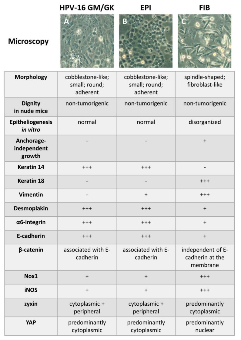
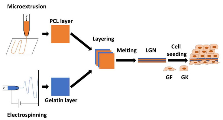
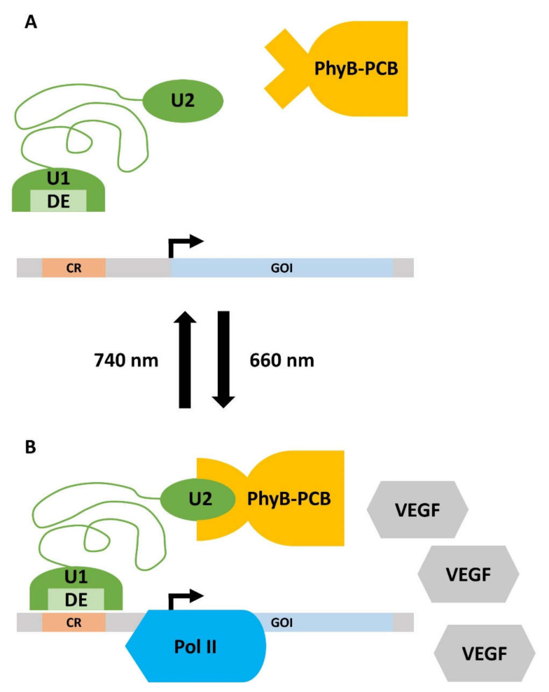
Oral diseases such as gingivitis, periodontitis, and oral cancer affect millions of people worldwide. Much research has been conducted to understand the pathogenetic mechanisms of these diseases and translate this knowledge into therapeutics. This review aims to take the reader on a journey from the initial molecular discoveries to complex regenerative issues in oral medicine. For this, a semi-systematic literature search was carried out in Medline and Web of Science databases to retrieve the primary literature describing oral cell models and biomaterial applications in oral regenerative medicine. First, an in vitro cell model of gingival keratinocytes is discussed, which illustrates patho- and physiologic principles in the context of oral epithelial homeostasis and carcinogenesis and represents a cellular tool to understand biomaterial-based approaches for periodontal tissue regeneration. Consequently, a layered gradient nonwoven (LGN) is described, which demonstrates that the key features of biomaterials serve as candidates for oral tissue regeneration. LGN supports proper tissue formation and obeys the important principles for molecular mechanotransduction. Furthermore, current biomaterial-based tissue regeneration trends, including polymer modifications, cell-based treatments, antimicrobial peptides and optogenetics, are introduced to represent the full spectrum of current approaches to oral disease mitigation and prevention. Altogether, this review is a foray through established and new concepts in oral regenerative medicine and illustrates the process of knowledge translation from basic molecular and cell biological research to future clinical applications.

Keywords: carcinogenesis; cell transformation; mechanotransduction; mesenchymal stem cells; non-woven; tissue homeostasis.


Conflict of interest statement

The authors declare no conflict of interest.

Figures

Figure 1
Figure 1
Summary of the morphological and molecular properties of HPV-16 GM (=GK), and the alcohol-treated cell lines EPI (epitheloid) and FIB (fibro-blastoid). The upper part of the figure depicts exemplary light microscopy pictures of the cell lines at the same magnification. (A) shows the GKs with their cobblestone morphology, which is typical for keratinocytes. (B) depicts EPI cells, which share morphological similarities with GKs but show more cytoplasmic inclusions and more pronounced nuclei. In (C), the fibroblast-like, spindle-shaped FIB cells are presented. When compared to GKs and EPI, FIB cells do not resemble normal keratinocytes and are characterized by multidirectional cytoplasmic extensions and a less dense growth pattern. The lower part of the figure summarizes the main cell behavioral and molecular properties of the three cell lines. The protein expression levels are designated as follows: +++ very strong expression, + strong expression, - no expression. Details are given in the main text. (Nox1 = NADPH Oxidase 1; iNOS = inducible Nitric Oxide Synthase; YAP = Yes-Associated Protein.)
Figure 2
Figure 2
Schematic representation of the manufacturing process of the layered gradient nonwoven (LGN). The polycaprolactone (PCL) layers were generated by 3D micro-extrusion (upper left part; orange). During this process, the polymer solution is extruded through a narrow orifice and deposited on a carrier platform. The gelatin layers were generated by electrospinning (lower left part; blue). The polymer solution is also extruded through an orifice but is additionally accelerated towards a collector (black bar) via an electric field. Alternating layers of PCL and gelatin were then briefly heated above the melting point of PCL, yielding the LGN. Subsequently, the cells (GF = gingival fibroblast; GK = gingival keratinocyte) were transferred on opposite sides of the LGN and incubated in culture medium. The resulting stratified epithelium and the layer of GFs is depicted on the right side. Details are given in the main text. (F = mechanical force).
Figure 3
Figure 3
Illustration of the polyethylene glycol (PEG)-based, pharmacologically tunable “smart” hydrogel. (A) Multi-arm PEG (green asterisk-like structure) was used as a backbone for the hydrogel. It was chemically coupled to Gyrase B (GyrB; blue hemicycle) and the arginine–glycine–aspartate binding motif (RGD motif; orange bar) from fibronectin. Upon the addition of coumermycin (red ellipse), GyrB molecules can dimerize, which leads to hydrogel assembly. Subsequently, cells (as exemplarily represented by gingiva keratinocytes (GKs)) can be seeded on the hydrogel, which can bind to the RGD motif via membrane-inherent integrin receptors (grey receptors). (B) Dissociation of the hydrogel is induced by adding novobiocin (brown hemicycle). Novobiocin competes with coumermycin for the GyrB binding sites. Contrary to coumermycin, novobiocin does not lead to dimerization of the GyrB molecules. Excess amounts of novobiocin, therefore, lead to the disassembly of the hydrogel. (C) When additionally coupled with the ZZ-domain of Protein A (green triangle), the multi-arm PEG can bind fibroblast growth factor 7 (FGF7), which is modified with Fc (crystallizable fragment of antibodies; yellow/grey construct). When adding novobiocin, the hydrogel disassembles and FGF7 can then bind to FGF7 receptors (FGF7-R; dark blue receptor) on cells such as GKs. Details are given in the main text. A graphic legend is included in the lower right corner.
Figure 4
Figure 4
Principles of the design of synthetic mimics of antimicrobial peptides (SMAMPS). The SMAMPS described in the main text are based on a poly(oxanorbornene) backbone. (A) shows the chemical structure of unit 1 (U1; see main text), which is a monomer from a poly(oxanorbornene) structure. The side chains are composed of a butyl residue (left side) and a positively charged ethylamine (right side). (B) Unit 2 (U2; see main text) differs from U1 in that the butyl chain was replaced by another ethylamine residue, yielding two positive charges per monomer. (C) Schematic representation of a complete SMAMP composed of 1 U1 and 9 U2 subunits, which, therefore, has a considerable number of positive charges and leads to the selective killing of various bacteria. Details are given in the main text.
Figure 5
Figure 5
Working principle of the optogenetic gene expression switch. (A) Upon irradiation with far-red light (740 nm), the gene expression is turned off. The phycocyanobilin B (PCB), which is bound to phytochromobilin (PhyB), is in a closed conformation and cannot bind to unit 2 (U2) of the split transcription factor (green). Thus, the distinct element (DE) of unit 1 (U1) of the transcription factor does not interact with the certain operator region (CR) on the response vector. Consequently, there is no detectable expression of the gene of interest (GOI). (B) Illumination with red light (660 nm) leads to the isomerization of PCB, which induces a conformational change in the PhyB-PCB complex. PhyB-PCB can now recognize U2, which activates the split transcription factor. DE recognizes CR and recruits RNA Polymerase II (Pol II) to the promotor of the response vector. The GOI, in this case, vascular endothelial growth factor (VEGF), is now expressed. Details are given in the main text.

Similar articles

Cited by

References

    1. Ahmad P., Rubbia Nawaz M.Q., Shaikh G.M., Mohamed R.N., Nagarajappa A.K., Asif J.A., Alam M.K. Risk factors associated with the mortality rate of oral squamous cell carcinoma patients: A 10-year retrospective study. Medicine. 2021;100:e27127. doi: 10.1097/MD.0000000000027127. - DOI - PMC - PubMed
    1. Wu L., Zhang S.Q., Zhao L., Ren Z.H., Hu C.Y. Global, regional, and national burden of periodontitis from 1990 to 2019: Results from the Global Burden of Disease study 2019. J. Periodontol. 2022 doi: 10.1002/JPER.21-0469. - DOI - PubMed
    1. Jepsen S., Caton J.G., Albandar J.M., Bissada N.F., Bouchard P., Cortellini P., Demirel K., de Sanctis M., Ercoli C., Fan J. Periodontal manifestations of systemic diseases and developmental and acquired conditions: Consensus report of workgroup 3 of the 2017 World Workshop on the Classification of Periodontal and Peri-Implant Diseases and Conditions. J. Clin. Periodontol. 2018;45:S219–S229. doi: 10.1111/jcpe.12951. - DOI - PubMed
    1. Muller S., Tilakaratne W.M. Update from the 5th Edition of the World Health Organization Classification of Head and Neck Tumors: Tumours of the Oral Cavity and Mobile Tongue. Head Neck Pathol. 2022;16:54–62. doi: 10.1007/s12105-021-01402-9. - DOI - PMC - PubMed
    1. Saccucci M., Di Carlo G., Bossù M., Giovarruscio F., Salucci A., Polimeni A. Autoimmune diseases and their manifestations on oral cavity: Diagnosis and clinical management. J. Immunol. Res. 2018;2018 doi: 10.1155/2018/6061825. - DOI - PMC - PubMed

Substances

LinkOut - more resources