Review
. 2022 Apr 20;11(9):1386.
doi: 10.3390/cells11091386.

Properties and Functions of Fibroblasts and Myofibroblasts in Myocardial Infarction

Affiliations
Review

Properties and Functions of Fibroblasts and Myofibroblasts in Myocardial Infarction

Harikrishnan Venugopal et al. Cells. .

Abstract

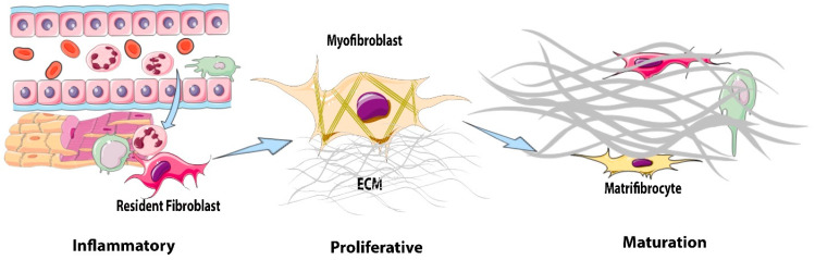
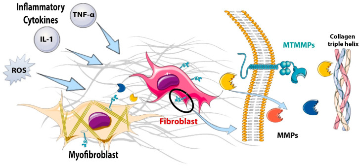
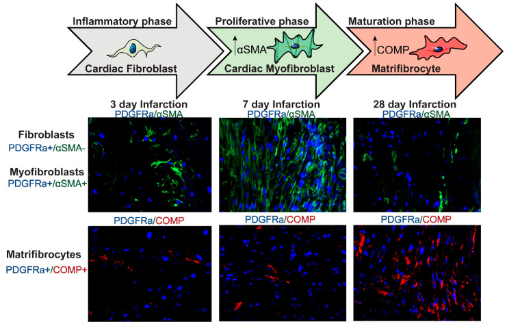
The adult mammalian heart contains abundant interstitial and perivascular fibroblasts that expand following injury and play a reparative role but also contribute to maladaptive fibrotic remodeling. Following myocardial infarction, cardiac fibroblasts undergo dynamic phenotypic transitions, contributing to the regulation of inflammatory, reparative, and angiogenic responses. This review manuscript discusses the mechanisms of regulation, roles and fate of fibroblasts in the infarcted heart. During the inflammatory phase of infarct healing, the release of alarmins by necrotic cells promotes a pro-inflammatory and matrix-degrading fibroblast phenotype that may contribute to leukocyte recruitment. The clearance of dead cells and matrix debris from the infarct stimulates anti-inflammatory pathways and activates transforming growth factor (TGF)-β cascades, resulting in the conversion of fibroblasts to α-smooth muscle actin (α-SMA)-expressing myofibroblasts. Activated myofibroblasts secrete large amounts of matrix proteins and form a collagen-based scar that protects the infarcted ventricle from catastrophic complications, such as cardiac rupture. Moreover, infarct fibroblasts may also contribute to cardiac repair by stimulating angiogenesis. During scar maturation, fibroblasts disassemble α-SMA+ stress fibers and convert to specialized cells that may serve in scar maintenance. The prolonged activation of fibroblasts and myofibroblasts in the infarct border zone and in the remote remodeling myocardium may contribute to adverse remodeling and to the pathogenesis of heart failure. In addition to their phenotypic plasticity, fibroblasts exhibit remarkable heterogeneity. Subsets with distinct phenotypic profiles may be responsible for the wide range of functions of fibroblast populations in infarcted and remodeling hearts.

Keywords: angiogenesis; cytokine; extracellular matrix; fibroblast; fibrosis; myocardial infarction; myofibroblast; remodeling.


Conflict of interest statement

The authors declare no conflict of interest.

Figures

Figure 1
Figure 1
Phenotypic transitions of cardiac fibroblasts in the infarcted myocardium. During the inflammatory phase of infarct healing, necrotic cardiomyocytes release damage-associated molecular patterns that activate a pro-inflammatory, matrix-degrading phenotype in cardiac fibroblasts and contribute to the recruitment of leukocytes to the injury site. The clearance of dead cardiomyocytes stimulates anti-inflammatory signals, leading to the transition to the proliferative phase of infarct healing. During the proliferative phase of infarct healing, resident cardiac fibroblasts proliferate and undergo myofibroblast conversion, incorporating α-smooth muscle actin (α-SMA) into cytoskeletal stress fibers. Myofibroblasts are the main matrix-synthetic cells in the infarcted heart and produce both structural and matricellular extracellular matrix (ECM) proteins. During scar maturation, fibroblasts exhibit the disassembly of stress fibers and transition to matrifibrocytes, specialized cells that may play a role in scar maintenance.
Figure 2
Figure 2
The matrix-degrading properties of fibroblasts in the infarcted myocardium. During the inflammatory phase of infarct healing, pro-inflammatory cytokines, such as interleukin (IL)-1β, tumor necrosis factor (TNF)-α, and IL-6 stimulate the expression and secretion of matrix metalloproteinases (MMPs) that degrade the extracellular matrix, setting the stage for the replacement of damaged tissue with a collagen-based scar. During the inflammatory and early proliferative phases of infarct healing, the induction of membrane-type matrix metalloproteinases (MT-MMPs) on the cell surface plays an important role in fibroblast migration, thus localizing the reparative fibroblasts to the area of infarction.
Figure 3
Figure 3
Dynamics of fibroblast activation during the proliferative and maturation phases of infarct healing. During the inflammatory phase of infarct healing, resident cardiac fibroblasts (identified as PDGFRα+ cells in PDGFRαEGFP reporter mice) do not express the myofibroblast marker a-smooth muscle actin (α-SMA). During the proliferative phase (3–14 days), many fibroblasts become activated, proliferate, and differentiate into cardiac myofibroblasts (PDGFRα+/α-SMA+) that secrete large amounts of extracellular matrix proteins. During the maturation phase of infarct healing (14 days–2 months), myofibroblasts disassemble the α-SMA+ stress fibers and convert to matrifibrocytes, specialized cells expressing bone-cartilage proteins, such as cartilage oligomeric matrix protein (COMP).

References

    1. Frangogiannis N.G. Pathophysiology of Myocardial Infarction. Compr. Physiol. 2015;5:1841–1875. doi: 10.1002/cphy.c150006. - DOI - PubMed
    1. Humeres C., Frangogiannis N.G. Fibroblasts in the Infarcted, Remodeling, and Failing Heart. JACC Basic Transl. Sci. 2019;4:449–467. doi: 10.1016/j.jacbts.2019.02.006. - DOI - PMC - PubMed
    1. Burke R.M., Burgos Villar K.N., Small E.M. Fibroblast contributions to ischemic cardiac remodeling. Cell Signal. 2021;77:109824. doi: 10.1016/j.cellsig.2020.109824. - DOI - PMC - PubMed
    1. Hanna A., Shinde A.V., Frangogiannis N.G. Validation of diagnostic criteria and histopathological characterization of cardiac rupture in the mouse model of nonreperfused myocardial infarction. Am. J. Physiol. Heart Circ. Physiol. 2020;319:H948–H964. doi: 10.1152/ajpheart.00318.2020. - DOI - PMC - PubMed
    1. Humeres C., Shinde A.V., Hanna A., Alex L., Hernandez S.C., Li R., Chen B., Conway S.J., Frangogiannis N.G. Smad7 effects on TGF-beta and ErbB2 restrain myofibroblast activation and protect from postinfarction heart failure. J. Clin. Investig. 2022;132:e146926. doi: 10.1172/JCI146926. - DOI - PMC - PubMed

Publication types

Substances