Mast Cells Meet Cytomegalovirus: A New Example of Protective Mast Cell Involvement in an Infectious Disease
- PMID: 35563708
- PMCID: PMC9101682
- DOI: 10.3390/cells11091402
Mast Cells Meet Cytomegalovirus: A New Example of Protective Mast Cell Involvement in an Infectious Disease
Abstract
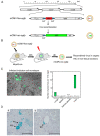
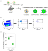
Cytomegaloviruses (CMVs) belong to the β-subfamily of herpesviruses. Their host-to-host transmission involves the airways. As primary infection of an immunocompetent host causes only mild feverish symptoms, human CMV (hCMV) is usually not considered in routine differential diagnostics of common airway infections. Medical relevance results from unrestricted tissue infection in an immunocompromised host. One risk group of concern are patients who receive hematopoietic cell transplantation (HCT) for immune reconstitution following hematoablative therapy of hematopoietic malignancies. In HCT patients, interstitial pneumonia is a frequent cause of death from hCMV strains that have developed resistance against antiviral drugs. Prevention of CMV pneumonia requires efficient reconstitution of antiviral CD8 T cells that infiltrate lung tissue. A role for mast cells (MC) in the immune control of lung infection by a CMV was discovered only recently in a mouse model. MC were shown to be susceptible for productive infection and to secrete the chemokine CCL-5, which recruits antiviral CD8 T cells to the lungs and thereby improves the immune control of pulmonary infection. Here, we review recent data on the mechanism of MC-CMV interaction, a field of science that is new for CMV virologists as well as for immunologists who have specialized in MC.
Keywords: CCL-5 chemokine; CD8 T cells; airway infection; antiviral protection; lung infection; mast cell (MC) degranulation; pneumonia; viral mitochondria-localized inhibitor of apoptosis (vMIA).
Conflict of interest statement
The authors declare no conflict of interest.
Figures


Similar articles
-
Mast cells: innate attractors recruiting protective CD8 T cells to sites of cytomegalovirus infection.Med Microbiol Immunol. 2015 Jun;204(3):327-34. doi: 10.1007/s00430-015-0386-1. Epub 2015 Feb 4. Med Microbiol Immunol. 2015. PMID: 25648117 Review.
-
The Anti-apoptotic Murine Cytomegalovirus Protein vMIA-m38.5 Induces Mast Cell Degranulation.Front Cell Infect Microbiol. 2020 Aug 25;10:439. doi: 10.3389/fcimb.2020.00439. eCollection 2020. Front Cell Infect Microbiol. 2020. PMID: 32984069 Free PMC article.
-
Mast cells expedite control of pulmonary murine cytomegalovirus infection by enhancing the recruitment of protective CD8 T cells to the lungs.PLoS Pathog. 2014 Apr 24;10(4):e1004100. doi: 10.1371/journal.ppat.1004100. eCollection 2014 Apr. PLoS Pathog. 2014. PMID: 24763809 Free PMC article.
-
Immunotherapy of cytomegalovirus infection by low-dose adoptive transfer of antiviral CD8 T cells relies on substantial post-transfer expansion of central memory cells but not effector-memory cells.PLoS Pathog. 2023 Nov 16;19(11):e1011643. doi: 10.1371/journal.ppat.1011643. eCollection 2023 Nov. PLoS Pathog. 2023. PMID: 37972198 Free PMC article.
-
Mouse Model of Cytomegalovirus Disease and Immunotherapy in the Immunocompromised Host: Predictions for Medical Translation that Survived the "Test of Time".Viruses. 2018 Dec 6;10(12):693. doi: 10.3390/v10120693. Viruses. 2018. PMID: 30563202 Free PMC article. Review.
Cited by
-
Immune-checkpoint expression in antigen-presenting cells (APCs) of cytomegaloviruses infection after transplantation: as a diagnostic biomarker.Arch Microbiol. 2023 Jul 10;205(8):280. doi: 10.1007/s00203-023-03623-8. Arch Microbiol. 2023. PMID: 37430000 Review.
References
-
- Thangam E.B., Jemima E.A., Singh H., Baig M.S., Khan M., Mathias C.B., Church M.K., Saluja R. The role of histamine and histamine receptors in mast cell-mediated allergy and inflammation: The hunt for new therapeutic targets. Front. Immunol. 2018;9:1873. doi: 10.3389/fimmu.2018.01873. - DOI - PMC - PubMed
Publication types
MeSH terms
Substances
LinkOut - more resources
Full Text Sources
Medical
Research Materials

