. 2022 Apr 25;11(9):1447.
doi: 10.3390/cells11091447.

Phytocomplex of a Standardized Extract from Red Orange (Citrus sinensis L. Osbeck) against Photoaging

Affiliations

Phytocomplex of a Standardized Extract from Red Orange (Citrus sinensis L. Osbeck) against Photoaging

Barbara Tomasello et al. Cells. .

Abstract

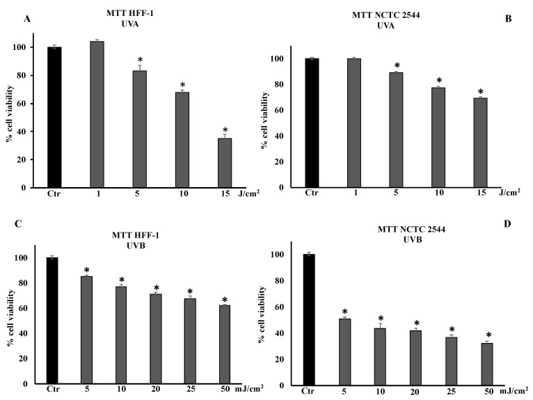
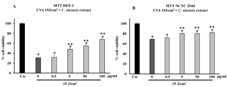
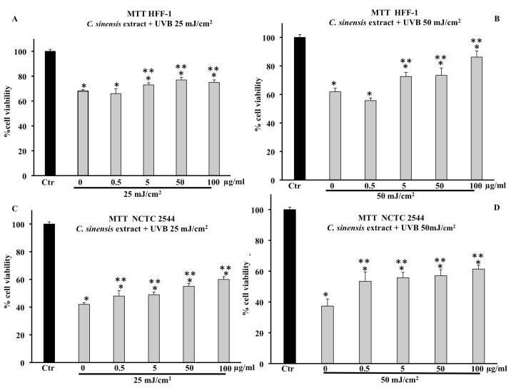
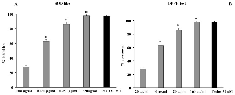
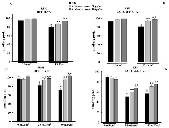
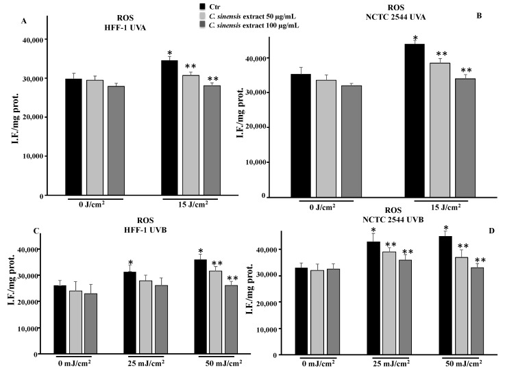
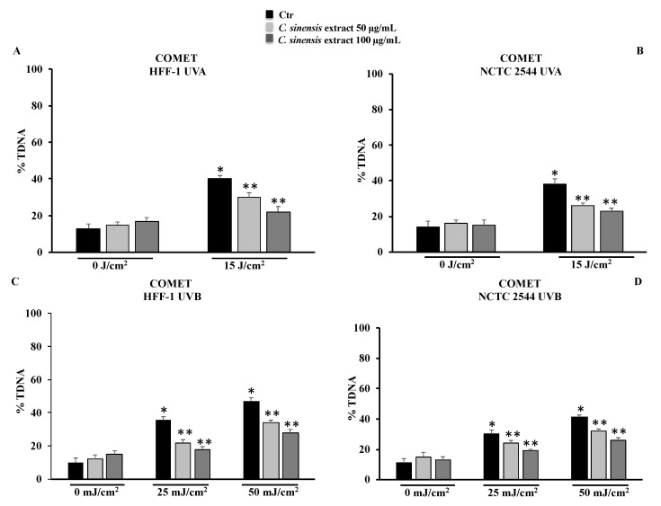
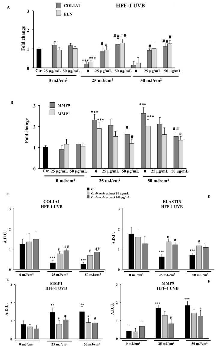
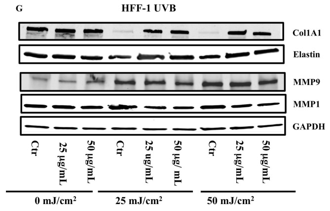
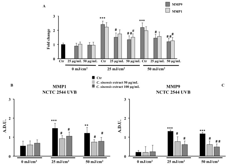
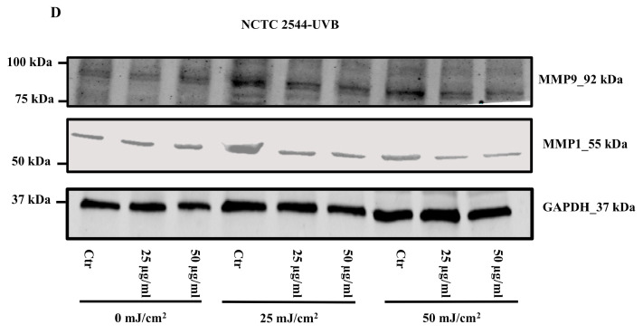
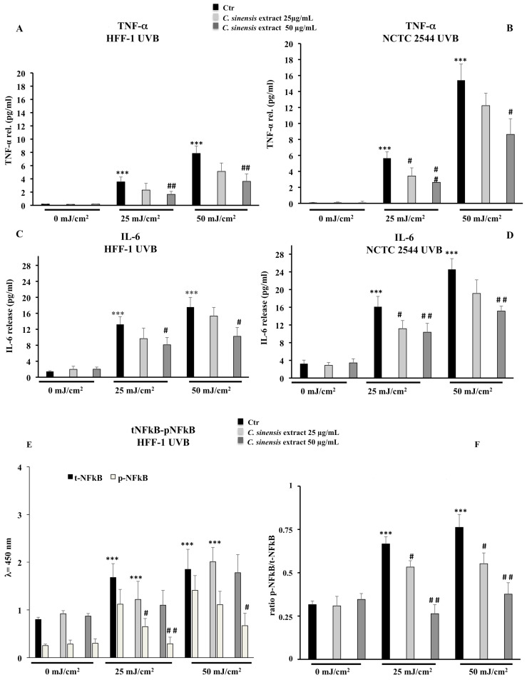
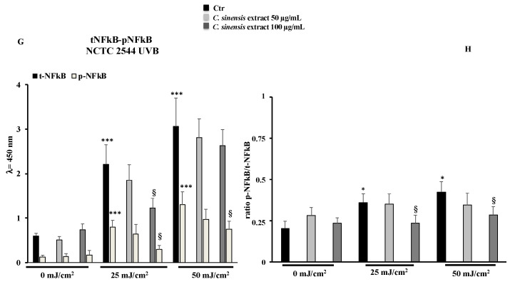
Excessive exposure to solar radiation is associated with several deleterious effects on human skin. These effects vary from the occasional simple sunburn to conditions resulting from chronic exposure such as skin aging and cancers. Secondary metabolites from the plant kingdom, including phenolic compounds, show relevant photoprotective activities. In this study, we evaluated the potential photoprotective activity of a phytocomplex derived from three varieties of red orange (Citrus sinensis (L.) Osbeck). We used an in vitro model of skin photoaging on two human cell lines, evaluating the protective effects of the phytocomplex in the pathways involved in the response to damage induced by UVA-B. The antioxidant capacity of the extract was determined at the same time as evaluating its influence on the cellular redox state (ROS levels and total thiol groups). In addition, the potential protective action against DNA damage induced by UVA-B and the effects on mRNA and protein expression of collagen, elastin, MMP1, and MMP9 were investigated, including some inflammatory markers (TNF-α, IL-6, and total and phospho NFkB) by ELISA. The obtained results highlight the capacity of the extract to protect cells both from oxidative stress—preserving RSH (p < 0.05) content and reducing ROS (p < 0.01) levels—and from UVA-B-induced DNA damage. Furthermore, the phytocomplex is able to counteract harmful effects through the significant downregulation of proinflammatory markers (p < 0.05) and MMPs (p < 0.05) and by promoting the remodeling of the extracellular matrix through collagen and elastin expression. This allows the conclusion that red orange extract, with its strong antioxidant and photoprotective properties, represents a safe and effective option to prevent photoaging caused by UVA-B exposure.

Keywords: IL-6; MMPs; ROS; TNF-α; antioxidant activity; collagen; comet assay; cyanidin; elastin; flavonoids.


Conflict of interest statement

The authors declare no conflict of interest.

Figures

Figure 1
Figure 1
Cell viability in HFF-1 and NCTC 2544 non-irradiated (Ctr) and irradiated with UVA (A,B) and UVB (C,D) at different doses. Values are the mean ± S.D. of five experiments in triplicate. * Significant vs. untreated control cells: p < 0.001.
Figure 2
Figure 2
Cell viability in HFF-1 (A) and NCTC 2544 (B) cells untreated (Ctr) and treated for 4 h with C. sinensis extract at different concentrations (0.5–5–50–100 μg/mL) and irradiated with 15 J/cm2 of UVA. Values are the mean ± S.D. of five experiments in triplicate. * Significant vs. untreated control cells: p < 0.001; ** Significant vs. untreated irradiated control cells: p < 0.001.
Figure 3
Figure 3
Cell viability in HFF-1 and NCTC 2544 cells untreated (Ctr) or treated for 4 h with C. sinensis extract at different concentrations (0.5–5–50–100 μg/mL) and irradiated with 25 mJ/cm2 (A,C) and 50 mJ/cm2 of UVB (B,D). Values are the mean ± S.D. of five experiments in triplicate. * Significant vs. untreated control cells: p < 0.001; ** Significant vs. untreated irradiated cells: p < 0.001.
Figure 4
Figure 4
Scavenger effect of C. sinensis extract by SOD-like (A) and DPPH assay (B). Superoxide anion results (A) are expressed as percentage of inhibition of NADH oxidation (rate of superoxide anion production was 4 nmol/min). Quenching effect (B) results are expressed as percentage of the decrease in absorbance at λ = 517 nm when compared with the control. Each value represents the mean ± S.D. of three independent experiments in triplicate. * Significant vs. p < 0.001 vs. previous concentration.
Figure 5
Figure 5
Total thiol groups in HFF-1 (A,C) and NCTC 2544 (B,D) cells untreated (Ctr) and treated for 4 h with C. sinensis extract at different concentrations (50–100 μg/mL) and irradiated with 15 J/cm2 of UVA and/or with 25 mJ/cm2 or 50 mJ/cm2 of UVB. Values are the mean ± S.D. of four experiments in triplicate. * Significant vs. untreated control cells: p < 0.001; ** Significant vs. untreated irradiated cells: p < 0.001.
Figure 6
Figure 6
ROS levels in HFF-1 (A,C) and NCTC 2544 (B,D) cells untreated (Ctr) and treated for 4 h with C. sinensis extract at different concentrations (50–100 μg/mL) and irradiated with 15 J/cm2 of UVA and/or with 25 mJ/cm2 or 50 mJ/cm2 of UVB. Values are the mean ± S.D. of four experiments in triplicate. * Significant vs. untreated control cells: p < 0.001; ** Significant vs. untreated irradiated cells: p < 0.001.
Figure 7
Figure 7
DNA damage was evaluated by alkaline comet assay in HFF-1 and in NCTC 2544 cells untreated and treated for 4 h with C. sinensis extract at different concentrations (50–100 μg/mL) and irradiated with UVA (A,B) and/or UVB (C,D). Results are expressed as percentage of DNA present in the comet tail (%TDNA). The data are reported as the mean ± S.D. from triplicate experiments. * Significant vs. untreated control cells: p ≤ 0.001; ** Significant vs. untreated irradiated cells: p ≤ 0.05.
Figure 8
Figure 8
Effects of C. sinensis extract pretreatment (25 and 50 μg/mL) for 4 h on the regulation of (A) mRNA expression of type 1 collagen (COL1A1) and elastin (ELN) and (B) mRNA expression of MMP9 and MMP1; protein levels of COL1A1 (C), ELN (D), MMP1 (E), and MPP9 (F) after exposure to UVB (25 and 50 mJ/cm2) in HFF1 cells (G). Representative immunoblotting image of each protein examined. The data are reported as the mean ± S.D. from two experiments performed in quadruplicates. Dashes to the right of the membrane bands show positions of prestained molecular mass markers. ** Significant vs. untreated control cells: p < 0.01; *** Significant vs. untreated control cells: p < 0.001; # Significant vs. untreated irradiated cells: p ≤ 0.05; ## Significant vs. untreated irradiated cells: p < 0.01.
Figure 8
Figure 8
Effects of C. sinensis extract pretreatment (25 and 50 μg/mL) for 4 h on the regulation of (A) mRNA expression of type 1 collagen (COL1A1) and elastin (ELN) and (B) mRNA expression of MMP9 and MMP1; protein levels of COL1A1 (C), ELN (D), MMP1 (E), and MPP9 (F) after exposure to UVB (25 and 50 mJ/cm2) in HFF1 cells (G). Representative immunoblotting image of each protein examined. The data are reported as the mean ± S.D. from two experiments performed in quadruplicates. Dashes to the right of the membrane bands show positions of prestained molecular mass markers. ** Significant vs. untreated control cells: p < 0.01; *** Significant vs. untreated control cells: p < 0.001; # Significant vs. untreated irradiated cells: p ≤ 0.05; ## Significant vs. untreated irradiated cells: p < 0.01.
Figure 9
Figure 9
Effects of C. sinensis extract pretreatment (25 and 50 μg/mL) for 4 h on the regulation of (A) the mRNA expression of MMP1 and MMP9; (B,C) protein levels of MMP1 and MMP9 after exposure to UVB (25 and 50 mJ/cm2) in NCTC 2544 cells. (D) Representative immunoblotting image of each protein examined. The data are reported as the mean ± S.D. from two experiments performed in quadruplicates. Dashes to the right of the membrane bands show positions of prestained molecular mass marker. ** Significant vs. untreated control cells: p < 0.01; *** Significant vs. untreated control cells: p < 0.001; # Significant vs. untreated irradiated cells: p < 0.05; ## Significant vs. untreated irradiated cells: p < 0.01.
Figure 9
Figure 9
Effects of C. sinensis extract pretreatment (25 and 50 μg/mL) for 4 h on the regulation of (A) the mRNA expression of MMP1 and MMP9; (B,C) protein levels of MMP1 and MMP9 after exposure to UVB (25 and 50 mJ/cm2) in NCTC 2544 cells. (D) Representative immunoblotting image of each protein examined. The data are reported as the mean ± S.D. from two experiments performed in quadruplicates. Dashes to the right of the membrane bands show positions of prestained molecular mass marker. ** Significant vs. untreated control cells: p < 0.01; *** Significant vs. untreated control cells: p < 0.001; # Significant vs. untreated irradiated cells: p < 0.05; ## Significant vs. untreated irradiated cells: p < 0.01.
Figure 10
Figure 10
Effects of C. sinensis extract pretreatment (25 and 50 μg/mL) for 4 h on the release of TNF-alpha (A,B), IL-6 (C,D) and the activation of NFkB (EH) after exposure to UVB (25 and 50 mJ/cm2) in HFF1 and NCT 2544 cells, respectively. The data are reported as the mean ± S.D. from triplicate experiments. * Significant vs. untreated control cells: p < 0.05; *** Significant vs. untreated control cells: p < 0.001; # Significant vs. untreated irradiated cells: p < 0.05; ## Significant vs. untreated irradiated cells: p < 0.01; § Significant vs. treated irradiated p ≤ 0.05.
Figure 10
Figure 10
Effects of C. sinensis extract pretreatment (25 and 50 μg/mL) for 4 h on the release of TNF-alpha (A,B), IL-6 (C,D) and the activation of NFkB (EH) after exposure to UVB (25 and 50 mJ/cm2) in HFF1 and NCT 2544 cells, respectively. The data are reported as the mean ± S.D. from triplicate experiments. * Significant vs. untreated control cells: p < 0.05; *** Significant vs. untreated control cells: p < 0.001; # Significant vs. untreated irradiated cells: p < 0.05; ## Significant vs. untreated irradiated cells: p < 0.01; § Significant vs. treated irradiated p ≤ 0.05.

Similar articles

Cited by

References

    1. Han A., Chien A.L., Kang S. Photoaging. Dermatol. Clin. 2014;32:291–299. doi: 10.1016/j.det.2014.03.015. - DOI - PubMed
    1. Griffiths H.R., Mistry P., Herbert K.E., Lunec J. Molecular and cellular effects of ultraviolet light-induced genotoxicity. Crit. Rev. Clin. Lab. Sci. 1998;35:189–237. doi: 10.1080/10408369891234192. - DOI - PubMed
    1. De Jager T.L., Cockrell A.E., Du Plessis S.S. Ultraviolet light induced generation of reactive oxygen species. Adv. Exp. Med. Biol. 2017;996:15–23. - PubMed
    1. Amaro-Ortiz A., Yan B., D’Orazio J.A. Ultraviolet radiation, aging and the skin: Prevention of damage by topical cAMP manipulation. Molecules. 2014;19:6202–6219. doi: 10.3390/molecules19056202. - DOI - PMC - PubMed
    1. Rinnerthaler M., Streubel M.K., Bischof J., Richter K. Skin aging, gene expression and calcium. Exp. Gerontol. 2015;68:59–65. doi: 10.1016/j.exger.2014.09.015. - DOI - PubMed

Publication types