Review
. 2022 Apr 20;12(9):1414.
doi: 10.3390/nano12091414.

Nanotechnology-Assisted Cell Tracking

Affiliations
Review

Nanotechnology-Assisted Cell Tracking

Alessia Peserico et al. Nanomaterials (Basel). .

Abstract

The usefulness of nanoparticles (NPs) in the diagnostic and/or therapeutic sector is derived from their aptitude for navigating intra- and extracellular barriers successfully and to be spatiotemporally targeted. In this context, the optimization of NP delivery platforms is technologically related to the exploitation of the mechanisms involved in the NP-cell interaction. This review provides a detailed overview of the available technologies focusing on cell-NP interaction/detection by describing their applications in the fields of cancer and regenerative medicine. Specifically, a literature survey has been performed to analyze the key nanocarrier-impacting elements, such as NP typology and functionalization, the ability to tune cell interaction mechanisms under in vitro and in vivo conditions by framing, and at the same time, the imaging devices supporting NP delivery assessment, and consideration of their specificity and sensitivity. Although the large amount of literature information on the designs and applications of cell membrane-coated NPs has reached the extent at which it could be considered a mature branch of nanomedicine ready to be translated to the clinic, the technology applied to the biomimetic functionalization strategy of the design of NPs for directing cell labelling and intracellular retention appears less advanced. These approaches, if properly scaled up, will present diverse biomedical applications and make a positive impact on human health.

Keywords: cell labelling; cell tracking; cell uptake; nanoparticles; stem cell homing.


Conflict of interest statement

The authors declare no conflict of interest.

Figures

Figure 1
Figure 1
Scientific community interest in tracking related research. Percentage of publications in Web of Science (WoS) database as function of publication year for the indicated combinations of keywords with the field tag «Topic». “OR” was used as Boolean operator to combine the keywords “object” and “particle tracking” and to lunch the search in WoS database. The plot shows the exponentially increasing interest in cell, particle, or object tracking (A) as well as tracking with nano systems (B) in the biomedical and related literature. The impact of the intrinsic growth of the number of publications was weighed and corrected by plotting percentages.
Figure 2
Figure 2
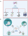
Direct cell tracking approaches suitable for imaging of cancer and cell transplantation for tissue regeneration, and related objectives. (A) Stem cells enclosing NPs can be used for monitoring transplantation events, as well as for directing therapeutic effects by releasing immunomodulatory factors into the tissue site requiring regeneration. (B) Stem cells or immune cells enclosing NPs can be adopted for homing in on tumors by allowing diagnosis and/or therapy. Figure created with BioRender. Available online: https://biorender.com/ (accessed on 25 March 2022).
Figure 3
Figure 3
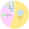
Classification of NPs based on the constitutive element. Classes and subclasses of nanoparticles based on their chemical properties (inorganic, polymeric, hybrid and lipid-based). Percentage (%) refers to the typology of NPs with respect to the total number of NPs identified in the literature survey. Figure created with BioRender. Available online: https://biorender.com/ (accessed on 25 March 2022).
Figure 4
Figure 4
Multimodal NP surface modifications applied for enhancing targeting, uptake, tracking, and therapy. Identification of different elements useful to improve labeling, targeting, therapy and cellular uptake. Figure created with BioRender. Available online: https://biorender.com/ (accessed on 25 March 2022).
Figure 5
Figure 5
Methods and devices for in vivo and in vitro imaging of NPs. (A) In vitro tracking of NPs. (B) In vivo tracking of NPs. Percentage (%) refers to different types of imaging tools for NP tracking based on our literature survey. Figure created with BioRender. Available online: https://biorender.com/ (accessed on 25 March 2022).
Figure 6
Figure 6
NP uptake mechanisms and strategies. Figure created with BioRender. Available online: https://biorender.com/ (accessed on 25 March 2022).
Figure 7
Figure 7
Cell sources adopted for efficient and safe NP delivery. Cell type subcategories and their relative percentage of utilization identified by the literature survey. Figure created with BioRender. Available online: https://biorender.com/ (accessed on 25 March 2022).
Figure 8
Figure 8
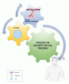
Essential ingredients for effective NP–cell tracking. NP design, cellular models and interaction mechanisms between NPs and cargo stem cells and/or target cells are key elements influencing NP-based cell tracking and delivery performances. Figure created with BioRender. Available online: https://biorender.com/ (accessed on 25 March 2022).

Similar articles

Cited by

References

    1. Zimmer C., Zhang B., Dufour A., Thebaud A., Berlemont S., Meas-Yedid V., Marin J.-C.O. On the digital trail of mobile cells. IEEE Signal Process. Mag. 2006;23:54–62. doi: 10.1109/MSP.2006.1628878. - DOI
    1. Meijering E., Smal I., Danuser G. Tracking in molecular bioimaging. IEEE Signal Process. Mag. 2006;23:46–53. doi: 10.1109/MSP.2006.1628877. - DOI
    1. Meijering E., Dzyubachyk O., Smal I., van Cappellen W.A. Tracking in cell and developmental biology. Semin. Cell Dev. Biol. 2009;20:894–902. doi: 10.1016/j.semcdb.2009.07.004. - DOI - PubMed
    1. Dorn J.F., Danuser G., Yang G. Computational processing and analysis of dynamic fluorescence image data. Methods Cell Biol. 2008;85:497–538. doi: 10.1016/S0091-679X(08)85022-4. - DOI - PubMed
    1. Jaqaman K., Danuser G. Computational image analysis of cellular dynamics: A case study based on particle tracking. Cold Spring Harb. Protoc. 2009;2009:pdb.top65. doi: 10.1101/pdb.top65. - DOI - PMC - PubMed

LinkOut - more resources