. 2022 Apr 27;12(9):1486.
doi: 10.3390/nano12091486.

Rapid Colorimetric pH-Responsive Gold Nanocomposite Hydrogels for Sensing Applications

Affiliations

Rapid Colorimetric pH-Responsive Gold Nanocomposite Hydrogels for Sensing Applications

Ahmed E Salih et al. Nanomaterials (Basel). .

Abstract

Surface functionalization of metallic nanoparticles (NPs) with external groups can be engineered to fabricate sensors that are responsive to various stimuli like temperature, pH, and numerous ions. Herein, we report the synthesis of gold nanoparticles (GNPs) functionalized with 3-mercaptopropionic acid (GNPs-MPA) and the doping of these nanoparticles into hydrogel materials using the breathing-in/breathing-out (BI-BO) method. MPA has a carboxyl group that becomes protonated and, thus, ionized at a pH below its pKa (4.32); hence, the GNPs-MPA solutions and gels were mostly pH-responsive in the range of 3-5. Optical properties were assessed through ultraviolet-visible (UV-Vis) spectroscopy, namely: transmission and absorption, and the parameters used to quantify the pH changes were the full width at half maximum (FWHM) and position of surface plasmon resonance (SPR). The solutions and gels gradually changed their colors from red to indigo with pH decrementation from 5 to 3, respectively. Furthermore, the solutions' and doped gels' highest FWHM sensitivities towards pH variations were 20 nm and 55 nm, respectively, while the SPR's position sensitivities were 18 nm and 10 nm, respectively. Also, transmission and scanning electron microscopy showed synchronized dispersion and aggregation of NPs with pH change in both solution and gel forms. The gel exhibited excellent repeatability and reversibility properties, and its response time was instantaneous, which makes its deployment as a colorimetric pH-triggered sensor practical. To the best of our knowledge, this is the first study that has incorporated GNPs into hydrogels utilizing the BI-BO method and demonstrated the pH-dependent optical and colorimetric properties of the developed nanocomposites.

Keywords: Biosensors; colorimetric sensing; nanocomposites; optical sensors; pH sensors.


Conflict of interest statement

The authors declare no conflict of interest.

Figures
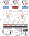
Figure 1
Figure 1
Gold-MPA nanocomposite hydrogel’s synthesis and mechanism of formation. (a) Breathing process utilized for loading the nanoparticles into the hydrogel matrix. Protonation of (b) MPA (at pKa = 4.32 and pKa = 10.20) and gold nanoparticles protected by MPA (at pKa = 4.32). (c) Illustration of pH-dependent reversible colorimetric change of gold colloidal NPs. (d) Synthesized gold nanoparticles protected by MPA. (i) Transmission and (ii) absorption spectra. Inset shows the calibration curve used to determine the concentration of the NPs. (iii) TEM of the synthesized gold nanoparticles (top) along with their corresponding size distribution (bottom).
Figure 2
Figure 2
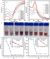
Optical characterization of GNPs-MPA solutions at different pHs ranging from 2 to 8: (a) Transmission and (b) Absorption spectra of the solutions at distinct pHs. (c) Images of the solutions. Extracted information from the optical spectra: (d) FWHM and (e) Position of SPR as a function of pH. Inset shows the calibration curves in the NPs’ pH-responsive region.
Figure 3
Figure 3
Imaging of the pH-responsive GNPs-MPA solutions: (i) TEM micrographs at different scales. (ii) Size distribution histograms for pH: (a) 3.62, (b) 4.34, (c) 4.84, and (d) 8.01. Inset shows few of the images used for size analysis in ImageJ.
Figure 4
Figure 4
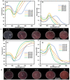
Utilization of BI-BO method to incorporate GNPs-MPA into hydrogels and their corresponding optical properties. (a) Transmission and (b) absorption spectra of the gels. Variation of the (c) transmission percentage, absorption at the surface plasmon, and (d) FWHM of the GNPs-MPA gels as a function of the number of BI-BO cycles. Images of the gel at each cycle are shown in the x-axis.
Figure 5
Figure 5
Optical properties of GNPs-MPA doped gels at different pHs: (a) Transmission, (b) absorption, and (c) images of a low concentrated gel (doped with 9 cycles) at different pHs. (d) Transmission, (e) absorption, and (f) images of high concentrated gel (doped with 15 BI-BO cycles) at distinct pH values.
Figure 6
Figure 6
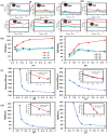
Analysis of the pH-responsiveness of GNPs-MPA doped (i) low and (ii) high concentrated gels. (a) RGB colorimetric analysis of the gel at different pHs using ImageJ. (b) RGB of the gel as a function of pH. (c) SPR’s position and (d) FWHM versus pH. Inset shows the gel’s linear behavior in the pH-responsive range (3–5).
Figure 7
Figure 7
Repeatability and time response of the GNPs-MPA doped gels: (a) Transmission and (b) absorption of the doped gels over three cycles switching between acidic (pH 2.16) and basic (pH 8.72) conditions (the number next to each curve denotes the number of the cycle). Time response of (c) SPR position and FWHM and (d) transmission and absorption values at SPR over three cycles. Images of the gel at the two distinct pHs are shown between (c,d).
Figure 8
Figure 8
Cross-sectional SEM images, at different magnifications, of the GNPs-MPA doped gel at (a) pH 8.72 and (b) pH 2.16.

Similar articles

References

    1. Amendola V., Pilot R., Frasconi M., Maragò O.M., Iatì M.A. Surface plasmon resonance in gold nanoparticles: A review. J. Phys. Condens. Matter Inst. Phys. J. 2017;29:203002. doi: 10.1088/1361-648X/aa60f3. - DOI - PubMed
    1. Sun C., Lee J.S.H., Zhang M. Magnetic nanoparticles in MR imaging and drug delivery. Adv. Drug Deliv. Rev. 2008;60:1252–1265. doi: 10.1016/j.addr.2008.03.018. - DOI - PMC - PubMed
    1. Sardar R., Funston A.M., Mulvaney P., Murray R.W. Gold Nanoparticles: Past, Present, and Future. Langmuir ACS J. Surf. Colloids. 2009;25:13840–13851. doi: 10.1021/la9019475. - DOI - PubMed
    1. Yougbare S., Chang T.-K., Tan S.-H., Kuo J.-C., Hsu P.-H., Su C.-Y., Kuo T.-R. Antimicrobial Gold Nanoclusters: Recent Developments and Future Perspectives. Int. J. Mol. Sci. 2019;20:2924. doi: 10.3390/ijms20122924. - DOI - PMC - PubMed
    1. Zahra Q.U.A., Luo Z., Ali R., Khan M.I., Li F., Qiu B. Advances in gold nanoparticles-based colorimetric aptasensors for the detection of antibiotics: An overview of the past decade. Nanomaterials. 2021;11:840. doi: 10.3390/nano11040840. - DOI - PMC - PubMed

LinkOut - more resources