

Human Adipose-Derived Stem Cell-Conditioned Medium Promotes Vascularization of Nanostructured Scaffold Transplanted into Nude Mice
- PMID: 35564230
- PMCID: PMC9100239
- DOI: 10.3390/nano12091521
Human Adipose-Derived Stem Cell-Conditioned Medium Promotes Vascularization of Nanostructured Scaffold Transplanted into Nude Mice
Abstract
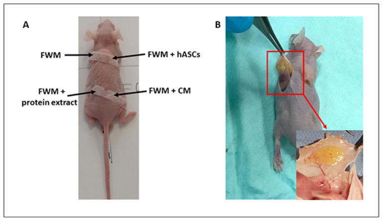
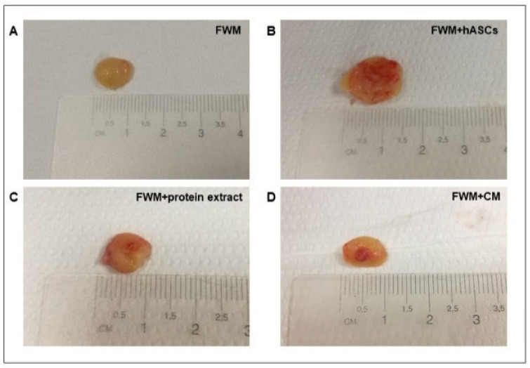
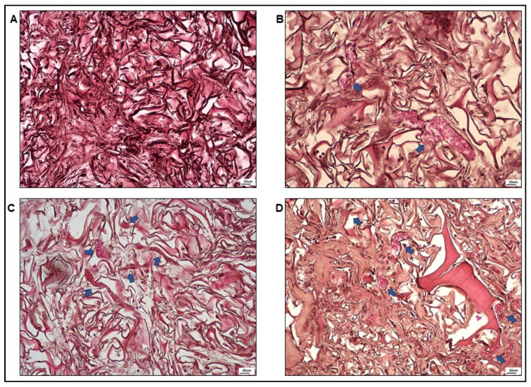
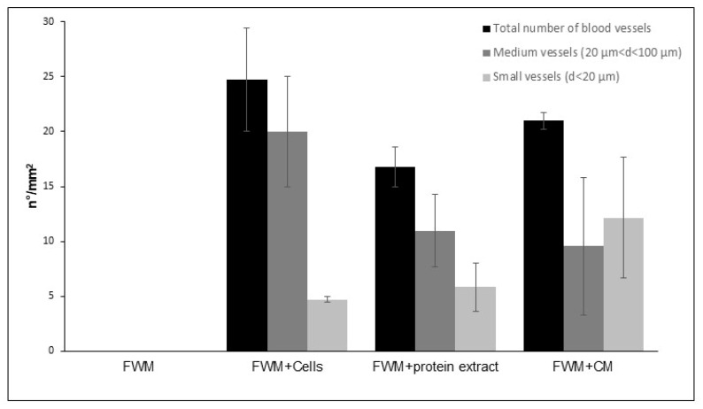
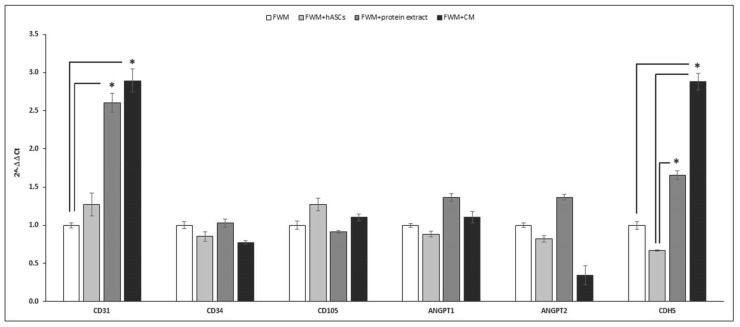
Several studies have been conducted on the interaction between three-dimensional scaffolds and mesenchymal stem cells for the regeneration of damaged tissues. Considering that stem cells do not survive for sufficient time to directly sustain tissue regeneration, it is essential to develop cell-free systems to be applied in regenerative medicine. In this work, by in vivo experiments, we established that a collagen-nanostructured scaffold, loaded with a culture medium conditioned with mesenchymal stem cells derived from adipose tissue (hASC-CM), exerts a synergic positive effect on angiogenesis, fundamental in tissue regeneration. To this aim, we engrafted athymic BALB-C nude mice with four different combinations: scaffold alone; scaffold with hASCs; scaffold with hASC crude protein extract; scaffold with hASC-CM. After their removal, we verified the presence of blood vessels by optical microscopy and confirmed the vascularization evaluating, by real-time PCR, several vascular growth factors: CD31, CD34, CD105, ANGPT1, ANGPT2, and CDH5. Our results showed that blood vessels were absent in the scaffold grafted alone, while all the other systems appeared vascularized, a finding supported by the over-expression of CD31 and CDH5 mRNA. In conclusion, our data sustain the capability of hASC-CM to be used as a therapeutic cell-free approach for damaged tissue regeneration.
Keywords: angiogenesis; cell culture; cell-free therapy; in vivo experiment; regenerative medicine; secretome; tissue engineering.
Conflict of interest statement
The authors declare no conflict of interest.
Figures
Similar articles
-
Human Dental Pulp Mesenchymal Stem Cell-Derived Soluble Factors Combined with a Nanostructured Scaffold Support the Generation of a Vascular Network In Vivo.Nanomaterials (Basel). 2023 Sep 2;13(17):2479. doi: 10.3390/nano13172479. Nanomaterials (Basel). 2023. PMID: 37686988 Free PMC article.
-
Engineering vascularized soft tissue flaps in an animal model using human adipose-derived stem cells and VEGF+PLGA/PEG microspheres on a collagen-chitosan scaffold with a flow-through vascular pedicle.Biomaterials. 2015 Dec;73:198-213. doi: 10.1016/j.biomaterials.2015.09.024. Epub 2015 Sep 18. Biomaterials. 2015. PMID: 26410787 Free PMC article.
-
Effect of Nanostructured Scaffold on Human Adipose-Derived Stem Cells: Outcome of In Vitro Experiments.Nanomaterials (Basel). 2020 Sep 12;10(9):1822. doi: 10.3390/nano10091822. Nanomaterials (Basel). 2020. PMID: 32932658 Free PMC article.
-
Human adipose-derived stem cells and three-dimensional scaffold constructs: a review of the biomaterials and models currently used for bone regeneration.J Biomed Mater Res B Appl Biomater. 2013 Jan;101(1):187-99. doi: 10.1002/jbm.b.32817. Epub 2012 Sep 21. J Biomed Mater Res B Appl Biomater. 2013. PMID: 22997152 Review.
-
Exosomes as Part of the Human Adipose-Derived Stem Cells Secretome- Opening New Perspectives for Cell-Free Regenerative Applications.Adv Exp Med Biol. 2021;1312:139-163. doi: 10.1007/5584_2020_588. Adv Exp Med Biol. 2021. PMID: 32986128 Review.
Cited by
-
The Role of the Extracellular Matrix in Inducing Cardiac Cell Regeneration and Differentiation.Cells. 2025 Jun 10;14(12):875. doi: 10.3390/cells14120875. Cells. 2025. PMID: 40558502 Free PMC article. Review.
-
Human Dental Pulp Mesenchymal Stem Cell-Derived Soluble Factors Combined with a Nanostructured Scaffold Support the Generation of a Vascular Network In Vivo.Nanomaterials (Basel). 2023 Sep 2;13(17):2479. doi: 10.3390/nano13172479. Nanomaterials (Basel). 2023. PMID: 37686988 Free PMC article.
-
Adipose mesenchymal stem cell-derived soluble factors, produced under hypoxic condition, efficiently support in vivo angiogenesis.Cell Death Discov. 2023 May 23;9(1):174. doi: 10.1038/s41420-023-01464-4. Cell Death Discov. 2023. PMID: 37221171 Free PMC article.
-
Dental pulp mesenchymal stem cell (DPSCs)-derived soluble factors, produced under hypoxic conditions, support angiogenesis via endothelial cell activation and generation of M2-like macrophages.J Biomed Sci. 2024 Nov 4;31(1):99. doi: 10.1186/s12929-024-01087-6. J Biomed Sci. 2024. PMID: 39491013 Free PMC article.
References
-
- Melocchi A., Uboldi M., Cerea M., Foppoli A., Maroni A., Moutaharrik S., Palugan L., Zema L., Gazzaniga A. A Graphical Review on the Escalation of Fused Deposition Modeling (FDM) 3D Printing in the Pharmaceutical Field. J. Pharm. Sci. 2020;109:2943–2957. doi: 10.1016/j.xphs.2020.07.011. - DOI - PubMed
-
- List of FDA Approved Stem Cell Therapies. [(accessed on 31 March 2021)]. Available online: https://ipscell.com/2021/03/list-of-fda-approved-stem-cell-therapies.
Grants and funding
LinkOut - more resources
Full Text Sources
Miscellaneous
