. 2022 May 3;12(9):1549.
doi: 10.3390/nano12091549.

Investigating the Morphology and Mechanics of Biogenic Hierarchical Materials at and below Micrometer Scale

Affiliations

Investigating the Morphology and Mechanics of Biogenic Hierarchical Materials at and below Micrometer Scale

Mohammad Soleimani et al. Nanomaterials (Basel). .

Abstract

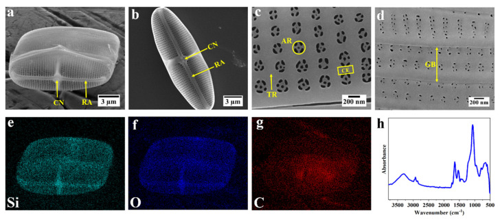
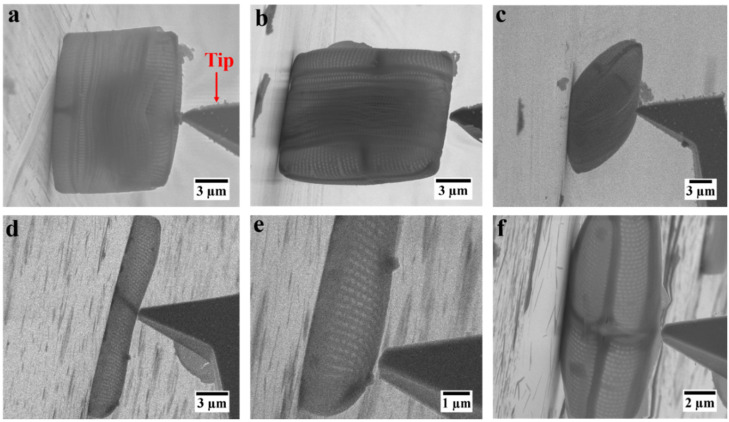
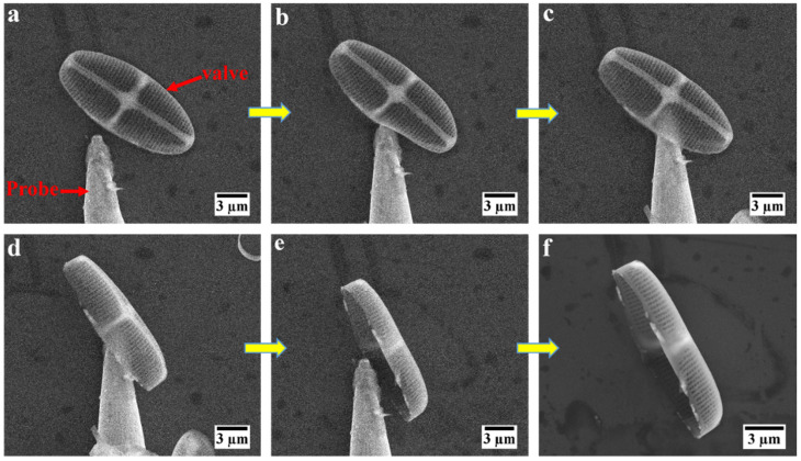
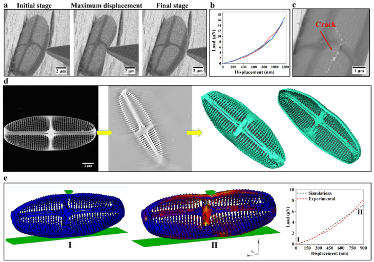
Investigating and understanding the intrinsic material properties of biogenic materials, which have evolved over millions of years into admirable structures with difficult to mimic hierarchical levels, holds the potential of replacing trial-and-error-based materials optimization in our efforts to make synthetic materials of similarly advanced complexity and properties. An excellent example is biogenic silica which is found in the exoskeleton of unicellular photosynthetic algae termed diatoms. Because of the complex micro- and nanostructures found in their exoskeleton, determining the intrinsic mechanical properties of biosilica in diatoms has only partly been accomplished. Here, a general method is presented in which a combination of in situ deformation tests inside an SEM with a realistic 3D model of the frustule of diatom Craspedostauros sp. (C. sp.) obtained by electron tomography, alongside finite element method (FEM) simulations, enables quantification of the Young's modulus (E = 2.3 ± 0.1 GPa) of this biogenic hierarchical silica. The workflow presented can be readily extended to other diatom species, biominerals, or even synthetic hierarchical materials.

Keywords: diatom frustule; electron tomography; hierarchical materials; in situ mechanical testing.


Conflict of interest statement

The authors declare no conflict of interest.

Figures

Figure 1
Figure 1
Morphology and elemental composition of the frustule of diatom C. sp. (ad) SEM images of frustule of C. sp. (a) intact frustule of C. sp.; (b) an isolated valve deposited on its concave orientation showing the interior surface of the valve (RA = raphe; CN = central nodule); (c) porous area of the valve (TR = transapical rib; CE = cross extension; AR = areola); (d) overlapping girdle bands (GB); (eg) EDS elemental mapping of the intact frustule; green = silicon, blue = oxygen, red = carbon; (h) FTIR spectra of frustule of C. sp.
Figure 2
Figure 2
SEM images of different orientations of intact frustule and isolated valves of C. sp. for in situ deformation tests: (a) intact frustule standing upright; (b) intact frustule standing on girdle bands; (c) intact frustule laying on the girdle bands; (d,e) deformation test at different locations of an isolated valve laying flat; (f) deformation test on isolated valve standing on its side.
Figure 3
Figure 3
Mechanical manipulation of an isolated valve of diatom C. sp. (af) SEM image sequence acquired during repositioning of an isolated valve from its flat orientation to standing on its side.
Figure 4
Figure 4
Overall workflow to quantify Young’s modulus of biosilica in the valve: (a) SEM images of an isolated valve at different stages during the in situ deformation test; (b) load–displacement curves for three valves obtained from one culture; (c) crack formed by deformation of the valve; (d) from left to right: STEM image of the valve, a slice of the reconstructed 3D intensity map of the valve, and 3D surface views of the valve; (e) initial undeformed (I) and final deformed (II) stages of the realistic valve model in FEM simulations and a comparison between experimental and simulated load–displacement curves.

Similar articles

Cited by

References

    1. Arakaki A., Shimizu K., Oda M., Sakamoto T., Nishimura T., Kato T. Biomineralization-inspired synthesis of functional organic/inorganic hybrid materials: Organic molecular control of self-organization of hybrids. Org. Biomol. Chem. 2015;13:974–989. doi: 10.1039/C4OB01796J. - DOI - PubMed
    1. Luz G.M., Mano J.F. Mineralized structures in nature: Examples and inspirations for the design of new composite materials and biomaterials. Compos. Sci. Technol. 2010;70:1777–1788. doi: 10.1016/j.compscitech.2010.05.013. - DOI
    1. Libonati F., Buehler M.J. Advanced structural materials by bioinspiration. Adv. Eng. Mater. 2017;19:1600787. doi: 10.1002/adem.201600787. - DOI
    1. Espinosa H.D., Rim J.E., Barthelat F., Buehler M.J. Merger of structure and material in nacre and bone–Perspectives on de novo biomimetic materials. Prog. Mater. Sci. 2009;54:1059–1100. doi: 10.1016/j.pmatsci.2009.05.001. - DOI
    1. Thula T.T., Rodriguez D.E., Lee M.H., Pendi L., Podschun J., Gower L.B. In vitro mineralization of dense collagen substrates: A biomimetic approach toward the development of bone-graft materials. Acta Biomater. 2011;7:3158–3169. doi: 10.1016/j.actbio.2011.04.014. - DOI - PMC - PubMed

LinkOut - more resources