. 2022 May 7;12(9):1585.
doi: 10.3390/nano12091585.

First Report of the Biosynthesis and Characterization of Silver Nanoparticles Using Scabiosa atropurpurea subsp. maritima Fruit Extracts and Their Antioxidant, Antimicrobial and Cytotoxic Properties

Affiliations

First Report of the Biosynthesis and Characterization of Silver Nanoparticles Using Scabiosa atropurpurea subsp. maritima Fruit Extracts and Their Antioxidant, Antimicrobial and Cytotoxic Properties

Badiaa Essghaier et al. Nanomaterials (Basel). .

Abstract

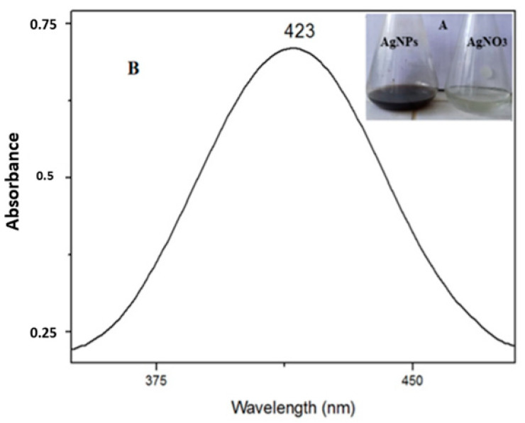
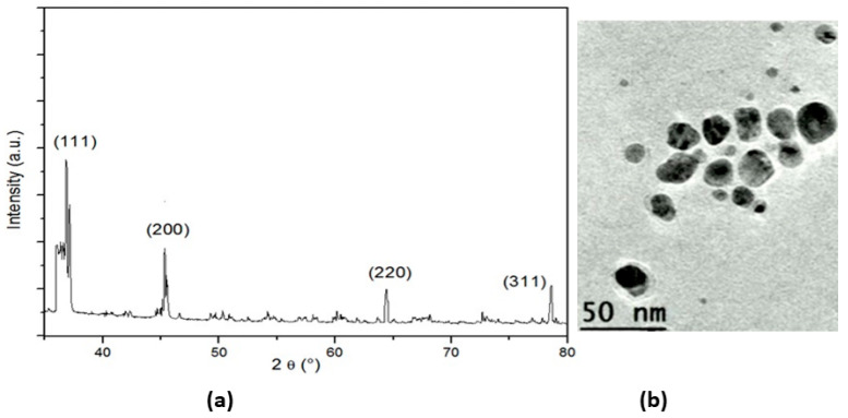
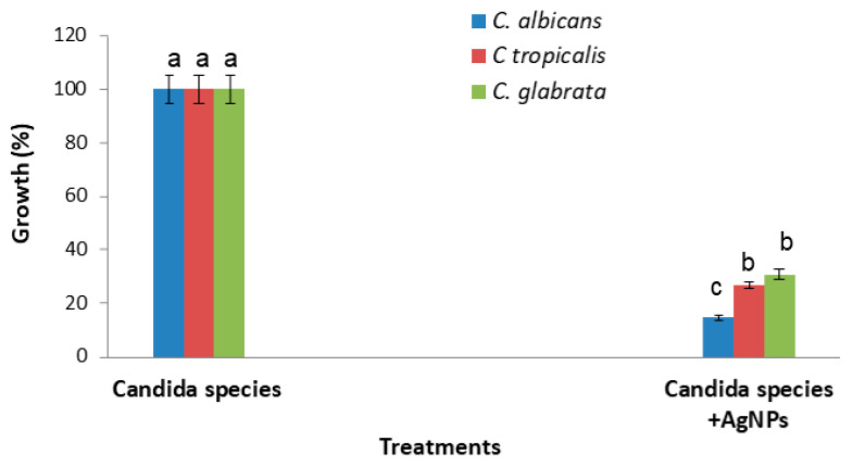
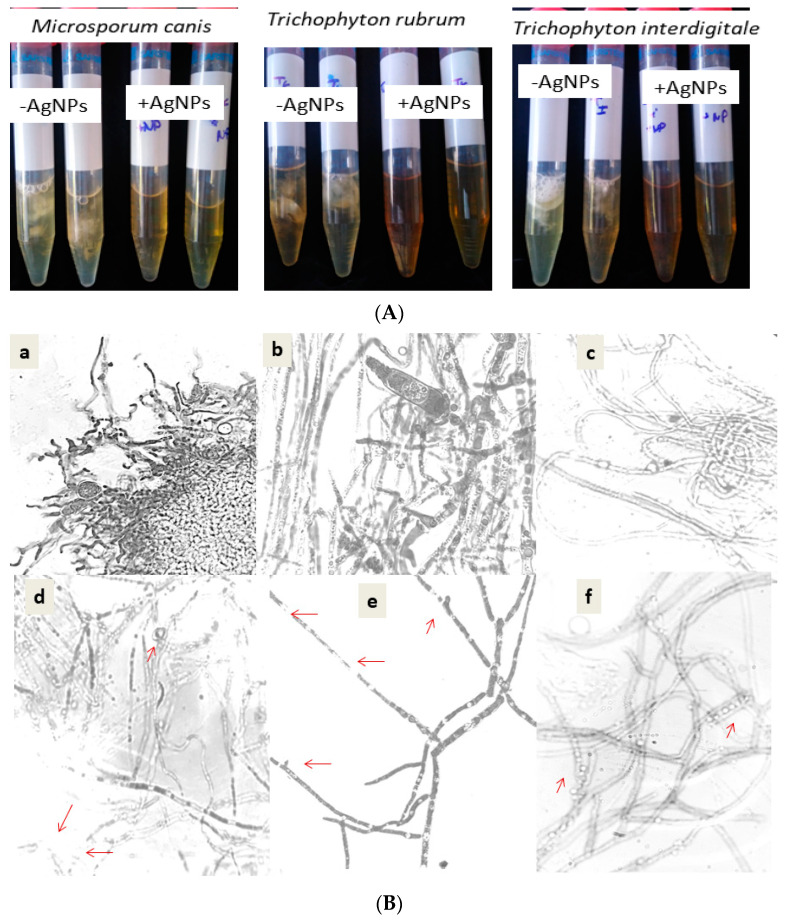
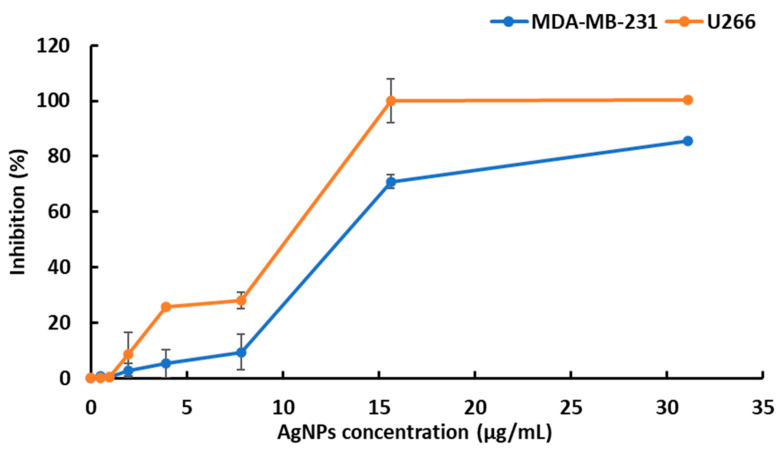
Candida and dermatophyte infections are difficult to treat due to increasing antifungal drugs resistance such as fluconazole, as well as the emergence of multi-resistance in clinical bacteria. Here, we first synthesized silver nanoparticles using aqueous fruit extracts from Scabiosa atropurpurea subsp. maritima (L.). The characterization of the AgNPs by means of UV, XRD, FTIR, and TEM showed that the AgNPs had a uniform spherical shape with average sizes of 40-50 nm. The biosynthesized AgNPs showed high antioxidant activity when investigated using 1,1-diphenyl-2-picryl-hydrazyl (DPPH) and ferric reducing antioxidant power (FRAP) assays. The AgNPs displayed strong antibacterial potential expressed by the maximum zone inhibition and the lowest MIC and MBC values. The AgNPs revealed a significant antifungal effect against the growth and biofilm of Candida species. In fact, the AgNPs were efficient against Trichophyton rubrum, Trichophyton interdigitale, and Microsporum canis. The antifungal mechanisms of action of the AgNPs seem to be due to the disruption of membrane integrity and a reduction in virulence factors (biofilm and hyphae formation and a reduction in germination). Finally, the silver nanoparticles also showed important cytotoxic activity against the human multiple myeloma U266 cell line and the human breast cancer cell line MDA-MB-231. Therefore, we describe new silver nanoparticles with promising biomedical application in the development of novel antimicrobial and anticancer agents.

Keywords: Scabiosa atropurpurea subsp. maritima (L.); anti-dermatophytes; antibacterial; antioxidants; bio-synthetized silver nanoparticles; cytotoxic activity.


Conflict of interest statement

The authors declare no conflict of interest.

Figures

Figure 1
Figure 1
(A). Change in the color of the silver solution after adding plant extract, from light brown (right) to dark brown (left) and (B). UV–Vis spectrum of synthesized AgNPs (black) and the AgNO3 solution (before adding the plant extract (red)).
Figure 2
Figure 2
(a) XRD pattern of AgNPs; (b)TEM image of silver nanoparticles.
Figure 3
Figure 3
AgNPs’ effect on Candida growth after incubation for 48 h at 37 °C, as compared to untreated Candida cells. Error bars represent SE of the mean (n = 3). Means followed by the same letter are not significantly different according to the SNK test.
Figure 4
Figure 4
(A) Visual observation of dermatophyte growth after 14 days at 28 °C in the absence of AgNPs (AgNPs) and in the presence of AgNPs (+AgNPs). (B) Comparative effect of silver nanoparticles on the dermatophyte morphology of Trichophyton interdigitale (d), Microsporum canis (e), and Trichophyton rubrum (f), as compared to their control cultures (ac), respectively. Red arrows indicate the morphological change by AgNPs.
Figure 5
Figure 5
Evaluation of silver nanoparticle-induced cytotoxic effects on U266 and MDA-MB-231 cell lines. The reported values are the means ± SD from three independent experiments. No statistically significant differences between the control (no treatment) and the vehicle were noticed.

Similar articles

Cited by

References

    1. Zhang X.F., Liu Z.G., Shen W., Gurunathan S. Silver nanoparticles: Synthesis, characterization, properties, applications, and therapeutic approaches. Int. J. Mol. Sci. 2016;17:1534. doi: 10.3390/ijms17091534. - DOI - PMC - PubMed
    1. Robinson J.R., Isikhuemhen O.S., Anike F.N. Fungal metal interactions: A review of toxicity and homeostasis. J. Fungi. 2021;7:225. doi: 10.3390/jof7030225. - DOI - PMC - PubMed
    1. Miranda R.R., Sampaio I., Zuculotto V. Exploring silver nanoparticles for cancer therapy and diagnosis. Colloids Surf. B Biointerfaces. 2022;210:112254. doi: 10.1016/j.colsurfb.2021.112254. - DOI - PubMed
    1. Gomes H.I.O., Martins C.S.M., Prior J.A.V. Silver nanoparticles as cariers of anticancer drugs for efficient target treatment of cancer cells. Nanomaterials. 2021;11:964. doi: 10.3390/nano11040964. - DOI - PMC - PubMed
    1. Renganathan S., Subramaniyan S., Karunanithi N., Vasanthakumar P., Kutzner A., Kim P.-S., Heese K. Antibacterial, Antifungal, and Antioxidant Activities of Silver Nanoparticles Biosynthesized from Bauhinia tomentosa Linn. Antioxidants. 2021;10:1959. doi: 10.3390/antiox10121959. - DOI - PMC - PubMed

LinkOut - more resources