

Expression of VEGF, EGF, and Their Receptors in Squamous Esophageal Mucosa, with Correlations to Histological Findings and Endoscopic Minimal Changes, in Patients with Different GERD Phenotypes
- PMID: 35564692
- PMCID: PMC9102479
- DOI: 10.3390/ijerph19095298
Expression of VEGF, EGF, and Their Receptors in Squamous Esophageal Mucosa, with Correlations to Histological Findings and Endoscopic Minimal Changes, in Patients with Different GERD Phenotypes
Abstract
Background: Gastroesophageal reflux disease (GERD) may present as nonerosive reflux disease (NERD), erosive esophagitis (EE), or be complicated by Barrett's esophagus (BE). The explanation as to what determines the phenotype of GERD is awaited. Therefore, we assessed the correlation between the growth factors expression and endoscopic as histologic findings in GERD patients.
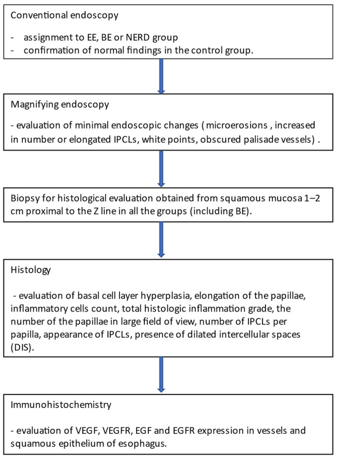
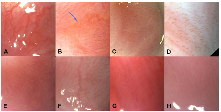
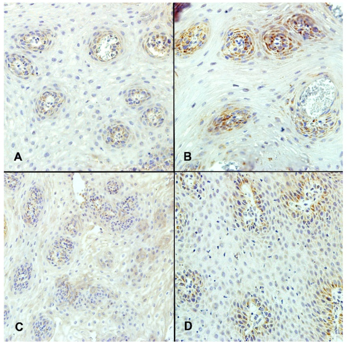
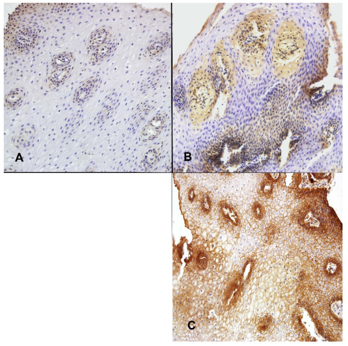
Methods: The squamous esophageal epithelium of 50 patients (20-NERD, 7-EE, 15-BE, 8 controls) was examined by: (1) magnification endoscopy with evaluation of minimal GERD changes such as: microerosions, white spots, palisade blood vessels visibility, and intrapapillary capillary loops (IPCLs) appearance, (2) histology, (3) immunohistochemistry with evaluation of the expression of vascular endothelial growth factor (VEGF), epidermal growth factor (EGF), and their receptors (VEGFR and EGFR).
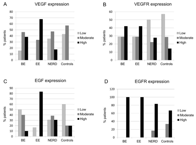
Results: The expression of VEGF, but not VEGFR, EGF, and EGFR, was significantly increased in EE patients compared to NERD patients and controls. VEGF levels correlated significantly with the presence of white spots, but not with other minimal endoscopic and histologic features. The EGFR expression correlated positively with basal cell hyperplasia and enlarged IPCLs.
Conclusions: Our findings suggest a correlation between growth factors expression and findings in conventional endoscopy, formation of endoscopic minimal changes, and histologic lesions.
Keywords: EGF; GERD; VEGF; growth factors; minimal change esophagitis.
Conflict of interest statement
The authors declare no conflict of interest. The funders had no role in the design of the study; in the collection, analyses, or interpretation of data; in the writing of the manuscript, or in the decision to publish the results.
Figures
References
-
- Ronkainen J., Aro P., Storskrubb T., Johansson S.E., Lind T., Bolling-Sternevald E., Graffner H., Vieth M., Stolte M., Engstrand L., et al. High prevalence of gastroesophageal reflux symptoms and esophagitis with or without symptoms in the general adult Swedish population: A Kalixanda study report. Scand. J. Gastroenterol. 2005;40:275–285. doi: 10.1080/00365520510011579. - DOI - PubMed
-
- Peery A.F., Crockett S.D., Barritt A.S., Dellon E.S., Eluri S., Gangarosa L.M., Jensen E.T., Lund J.L., Pasricha S., Runge T., et al. Burden of Gastrointestinal, Liver, and Pancreatic Diseases in the United States. Gastroenterology. 2015;149:1731–1741.e3. doi: 10.1053/j.gastro.2015.08.045. - DOI - PMC - PubMed
-
- Mastracci L., Grillo F., Parente P., Unti E., Battista S., Spaggiari P., Campora M., Scaglione G., Fassan M., Fiocca R. Gastro-esophageal reflux disease and Barrett’s esophagus: An overview with an histologic diagnostic approach. Pathologica. 2020;112:117–127. doi: 10.32074/1591-951X-162. - DOI - PMC - PubMed
Publication types
MeSH terms
Substances
LinkOut - more resources
Full Text Sources
Medical
Research Materials
Miscellaneous
