

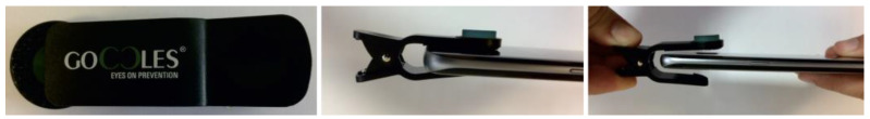
Clinical Evaluation of the Optical Filter for Autofluorescence Glasses for Oral Cancer Curing Light Exposed (GOCCLES®) in the Management of Potentially Premalignant Disorders: A Retrospective Study
- PMID: 35564975
- PMCID: PMC9100244
- DOI: 10.3390/ijerph19095579
Clinical Evaluation of the Optical Filter for Autofluorescence Glasses for Oral Cancer Curing Light Exposed (GOCCLES®) in the Management of Potentially Premalignant Disorders: A Retrospective Study
Abstract
Background: Any oral potentially malignant disorders (OPMDs) must be regularly monitored through clinical examination to detect any possible malignant transformation. Conventional intraoral exams, however, can be difficult because these conditions may resemble benign lesions. For this reason, several non-invasive diagnostic technologies have been developed to help the clinician in detecting and distinguishing between cancerous and benign lesions. Epithelial dysplasia can be considered the most important predictor of malignant evolution. Therefore, in this study we aim to evaluate the ability of an optical filter for autofluorescence Glasses for Oral Cancer Curing Light Exposed (GOCCLES®) and of toluidine blue staining in identifying dysplastic areas in patients with OPMDs.
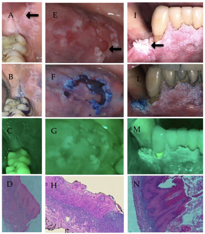
Methods: In this retrospective study, medical records, photographs and videos of 25 patients with oral lesions were analyzed. Forty-two biopsy samples in 25 patients with OPMDs and at least one suspicious oral mucosa lesion that were evaluated in white light, autofluorescence with optical filter GOCCLES®, toluidine blue staining and then biopsied with histopathological analysis were analyzed.
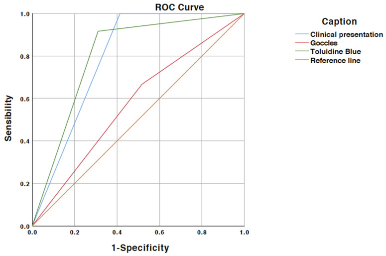
Results: The sensitivity and specificity for the autofluorescence evaluation with GOCCLES® for identifying dysplasia or carcinoma were 66% and 48%, respectively. The positive and negative predictive values were 34% and 77%, respectively, and the accuracy was 53%. The sensitivity and specificity for toluidine blue staining were 91% and 68%, respectively. The positive and negative predictive values were 55% and 95%, respectively, and the accuracy was 75%.
Conclusions: The optical filter for autofluorescence (GOCCLES®) and toluidine blue staining are simple, inexpensive, rapid and non-invasive procedures that can assist the clinician in distinguishing OPMDs from healthy mucosa but they are not able to distinguish benign and malignant lesions.
Keywords: autofluorescence; biopsy; dysplasia; oral cancer; oral potentially malignant disorders; retrospective study; toluidine blue.
Conflict of interest statement
The authors declare no conflict of interest.
Figures
Similar articles
-
Comparing the efficacy of chemiluminescence with lugol's iodine versus toluidine blue in the diagnosis of dysplasia in tobacco associated oral lesions - A diagnostic study.Indian J Dent Res. 2021 Oct-Dec;32(4):459-466. doi: 10.4103/ijdr.IJDR_1004_20. Indian J Dent Res. 2021. PMID: 35645072 Clinical Trial.
-
Reliability of toluidine blue application in the detection of oral epithelial dysplasia and in situ and invasive squamous cell carcinomas.Oral Surg Oral Med Oral Pathol Oral Radiol Endod. 2001 May;91(5):535-40. doi: 10.1067/moe.2001.112949. Oral Surg Oral Med Oral Pathol Oral Radiol Endod. 2001. PMID: 11346731
-
The GOCCLES® medical device is effective in detecting oral cancer and dysplasia in dental clinical setting. Results from a multicentre clinical trial.Acta Otorhinolaryngol Ital. 2015 Dec;35(6):449-54. doi: 10.14639/0392-100X-922. Acta Otorhinolaryngol Ital. 2015. PMID: 26900252 Free PMC article. Clinical Trial.
-
Autofluorescence imaging to identify oral malignant or premalignant lesions: Systematic review and meta-analysis.Head Neck. 2020 Dec;42(12):3735-3743. doi: 10.1002/hed.26430. Epub 2020 Aug 31. Head Neck. 2020. PMID: 32866310
-
Oral potentially malignant disorders: A comprehensive review on clinical aspects and management.Oral Oncol. 2020 Mar;102:104550. doi: 10.1016/j.oraloncology.2019.104550. Epub 2020 Jan 22. Oral Oncol. 2020. PMID: 31981993
Cited by
-
Accuracy of special histochemical staining methods in diagnosis of oral pathology: A systematic review and meta-analysis.Dent Res J (Isfahan). 2024 Jul 12;21:34. eCollection 2024. Dent Res J (Isfahan). 2024. PMID: 39188398 Free PMC article. Review.
-
Tissue autofluorescence system as an aid for Dental Hygienist to screening oral mucosa alterations during supportive periodontal care. An observational single-arm study.Clin Oral Investig. 2025 Feb 21;29(3):144. doi: 10.1007/s00784-025-06220-9. Clin Oral Investig. 2025. PMID: 39982515
-
Efficacy of Autofluorescence in Detection of Tobacco-associated Oral Mucosal Lesions - A Systematic Review.Ann Maxillofac Surg. 2024 Jul-Dec;14(2):212-220. doi: 10.4103/ams.ams_128_24. Epub 2024 Nov 18. Ann Maxillofac Surg. 2024. PMID: 39957885 Free PMC article.
-
Chemical, optical, and morphological properties of TPU and PET-G samples after aging in artificial saliva: an in vitro study.BMC Oral Health. 2025 Apr 11;25(1):533. doi: 10.1186/s12903-025-05863-0. BMC Oral Health. 2025. PMID: 40217188 Free PMC article.
-
Effects of Thermoforming on the Mechanical, Optical, Chemical, and Morphological Properties of PET-G: In Vitro Study.Polymers (Basel). 2024 Jan 10;16(2):203. doi: 10.3390/polym16020203. Polymers (Basel). 2024. PMID: 38257002 Free PMC article.
References
-
- The Global Cancer Observatory IARC. WHO lip, Oral Cavity Cancer. [(accessed on 22 April 2021)]. Available online: https://gco.iarc.fr/today/data/factsheets/cancers/1-Lip-oral-cavity-fact...
-
- Warnakulasuriya S., Greenspan J.S. Textbook of oral cancer: Prevention, diagnosis and management. 1st ed. Springer; New York, NY, USA: 2020.
-
- Romano A., Di Stasio D., Petruzzi M., Fiori F., Lajolo C., Santarelli A., Lucchese A., Serpico R., Contaldo M. Noninvasive imaging methods to improve the diagnosis of oral carcinoma and its precursors: State of the art and proposal of a three-step diagnostic process. Cancers. 2021;13:2864. doi: 10.3390/cancers13122864. - DOI - PMC - PubMed
MeSH terms
Substances
LinkOut - more resources
Full Text Sources
Medical
