. 2022 Apr 19;14(9):2050.
doi: 10.3390/cancers14092050.

The Effect of Hyperthermia and Radiotherapy Sequence on Cancer Cell Death and the Immune Phenotype of Breast Cancer Cells

Affiliations

The Effect of Hyperthermia and Radiotherapy Sequence on Cancer Cell Death and the Immune Phenotype of Breast Cancer Cells

Azzaya Sengedorj et al. Cancers (Basel). .

Abstract

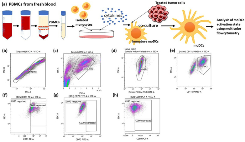
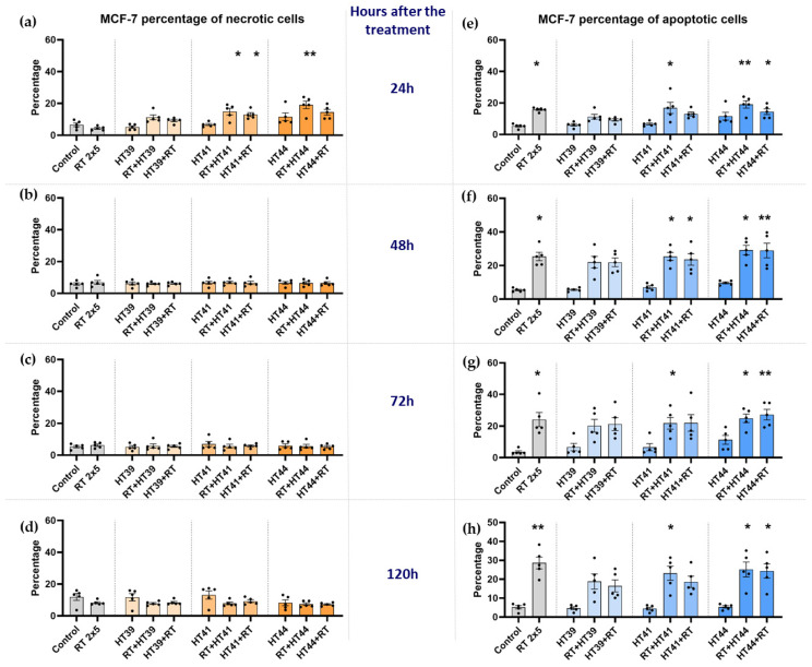
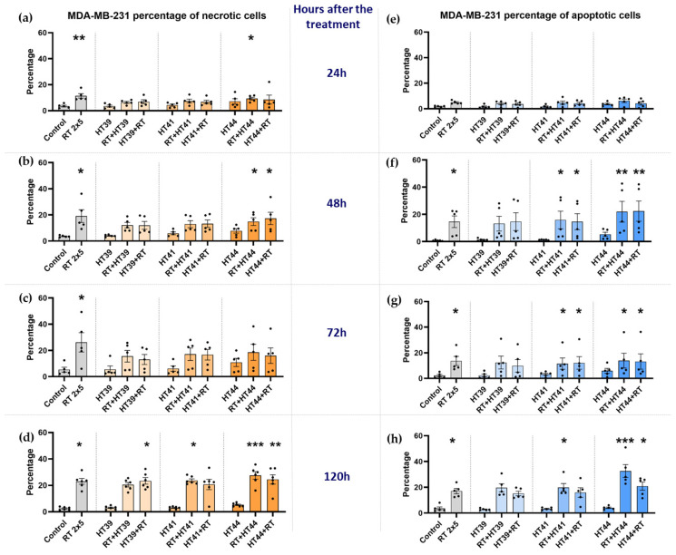
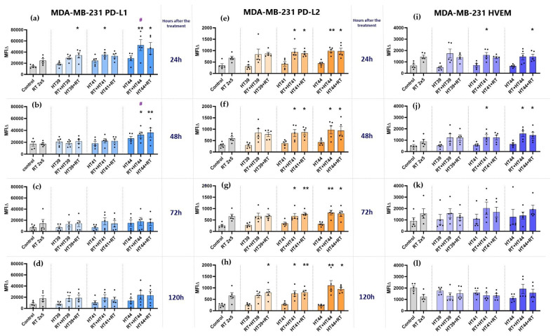
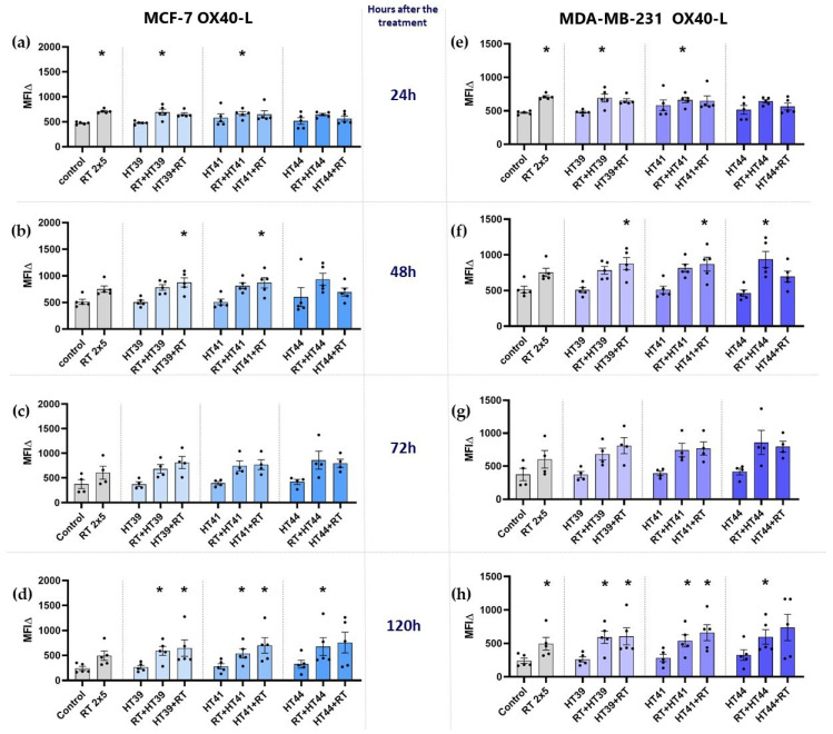
Hyperthermia (HT) is an accepted treatment for recurrent breast cancer which locally heats the tumor to 39-44 °C, and it is a very potent sensitizer for radiotherapy (RT) and chemotherapy. However, currently little is known about how HT with a distinct temperature, and particularly, how the sequence of HT and RT changes the immune phenotype of breast cancer cells. Therefore, human MDA-MB-231 and MCF-7 breast cancer cells were treated with HT of different temperatures (39, 41 and 44 °C), alone and in combination with RT (2 × 5 Gy) in different sequences, with either RT or HT first, followed by the other. Tumor cell death forms and the expression of immune checkpoint molecules (ICMs) were analyzed by multicolor flow cytometry. Human monocyte-derived dendritic cells (moDCs) were differentiated and co-cultured with the treated cancer cells. In both cell lines, RT was the main stressor for cell death induction, with apoptosis being the prominent cell death form in MCF-7 cells and both apoptosis and necrosis in MDA-MB-231 cells. Here, the sequence of the combined treatments, either RT or HT, did not have a significant impact on the final outcome. The expression of all of the three examined immune suppressive ICMs, namely PD-L1, PD-L2 and HVEM, was significantly increased on MCF-7 cells 120 h after the treatment of RT with HT of any temperature. Of special interest for MDA-MB-231 cells is that only combinations of RT with HT of both 41 and 44 °C induced a significantly increased expression of PD-L2 at all examined time points (24, 48, 72, and 120 h). Generally, high dynamics of ICM expression can be observed after combined RT and HT treatments. There was no significant difference between the different sequences of treatments (either HT + RT or RT + HT) in case of the upregulation of ICMs. Furthermore, the co-culture of moDCs with tumor cells of any treatment had no impact on the expression of activation markers. We conclude that the sequence of HT and RT does not strongly affect the immune phenotype of breast cancer cells. However, when HT is combined with RT, it results in an increased expression of distinct immune suppressive ICMs that should be considered by including immune checkpoint inhibitors in multimodal tumor treatments with RT and HT. Further, combined RT and HT affects the immune system in the effector phase rather than in the priming phase.

Keywords: breast cancer; dendritic cell activation; hyperthermia; hyperthermia treatment sequence; immune checkpoint molecules; immune phenotype; radiotherapy.


Conflict of interest statement

The authors declare no conflict of interest with regard to the work presented here.

Figures

Figure 1
Figure 1
Graphical illustration of the heating device. The heating device was mostly made of stainless steel. The device consists of a temperature control unit, heating wire, temperature sensor, and the connection box for the heating wire. The heating chamber is automatically self-controlled and the target temperature was set to 39 °C, 41 °C, or 44 °C. The temperature deviation was not more than ±0.1 °C.
Figure 2
Figure 2
Treatment set-up. On the day before the start of the treatment, the cells of the respective cell line (either MDA-MB-231 or MCF-7) were seeded (displayed with the cell culture flasks). In hyperthermia-only treatment (HT), or HT followed by radiotherapy (HT + RT), HT treatment was performed on day 0 in the heating chamber system for 60 min with three different respective temperatures (39 °C, 41 °C, and 44 °C). After the HT treatment, irradiation was performed at the latest within 2 h for HT + RT. For RT + HT, the respective treatments were performed in the same manner but in reverse order. RT was performed in clinically relevant doses of 2 × 5 Gy. Irradiation in the respective RT + HT arms was always performed 2 h after the initial treatment at the latest. Sampling in all arms was performed on day 1 (24 h), d2 (48 h), d3 (72 h) and d5 (120 h) after the last irradiation.
Figure 3
Figure 3
Gating strategy for the detection of cell death forms by AnnexinV/PI staining. Exemplarily shown are data of MCF-7 breast cancer cells. The cells were first gated on singlets (a,d) by FSC-A vs. FSC-H gating, followed by the exclusion of debris in the FSC/SSC plot (b,e). Viable cells were defined as Annexin negative/PI negative, apoptotic cells as Annexin positive/PI negative and necrotic cells as Annexin positive/PI positive (c,f). Data of cultured control samples (ac) and of 2 × 5 Gy irradiated cells (df) are shown exemplarily.
Figure 4
Figure 4
Generation of human monocyte-derived DCs (moDCs) from PBMCs and the detection of DC activation markers after co-incubation with treated cancer cells. (a) PBMCs were isolated from buffy coat and seeded into an IgG pre-coated cell culture dish. On day 6 after differentiation, moDCs were co-cultured with differently treated MCF-7 breast cancer cells. After 24 h and 48 h of co-incubation, the activation markers of the moDCs were analysed using multicolor flow cytometry. The gating strategies for flow cytometry are shown (bh). (b) After pre-gating on the singlets, the viable cells were detected (c,d). Then, gating on CD11c positive cells identified moDCs (e). Dot plots of CD83 (f), CD70 (g) and CD80 (h) expression on the cell surface of moDCs are exemplarily presented.
Figure 5
Figure 5
Radiotherapy alone and in combination with hyperthermia regardless of the treatment sequence induces apoptosis in MCF-7 breast cancer cells. The percentage of necrotic MCF-7 cells are shown in graphs (a) 24 h, (b) 48 h, (c) 72 h and (d) 120 h after the treatment. The percentage of apoptotic MCF-7 cells is shown in graphs (e) 24 h, (f) 48 h, (g) 72 h and (h) 120 h after the treatment. MCF-7 cells were irradiated 2 times with 5 Gy (RT) or treated with HT of different temperatures (39 °C, 41 °C, 44 °C) and combinations of both, either HT followed by RT (HT (39 °C, 41 °C, 44 °C) + RT) or vice versa (RT + HT (39 °C, 41 °C, 44 °C)). The time interval between HT and RT was less than 2 h. The cell death forms were analyzed by AnxV/PI staining using multicolor flow cytometry. Mean ± SD are presented from at least five independent experiments. Statistical significance is calculated by using a Kruskal–Wallis test with Dunn’s correction to compare the percentage of necrotic and apoptotic cells of each group of a respective temperature to the untreated control, and a Mann–Whitney U test to compare the different sequences of HT and RT. * (p < 0.1), ** (p < 0.01) for Kruskal–Wallis test with Dunn’s correction.
Figure 6
Figure 6
Radiotherapy alone and in combination with hyperthermia regardless of the treatment sequence significantly induces apoptosis and necrosis in MDA-MB-231 breast cancer cells. The percentage of necrotic MDA-MB-231 cells are shown in graphs (a) 24 h, (b) 48 h, (c) 72 h and (d) 120 h after the treatment. The percentage of apoptotic MDA-MB-231 cells is shown in graphs (e) 24 h, (f) 48 h, (g) 72 h and (h) 120 h after the treatment. MDA-MB-231 breast cancer cells were irradiated 2 times with 5 Gy (RT) or treated with HT of different temperatures (39 °C, 41 °C, 44 °C) and combinations of both, either HT followed by RT (HT (39 °C, 41 °C, 44 °C) + RT) or vice versa (RT + HT (39 °C, 41 °C, 44 °C)). Mean ± SD are presented from at least five independent experiments. Statistical significance was calculated by using a Kruskal–Wallis test with Dunn’s correction to compare the percentage of necrotic and apoptotic cells of each group of a respective temperature to the untreated control, and a Mann–Whitney U test to compare the different sequences of HT and RT. * (p < 0.1), ** (p < 0.01), *** (p < 0.001) for Kruskal–Wallis test with Dunn’s correction.
Figure 7
Figure 7
Hyperthermia in combination with radiotherapy affects the expression of inhibitory immune checkpoint molecules (PD-L1, PD-L2, and HVEM) on MCF-7 breast cancer cells. MCF-7 cells were irradiated 2 times with 5 Gy (RT) or treated with HT of different temperatures (39 °C, 41 °C, 44 °C) and combination of both, either HT followed by RT (HT (39 °C, 41 °C, 44 °C) + RT) or vice versa (RT + HT (39 °C, 41 °C, 44 °C)). The time interval between HT and RT was less than 2 h. The expression of ICMs ((ad): PD-L1, (eh): PD-L2 and (il): HVEM) were analyzed by multicolor flow cytometry. The mean fluorescence intensity (ΔMFI) was calculated by subtracting the fluorescence intensity of unstained samples from stained samples. Mean ± SD are presented from at least five independent experiments. Statistical significance is calculated by using Kruskal–Wallis tests with Dunn’s correction by comparing the ΔMFI of cells after the treatment to untreated control of the corresponding timepoint, and Mann–Whitney U tests to compare the ΔMFI of different sequences of HT and RT. * (p < 0.1), ** (p < 0.01). Further, RT alone was compared with combinational treatments (HT + RT and RT + HT); # (p < 0.1).
Figure 8
Figure 8
Hyperthermia in combination with radiotherapy affects the expression of inhibitory immune checkpoint molecules (PD-L1, PD-L2, and HVEM) on MDA-MB-231 breast cancer cells. MDA-MB-231 cells were irradiated 2 times with 5 Gy (RT) or treated with HT of different temperatures (39 °C, 41 °C, 44 °C) and a combination of both, either HT followed by RT (HT (39 °C, 41 °C, 44 °C) +RT) or vice versa (RT + HT (39 °C, 41 °C, 44 °C)). The time interval between HT and RT was less than 2 h. The expression of ICMs ((ad): PD-L1, (eh): PD-L2 and (il): HVEM) was analyzed by multicolor flow cytometry. The mean fluorescence intensity (ΔMFI) was calculated by subtracting the fluorescence intensity of unstained samples from stained samples. Mean ± SD are presented from at least five independent experiments. Statistical significance is calculated by using Kruskal–Wallis tests with Dunn’s correction by comparing the ΔMFI of cells after the treatment to untreated control of the corresponding timepoint, and Mann–Whitney U tests to compare the ΔMFI of different sequences of HT and RT. * (p < 0.1), ** (p < 0.01). RT alone was compared with combinational treatments (HT + RT and RT + HT); # (p < 0.1).
Figure 9
Figure 9
Expression of the immune stimulatory ICM OX40-L on MCF-7 and MDA-MB-231 cells at different timepoints after the treatment. (ad) MCF-7 and (eh) MDA-MB-231 cells were irradiated 2 times with 5 Gy (RT) or treated with HT of different temperatures (39 °C, 41 °C, 44 °C) and combinations of both, either HT followed by RT (HT (39 °C, 41 °C, 44 °C) + RT) or vice versa (RT + HT (39 °C, 41 °C, 44 °C)). The time interval between HT and RT was less than 2 h. The expression of OX40-L was analyzed by multicolor flow cytometry (a,e) 24 h, (b,f) 48 h, (c,g) 72 h, or (d,h) 120 h later. The mean fluorescence intensity (ΔMFI) was calculated by subtracting the fluorescence intensity of unstained samples from stained samples. Mean ± SD are presented from at least five independent experiments. Statistical significance is calculated by using Kruskal–Wallis tests with Dunn’s correction by comparing the ΔMFI of cells after the treatment to untreated control of the corresponding timepoint, and Mann–Whitney U tests to compare the ΔMFI of different sequences of HT and RT. * (p < 0.1).
Figure 10
Figure 10
Expression of activation markers on moDCs after contact with hyperthermia- and radiotherapy treated MCF-7 breast cancer cells. Displayed is the expression of DC activation markers after 24 h (a)—CD83, (b)—CD70, (c)—CD80, and 48 h (d)—CD83, (e)—CD70, (f)—CD80 after co-incubation of immature moDCs with untreated MCF-7 tumor cells or with differently treated MCF-7 tumor cells. The tumor cells were treated with 2 × 5 Gy RT, HT of 44 °C, and first HT of 44 °C and then RT or RT followed by HT of 44 °C. As a positive control, immature moDCs were activated with the standard maturation cocktail (MC), and as negative control, immature moDCs were kept in moDC medium without the maturation cocktail (w/o MC). The expression of DC activation markers was analyzed by multicolor flow cytometry. The mean fluorescence intensity (ΔMFI) was calculated by subtracting the fluorescence intensity of unstained samples from stained samples. Mean ± SD are presented from at least four independent experiments. Statistical significance is calculated by using Kruskal–Wallis tests with Dunn’s correction by comparing the ΔMFI of the treatments to untreated controls at the corresponding timepoint, and Mann–Whitney U tests to compare the ΔMFI of different sequence of HT and RT. * (p < 0.1).The arm without maturation cocktail was compared to the arm with maturation cocktail with a Mann–Whitney U test # (p < 0.1), ## (p < 0.01).

Similar articles

Cited by

References

    1. Jemal A., Bray F., Center M.M., Ferlay J., Ward E., Forman D. Global cancer statistics. CA Cancer J. Clin. 2011;61:69–90. doi: 10.3322/caac.20107. - DOI - PubMed
    1. O’Shaughnessy J. Extending Survival with Chemotherapy in Metastatic Breast Cancer. Oncologist. 2005;10:20–29. doi: 10.1634/theoncologist.10-90003-20. - DOI - PubMed
    1. Villacampa G., Tolosa P., Salvador F., Sánchez-Bayona R., Villanueva L., Dienstmann R., Ciruelos E., Pascual T. Addition of immune checkpoint inhibitors to chemotherapy versus chemotherapy alone in first-line metastatic triple-negative breast cancer: A systematic review and meta-analysis. Cancer Treat. Rev. 2022;104:102352. doi: 10.1016/j.ctrv.2022.102352. - DOI - PubMed
    1. Frey B., Rückert M., Deloch L., Rühle P.F., Derer A., Fietkau R., Gaipl U.S. Immunomodulation by ionizing radiation-impact for design of radio-immunotherapies and for treatment of inflammatory diseases. Immunol. Rev. 2017;280:231–248. doi: 10.1111/imr.12572. - DOI - PubMed
    1. Issels R., Kampmann E., Kanaar R., Lindner L. Hallmarks of hyperthermia in driving the future of clinical hyperthermia as targeted therapy: Translation into clinical application. Int. J. Hyperth. 2016;32:89–95. doi: 10.3109/02656736.2015.1119317. - DOI - PubMed