. 2022 Apr 23;14(9):2110.
doi: 10.3390/cancers14092110.

BRAF Modulates Lipid Use and Accumulation

Affiliations

BRAF Modulates Lipid Use and Accumulation

Jacqueline A Turner et al. Cancers (Basel). .

Abstract

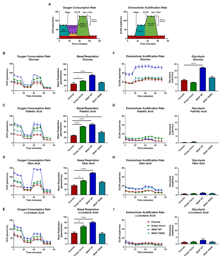
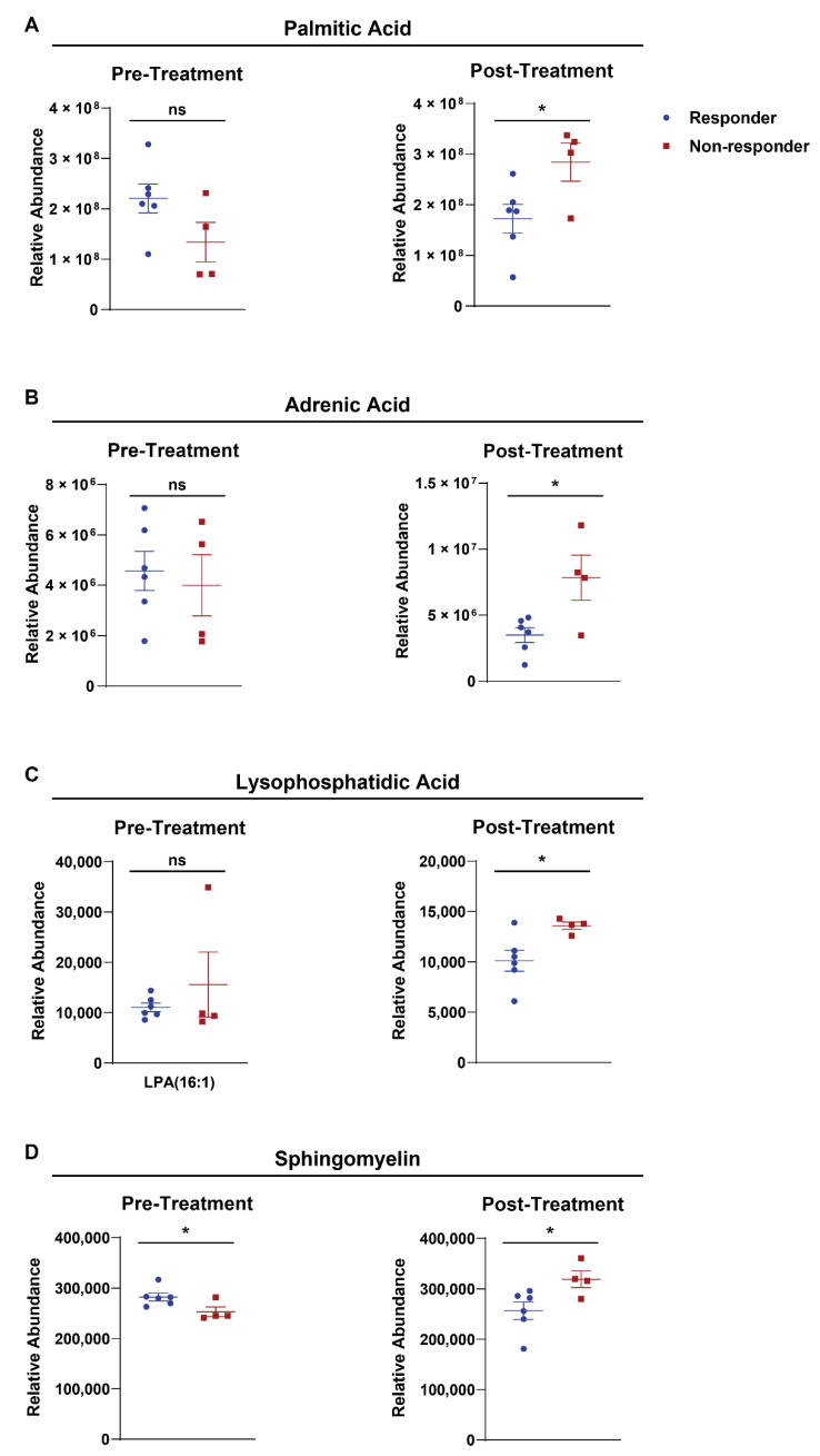
There is increasing evidence that oxidative metabolism and fatty acids play an important role in BRAF-driven tumorigenesis, yet the effect of BRAF mutation and expression on metabolism is poorly understood. We examined how BRAF mutation and expression modulates metabolite abundance. Using the non-transformed NIH3T3 cell line, we generated cells that stably overexpressed BRAF V600E or BRAF WT. We found that cells expressing BRAF V600E were enriched with immunomodulatory lipids. Further, we found a unique transcriptional signature that was exclusive to BRAF V600E expression. We also report that BRAF V600E mutation promoted accumulation of long chain polyunsaturated fatty acids (PUFAs) and rewired metabolic flux for non-Warburg behavior. This cancer promoting mutation further induced the formation of tunneling nanotube (TNT)-like protrusions in NIH3T3 cells that preferentially accumulated lipid droplets. In the plasma of melanoma patients harboring the BRAF V600E mutation, levels of lysophosphatidic acid, sphingomyelin, and long chain fatty acids were significantly increased in the cohort of patients that did not respond to BRAF inhibitor therapy. Our findings show BRAF V600 status plays an important role in regulating immunomodulatory lipid profiles and lipid trafficking, which may inform future therapy across cancers.

Keywords: BRAF; lipids; melanoma; polyunsaturated.


Conflict of interest statement

The authors of this paper declare no conflict of interest.

Figures

Figure 1
Figure 1
BRAF V600 status and expression modulates the metabolic profile. Global metabolomic data was collected by mass spectrometry for each biological replicate and evaluated by (A) unsupervised principal component analysis (PCA) and (B) and hierarchical clustering analysis. (C) Class and frequency of the top 50 metabolites present in BRAF V600E compared to BRAF WT. Normalized % refers to normalized abundancy based on the total number of metabolites in each class. (D) Heatmap representing relative metabolite abundancies with averaged triplicates normalized to the parental control. Z-scores represent highly abundant metabolites in red and less abundant metabolites in blue. Heatmap was generated using http://heatmapper.ca/ and was accessed on 19 February 2019.
Figure 2
Figure 2
Cells expressing BRAF WT are more sensitive to nutrient availability than cells expressing BRAF V600E. (A) Schematics of Seahorse metabolic flux analysis for both oxygen consumption rate (OCR) and extracellular acidification rate (ECAR). The poisons used (10 µM each) are oligomycin (oligo), (4-(trifluoromethoxy) phenyl) carbonohydrazonoyl dicyanide (FCCP), antimycin (ant), and rotenone (rot). In OCR (left), red represents non-mitochondrial respiration, teal represents basal respiration, purple represents ATP-linked production, yellow represents proton leak, green represents maximal respiratory capacity, and the reserve respiratory capacity is calculated from [maximal respiratory capacity] − [basal respiration]. In ECAR (right), red represents non-glycolytic acidification, teal represents glycolysis, green represents glycolytic capacity, and the reserve glycolytic capacity is calculated from [glycolytic capacity] − [glycolysis]. OCR and ECAR were measured one hour after BRAF WT (blue lines), BRAF V600E (teal lines), control (green lines), and parental (red lines) cells were cultured in media supplemented with either (B,F) 25 mM glucose, (C,G) 25 µM palmitic acid, (D,H) 25 µM oleic acid, or (E,I) 25 µM α-linolenic acid. The ANOVA statistical test with post-hoc analysis was performed where * p < 0.05, ** p < 0.005, **** p < 0.00005 indicates the level of statistical significance.
Figure 3
Figure 3
BRAF V600E overexpressing cells accumulate lipids in tunneling nanotube (TNT)-like structures. (A) Immunofluorescence staining for lipid droplets using Nile red (red), F-actin using phalloidin (green), and nuclei using DAPI (blue). Scale bars represent 20 µm. (B) Quantified total fluorescence in the red channel normalized to cell number. (C) Aspect ratios were determined by measuring the length and width of individual cells. (D) Annotated immunofluorescence staining from the experiment shown in panel (A) with white arrows highlighting TNT-like protrusions. (E,F) Regions of interest were quantified using fixed areas to measure fluorescence in the red channel. All quantifications were performed using FIJI and statistics were performed with GraphPad software. The ANOVA statistical test with post-hoc analysis was performed where * p < 0.05, ** p < 0.005, *** p < 0.0005, **** p < 0.00005 indicates the level of statistical significance.
Figure 4
Figure 4
Immunomodulatory lipid enrichment and a unique transcriptional signature are exclusive characteristics of cells expressing BRAF V600E. (A) Schematic of polyunsaturated fatty acid metabolism. LA, linoleic acid; αLA, α-linolenic acid; AA, arachidonic acid; EPA, eicosapentaenoic acid; HETE, hydroxyeicosatetraenoic acids; LxA4, lipoxin A4; PGE2, prostaglandin E2; DHA, docosahexaenoic acid; RvE1, resolvin E1. Fold-change in abundancy of (B) intracellular and (C) extracellular LA, αLA, AA, and EPA in BRAF V600E, BRAF WT, and control cells normalized to parental cells. (DF) Relative intracellular abundancies of (D) 9-hydroxyeicosatetraenoic acid (9-HETE), (E) docosahexaenoic acid (DHA), and (F) prostaglandin E2 (PGE2) in BRAF V600E, BRAF WT, and control cells normalized to parental cells. (G) Heatmap of 2 ^ (average of -dCT) values. Z-scores represent highly expressed mRNA in red and less expressed mRNA in blue. Heatmap was generated using http://heatmapper.ca/ accessed on 19 February 2019. Quantified mRNA expression for (H) FAS, (I) SCL27a1, (J) CPT1a, and (K) PPARγ by qRT-PCR analysis in BRAF V600E, BRAF WT, and control cells normalized to parental cells. Expression was normalized to GAPDH and the standard error of the mean of triplicates are represented by the error bars. Individual graphs represent the top four upregulated genes in cells expressing BRAF V600E. The ANOVA statistical test with post-hoc analysis was performed where * p < 0.05, ** p < 0.005, *** p < 0.0005, **** p < 0.00005 indicates the level of statistical significance.
Figure 5
Figure 5
Plasma long chain fatty acid levels vary in response BRAF/MAPK inhibitor therapy in advanced stage melanoma patients. Relative abundance of (A) palmitic acid, (B) adrenic acid, (C) lysophosphatidic acid, and (D) sphingomyelin in responder patients (blue symbols; complete response and partial response) or non-responders (red symbols; stable disease and progressive disease) measured both pre-(left) and post-treatment (right). The unpaired Student’s t test analysis was performed where * p < 0.05 and ns designates not statistically significant.

References

    1. Liu F., Yang X., Geng M., Huang M. Targeting ERK, an Achilles’ Heel of the MAPK pathway, in cancer therapy. Acta Pharm. Sin. B. 2018;8:552–562. doi: 10.1016/j.apsb.2018.01.008. - DOI - PMC - PubMed
    1. Yaeger R., Corcoran R.B. Targeting Alterations in the RAF–MEK Pathway. Cancer Discov. 2019;9:329–341. doi: 10.1158/2159-8290.CD-18-1321. - DOI - PMC - PubMed
    1. Gardner A.M., Vaillancourt R.R., Lange-Carter C.A., Johnson G.L. MEK-1 phosphorylation by MEK kinase, Raf, and mitogen-activated protein kinase: Analysis of phosphopeptides and regulation of activity. Mol. Biol. Cell. 1994;5:193–201. doi: 10.1091/mbc.5.2.193. - DOI - PMC - PubMed
    1. Graves J.D., Campbell J.S., Krebs E.G. Protein Serine/Threonine Kinases of the MAPK Cascade. Ann. N. Y. Acad. Sci. 1995;766:320–343. doi: 10.1111/j.1749-6632.1995.tb26684.x. - DOI - PubMed
    1. Ahn N.G. The MAP kinase cascade. Discovery of a new signal transduction pathway. Mol. Cell. Biochem. 1993;127–128:201–209. doi: 10.1007/BF01076771. - DOI - PubMed