. 2022 Apr 24;14(9):2120.
doi: 10.3390/cancers14092120.

Potential Role of Inflammation-Promoting Biliary Microbiome in Primary Sclerosing Cholangitis and Cholangiocarcinoma

Affiliations

Potential Role of Inflammation-Promoting Biliary Microbiome in Primary Sclerosing Cholangitis and Cholangiocarcinoma

Katsuyuki Miyabe et al. Cancers (Basel). .

Abstract

Background: Primary sclerosing cholangitis (PSC) is a major risk factor for cholangiocarcinoma (CCA). We investigated biliary and fecal microbiota to determine whether specific microbes in the bile or stool are associated with PSC or CCA.

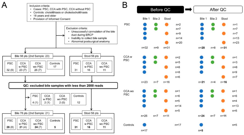
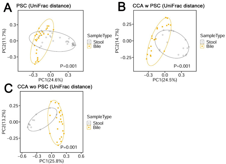
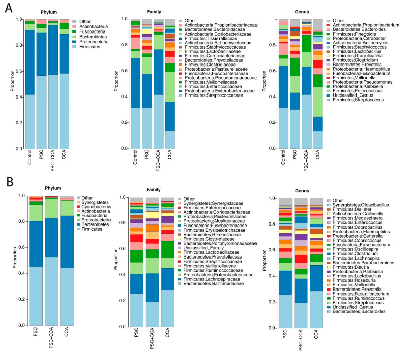
Methods: Bile was obtained from 32 patients with PSC, 23 with CCA with PSC, 26 with CCA without PSC, and 17 controls. Over 90% of bile samples were from patients with perihilar CCA. Stool was obtained from 31 patients with PSC (11 were matched to bile), 16 with CCA with PSC (10 matched to bile), and 11 with CCA without PSC (6 matched to bile). Microbiota composition was assessed using 16SrRNA-marker-based sequencing and was compared between groups.

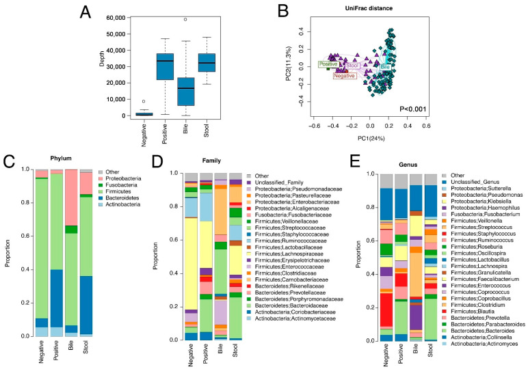
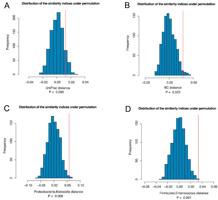
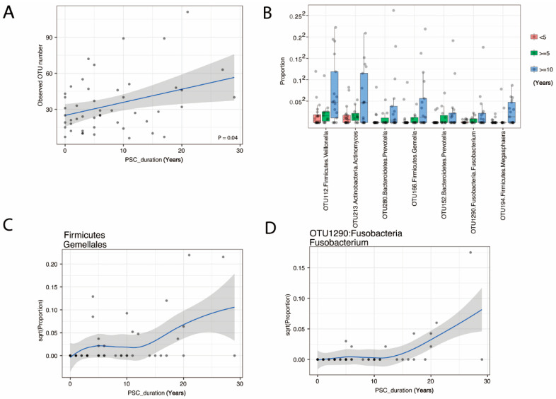
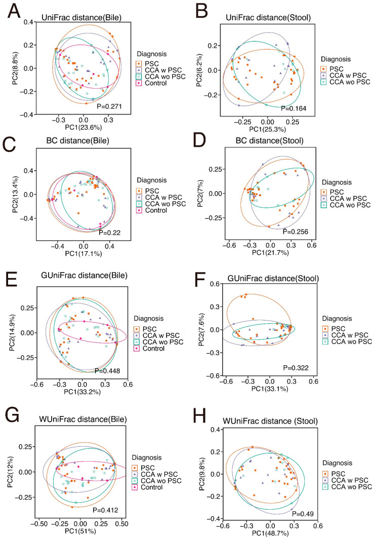
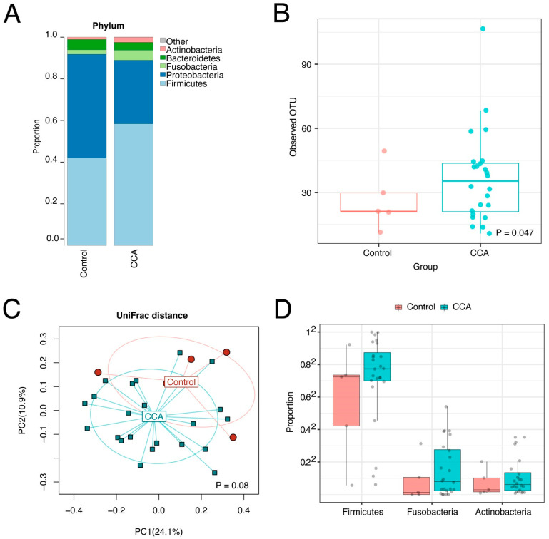
Results: Bile has a unique microbiota distinguished from negative DNA controls and stool. Increased species richness and abundance of Fusobacteria correlated with duration of PSC and characterized the biliary microbiota in CCA. Stool microbiota composition showed no significant differences between groups.

Conclusions: We identified a unique microbial signature in the bile of patients with increased duration of PSC or with CCA, suggesting a role for microbiota-driven inflammation in the pathogenesis and or progression to perihilar CCA. Further studies are needed to test this hypothesis.

Keywords: bile microbiome; cholangiocarcinoma; primary sclerosing cholangitis.


Conflict of interest statement

L.R.R. has received grant funding from Bayer, Boston Scientific, Exact Sciences, Gilead Sciences, Glycotest, Redhill Biopharma, TARGET PharmaSolutions, and FUJIFILM Medical Systems and has served on advisory boards for AstraZeneca, Bayer, Eisai, Exact Sciences, Gilead Sciences, and QED Therapeutics.

Figures

Figure 1
Figure 1
(A) Flow chart of the study with quality control after sample collection. (B) Outline of sample collection. PSC, primary sclerosing cholangitis; CCA, cholangiocarcinoma; QC, quality control; CCA w PSC, CCA with PSC; CCA wo PSC, CCA without PSC.
Figure 2
Figure 2
Microbiome variation across bile, stool, negative and positive controls. The negative controls were empty tubes processed alongside the DNA extractions for the samples, and positive controls were derived from stool samples pooled from 20 healthy subjects. (A) Sequence depth distribution shows that bile samples have a higher sequence depth than negative controls. (B) Ordination plot based on unweighted UniFrac distance reveals a distinct microbiota structure for bile samples. The statistical significance is confirmed by PERMANOVA (p < 0.001, bile vs. other sample types). (CE) Average microbiota profiles in each sample type show that bile has a unique microbiota profile different from negative controls.
Figure 3
Figure 3
β-diversity metrics of PSC (A), CCA w PSC (B), and CCA wo PSC (C) samples. UniFrac distance consists of unweighted UniFrac distance.
Figure 4
Figure 4
The similarity between the bile and stool microbiome from the same subject. We compared the average distance between the bile and stool samples from the same subjects (Dw) to the average distance between the bile and stool samples from different subjects (Db) based on UniFrac distance (A) and Bray–Curtis distance (B). If Db > Dw, it indicates that the bile and stool from the same subject is correlated. Thus, the test statistic Ts = (Db–Dw) can be interpreted as the similarity index. To establish significance, the observed similarity index (red vertical line) was compared to that under permutation (no correlation) to establish statistical significance. Two genera of Klebsiella (C) and Enterococcus (D) were identified to drive the “similarity” at FDR < 20%.
Figure 5
Figure 5
Changes in bile microbiome with PSC duration in years. Scatter plot of OTU number (A) shows an increase over PSC duration in years. Potential pathogens from 7 OTU (B) and the Order Firmicute; Gemellales (C) increase with PSC duration (FDR < 20%). An example detail from OTU1290:Fusobacteria Fusobacterium is shown in (D).
Figure 6
Figure 6
Average bile (A) and stool (B) microbiome profiles in PSC, CCA w PSC, and CCA wo PSC.
Figure 7
Figure 7
β-diversity metrics of all groups in bile (A,C,E,G) and stool (B,D,F,H) samples. UniFrac distance consists with unweighed UniFrac distance. BC, Bray–Cutis; GUniFrac, generalized UniFrac; WUniFrac, weighted UniFrac.
Figure 8
Figure 8
Bile microbiome profiles of CCA and controls. The bile microbiome profiles (A), the boxplots (B), and ordination plot analyzed by unweighted UniFrac distance (C) of CCA and controls shows species richness in CCA bile samples compared to controls. The bile microbiome boxplots (D) show increases in Firmicutes, Fusobacteria, and Actinobacteria in bile from CCA patients.

References

    1. Tabibian J.H., Lindor K.D. Primary sclerosing cholangitis: A review and update on therapeutic developments. Expert Rev. Gastroenterol. Hepatol. 2013;7:103–114. doi: 10.1586/egh.12.80. - DOI - PubMed
    1. Gupta A., Bowlus C.L. Primary sclerosing cholangitis: Etiopathogenesis and clinical management. Front. Biosci. 2012;4:1683–1705. doi: 10.2741/e490. - DOI - PubMed
    1. Dyson J.K., Beuers U., Jones D.E.J., Lohse A.W., Hudson M. Primary sclerosing cholangitis. Lancet. 2018;391:2547–2559. doi: 10.1016/S0140-6736(18)30300-3. - DOI - PubMed
    1. Rizvi S., Gores G.J. Pathogenesis, diagnosis, and management of cholangiocarcinoma. Gastroenterology. 2013;145:1215–1229. doi: 10.1053/j.gastro.2013.10.013. - DOI - PMC - PubMed
    1. Banales J.M., Marin J.J.G., Lamarca A., Rodrigues P.M., Khan S.A., Roberts L.R., Cardinale V., Carpino G., Andersen J.B., Braconi C., et al. Cholangiocarcinoma 2020: The next horizon in mechanisms and management. Nat. Rev. Gastroenterol. Hepatol. 2020;17:557–588. doi: 10.1038/s41575-020-0310-z. - DOI - PMC - PubMed

LinkOut - more resources