Review
. 2022 Apr 26;14(9):2160.
doi: 10.3390/cancers14092160.

Malignant Superficial Mesenchymal Tumors in Children

Affiliations
Review

Malignant Superficial Mesenchymal Tumors in Children

Philippe Drabent et al. Cancers (Basel). .

Abstract

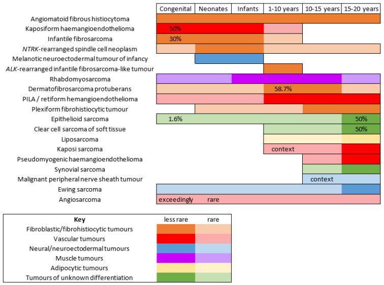
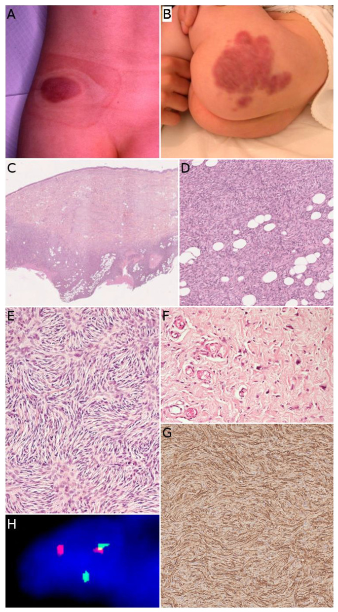
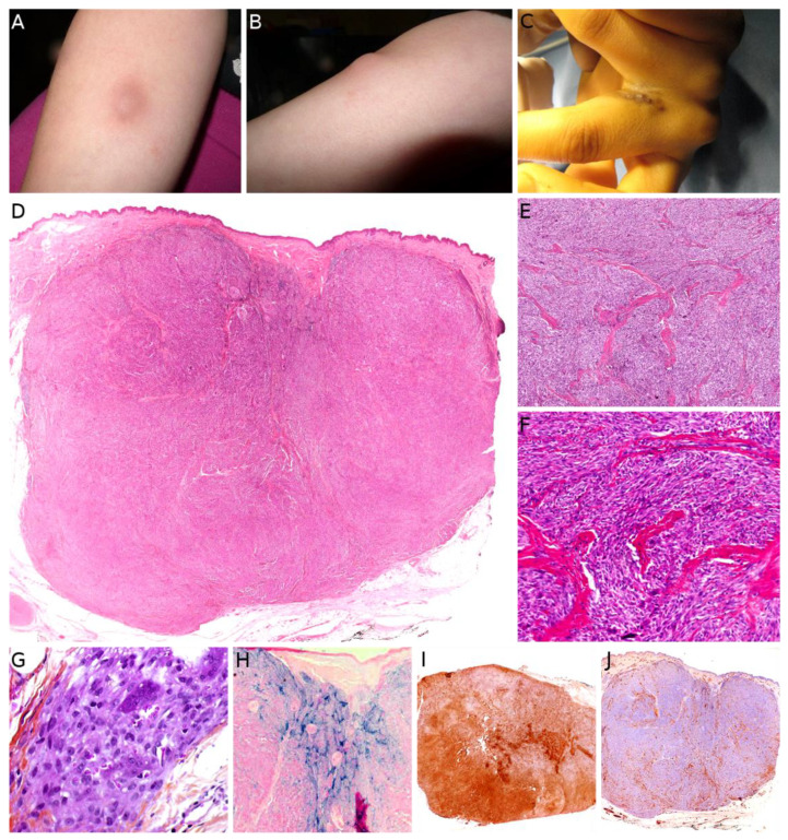
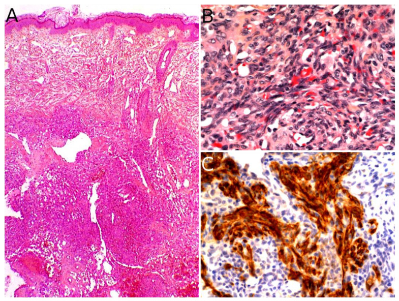
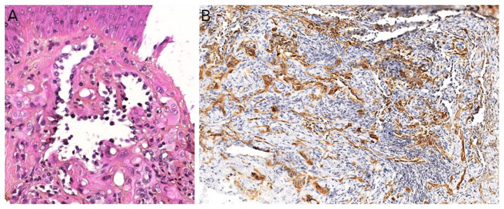
Malignant superficial mesenchymal tumors are a very diverse group of neoplasms with few clinical and radiological discriminatory factors. Hence, some of these cancers are rarely suspected based on clinical and radiological grounds, others may be easily misdiagnosed, and the histological analysis of a biopsy or resection is central in the diagnostic process. In children, the age at presentation is a major element of the differential diagnosis. Some tumors have a very distinct epidemiology, while others may be seen at any age. More recently, the advances in molecular biology have greatly improved the diagnosis of mesenchymal tumors and new entities are still being described. In the present review, we provide an overview of the diversity of malignant superficial mesenchymal tumors in children, including new and/or rare entities. We discuss the important diagnostic features, be they clinical, histological, or molecular. Special attention was given to the genetic features of these tumors, particularly when they were helpful for the diagnosis or treatment.

Keywords: children; diagnosis; genetics; histology; mesenchymal tumors; sarcoma; skin.


Conflict of interest statement

The authors declare no conflict of interest.

Figures

Figure 1
Figure 1
Malignant superficial mesenchymal tumors of the child by age range.
Figure 2
Figure 2
Angiomatoid fibrous histiocytoma: (A) Pseudo-angiomatous spaces (×25); (B) blood-filled pseudo-angiomatous space (×25); (C) dense lymphocytic infiltrate at the periphery of the tumor (×25); (D) histiocytoid tumor cells and hemosiderin deposits (×200); (E) positivity for desmin (×25) and (F) for CD68 (×25).
Figure 3
Figure 3
Infantile fibrosarcoma: (A) Large vascularized tumor on the arm of a newborn; (B) large surgical biopsy of a dermo-hypodermal multi-lobulated tumor with high cellularity (×25); (C) core-needle biopsy of another case (×25); (D) cellular, monomorphic spindle cell proliferation arranged in compact sheets or long fascicles (×100).
Figure 4
Figure 4
NTRK-rearranged spindle cell neoplasm: (A) Infiltrative growth pattern within the subcutaneous fat, with spindle cells haphazardly arranged or in fascicles (×100); (B) mild atypia and hyperchromasia (×100); (C) diffuse positivity for pan-TRK (×25); (D) fusion signals involving NTRK on FISH.
Figure 5
Figure 5
Dermatofibrosarcoma protuberans: (A) Plaque-like lesion on the flank of a teenager; (B) irregular plaque-like lesion on the buttock of neonate (congenital lesion); (C) dense cellular proliferation infiltrating into the subcutaneous fat (×25); (D) infiltration of the subcutaneous adipose tissue (×100); (E) spindle cells in a typical storiform pattern (×200); (F) giant cell fibroblastoma: less cellular tumor with giant cells and a loose stroma (×100); (G) diffuse strong CD34 positivity (×100); (H) COL1A1-PDGFB fusion on FISH.
Figure 6
Figure 6
Plexiform fibrohistiocytic tumor: (A,B) Subcutaneous nodule of the leg in a girl; (C) irregular indurated plaque of the inter-digital space in a teenager; (D) dense cellular, subcutaneous, nodular proliferation extending in the dermis (×25); (E) plexiform pattern of growth (×100); (F) plexiform pattern with some multinucleated giant cells (×100); (G) histiocyte-like cells in a fascicle with a multinucleated giant cell (×200); (H) hemosiderin deposition (Perls ×25); (I) diffuse strong CD163 positivity (×25); (J) partial SMA positivity (×25).
Figure 7
Figure 7
Kaposiform hemangioendothelioma: (A) Deep dermal and subcutaneous, infiltrating proliferation of spindle cells arranged in nodules (×25); (B) spindle endothelial cells with no mitoses (×200); (C) positivity for the lymphatic marker podoplanin in the spindle cells (×200).
Figure 8
Figure 8
Papillary intralymphatic angioendothelioma: (A) Vascular proliferation with papillary tufts protruding from the vessel wall, lined by hobnail endothelial cells with large hyperchromatic nuclei (×200); (B) partial positivity for the lymphatic marker podoplanin (×100).
Figure 9
Figure 9
Pseudomyogenic hemangioendothelioma: (A) Dense cellular proliferation with an infiltrating pattern of growth (×25); (B) sheets of epithelioid cells or plump spindle cells with a deeply eosinophilic cytoplasm (×100); (C) rhabdomyoblast-like cells with moderate nuclear atypia and some neutrophils in the stroma (×200).
Figure 10
Figure 10
Angiosarcoma: (A) Ulcerated nodule (×25); (B) atypical epithelioid cells with numerous mitoses and a tripolar mitosis (center of the panel) (×200); (C) strong CD31 positivity (×200); (D) partial CD34 positivity on small dystrophic vascular structures (×200); (E) strong nuclear ERG positivity (×200); (F) partial positivity for the cytokeratin marker AE1/AE3 (×200); (G) high proliferation index and multiple mitoses (Ki67 ×200).
Figure 11
Figure 11
Kaposi sarcoma: (A) Multiple vascular nodules on the face of a child with Wiscott–Aldrich syndrome; (B) rare spindle cells infiltrating between the collagen bundles in a clinically macular lesion (×100); (C) irregular vascular spaces and lymphocytic infiltrate in a clinically papular lesion (×100); (D) nodular spindle-cell proliferation in a clinically nodular lesion (×25); (E) spindle-cell proliferation admixed with lymphocytes in a nodular stage lesion (×200).
Figure 12
Figure 12
Rhabdomyosarcoma of the ear in a young boy: solitary erythematous nodule.
Figure 13
Figure 13
Melanotic neuroectodermal tumor of infancy: (A) Nests of small round blue cells surrounded by pigmented cells in a fibrous stroma (×25); (B) biphasic population of neuroblast-like cells and pigmented epithelioid cells (×100); (C) higher view of the epithelioid cells producing melanin (×200); (D) higher view of the neuroblast-like cells arranged in a small nest (×200); (E) HMB45 positivity on the epithelioid cells (×100); (F) synaptophysin positivity on the small neuroblast-like cells (×100).
Figure 14
Figure 14
Malignant peripheral nerve sheath tumor (MPNST): (A) MPNST arising on a pre-existing plexiform neurofibroma in a child with NF1, note the two highly cellular areas among the otherwise poorly cellular plexiform neurofibroma (×25); (B) higher view of one of the MPNST areas showing nuclear atypia and numerous mitoses (×100); (C) partial loss of S100 expression with only sparse positive cells (×200).
Figure 15
Figure 15
Epithelioid sarcoma: (A) Dermal nodule with central necrosis in an acral skin (note the thick stratum corneum) (×25); (B) epithelioid cells in a dense fibrous stroma (×100); (C) epithelioid cells and calcifications (×100); (D) loss of INI1 in the tumor cells (×100).
Figure 16
Figure 16
Clear cell sarcoma of soft tissue: (A) Densel cellular dermal and subcutaneous proliferation (×25); (B) uniform clear cells in fascicles (×100); (C) clear cells with no or slight nuclear atypia (×100); (D) diffuse HMB45 positivity (×100).

Similar articles

Cited by

References

    1. Liszewski W., Maguiness S., Greengard E., Boull C. The Incidence of Pediatric Malignant Soft Tissue Tumors of the Skin and Subcutaneous Tissue. Pediatr. Dermatol. 2018;35:e427–e429. doi: 10.1111/pde.13669. - DOI - PubMed
    1. Amadeo B., Penel N., Coindre J.-M., Ray-Coquard I., Ligier K., Delafosse P., Bouvier A.-M., Plouvier S., Gallet J., Lacourt A., et al. Incidence and Time Trends of Sarcoma (2000–2013): Results from the French Network of Cancer Registries (FRANCIM) BMC Cancer. 2020;20:190. doi: 10.1186/s12885-020-6683-0. - DOI - PMC - PubMed
    1. Bohelay G., Kluger N., Battistella M., Biaggi-Frassati A., Plantier F., Harraudeau A., Avril M.-F., Pedeutour F., Fraitag S. Angiomatoid fibrous histiocytoma in children: 6 cases. Ann. Dermatol. Venereol. 2015;142:541–548. doi: 10.1016/j.annder.2015.07.007. - DOI - PubMed
    1. Thway K., Fisher C. Angiomatoid Fibrous Histiocytoma: The Current Status of Pathology and Genetics. Arch. Pathol. Lab. Med. 2015;139:674–682. doi: 10.5858/arpa.2014-0234-RA. - DOI - PubMed
    1. Saito K., Kobayashi E., Yoshida A., Araki Y., Kubota D., Tanzawa Y., Kawai A., Yanagawa T., Takagishi K., Chuman H. Angiomatoid Fibrous Histiocytoma: A Series of Seven Cases Including Genetically Confirmed Aggressive Cases and a Literature Review. BMC Musculoskelet. Disord. 2017;18:31. doi: 10.1186/s12891-017-1390-y. - DOI - PMC - PubMed

LinkOut - more resources