Review
. 2022 Apr 27;14(9):2183.
doi: 10.3390/cancers14092183.

Targeting Sphingolipid Metabolism as a Therapeutic Strategy in Cancer Treatment

Affiliations
Review

Targeting Sphingolipid Metabolism as a Therapeutic Strategy in Cancer Treatment

Alhaji H Janneh et al. Cancers (Basel). .

Abstract

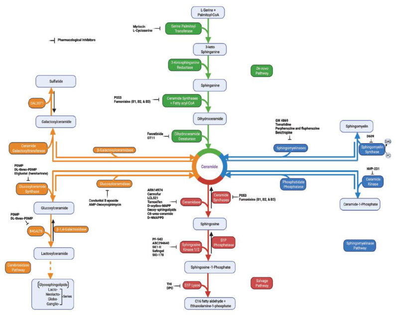
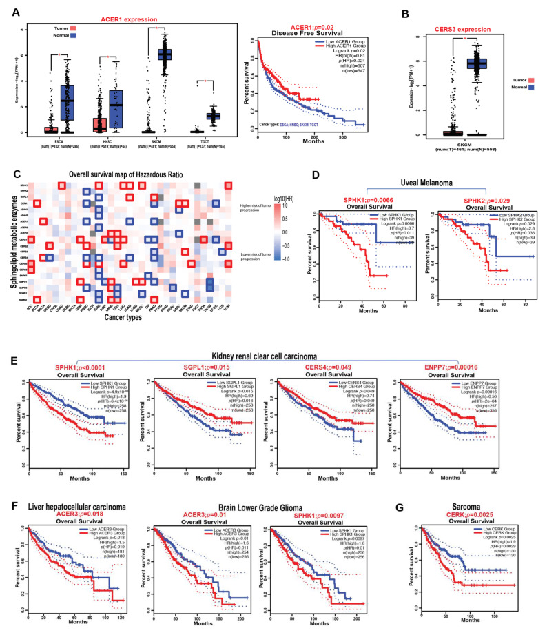
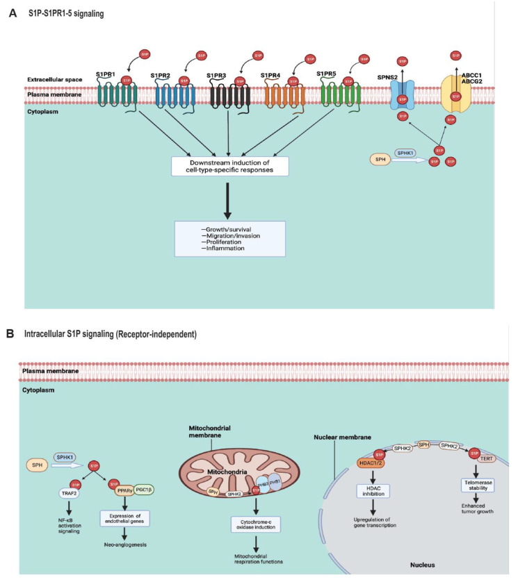
Sphingolipids are bioactive molecules that have key roles in regulating tumor cell death and survival through, in part, the functional roles of ceramide accumulation and sphingosine-1-phosphate (S1P) production, respectively. Mechanistic studies using cell lines, mouse models, or human tumors have revealed crucial roles of sphingolipid metabolic signaling in regulating tumor progression in response to anticancer therapy. Specifically, studies to understand ceramide and S1P production pathways with their downstream targets have provided novel therapeutic strategies for cancer treatment. In this review, we present recent evidence of the critical roles of sphingolipids and their metabolic enzymes in regulating tumor progression via mechanisms involving cell death or survival. The roles of S1P in enabling tumor growth/metastasis and conferring cancer resistance to existing therapeutics are also highlighted. Additionally, using the publicly available transcriptomic database, we assess the prognostic values of key sphingolipid enzymes on the overall survival of patients with different malignancies and present studies that highlight their clinical implications for anticancer treatment.

Keywords: apoptosis; cancer; cell growth; ceramide; sphingolipids; sphingosine-1-phosphate (S1P); therapeutics.


Conflict of interest statement

The authors declare no conflict of interest.

Figures

Figure 1
Figure 1
Sphingolipid metabolic pathways with selected inhibitors targeting enzymes. Ceramide, which is the intermediate molecule in sphingolipid metabolic pathway, can be formed either through de novo synthesis (green), sphingomyelin hydrolysis (blue), cerebrosides (orange), or salvage pathway (red). De novo synthesis starts with the functions of serine palmitoyltransferase (generates 3-keto sphinganine), 3-ketosphinganine reductase (generates sphinganine), (dihydro)ceramide synthases (generates dihydroceramide), and dihydroceramide desaturase (generates ceramide). The hydrolysis of sphingomyelin by the functions of sphingomyelinases can also generate ceramide (blue). Glucosylceramidase and β-galactosylceramidase can break down glucosylceramide and galactosylceramide, respectively, to generate ceramide (orange path). In the salvage pathway, ceramide synthases again can convert sphingosine to ceramide. In reverse, ceramide can be metabolized by ceramidases to generate sphingosine, which can then be phosphorylated to produce sphingosine-1-phosphate (S1P) by the functions of sphingosine kinases. S1P is broken down by the actions of S1P phosphatase to restore sphingosine or by S1P lyase functions, yielding ethanolamine 1-phosphate and C16 fatty aldehyde to exit the sphingolipid metabolic pathway. Sphingomyelin synthase transfers phosphorylcholine to ceramide from phosphatidylcholine (PC) to generate sphingomyelin and, thus, releasing diacylglycerol (DAG) [8]. Additionally, ceramide kinase functions to converts ceramide into ceramide-1-phosphate, while phosphatidate phosphatase functions to restore ceramide from ceramide-1-phosphate. In the generation of complex sphingolipids from ceramide, glucosylceramide synthase and ceramide galactosyltransferase produce glucosylceramide and galactosylceramide, respectively. Generation of glycosphingolipid series requires the synthesis of lactosylceramide from glucosylceramide (orange, dotted arrows). The enzymes can be inhibited by pharmacological inhibitors to regulate the sphingolipid metabolic pathway in both in vivo and in vitro studies. B4GALT6, beta-1,4-galactosyltransferase 6 [14]; GAL3ST1, galactosylceramide sulfotransferase; PDMP, 1-phenyl-2-decanoylamino-3-morpholino-1-propanol [15]; THI, 2-acetyl-5-tetrahydroxybutyl imidazole; DPO, 4-deoxy pyridoxine.
Figure 2
Figure 2
Sphingolipid structures.
Figure 3
Figure 3
Expression effects of sphingolipid metabolic enzymes on the survival outcomes of cancer patients. (A) Box plots indicating the differential expression of ACER1 in ESCA, HNSC, SKCM, and TGCT patients compared to healthy controls. The Kaplan–Meier survival curve shows the overall survival impact of ACER1 expression in ESCA, HNSC, SKCM, and TGCT tumors combined. Tumor group, (T); normal group, (N). * p-Value < 0.05. The differential expression is calculated by the mean value of log2(TPM + 1). TPM, transcript per million. (B) Box plot indicating the differential expression of CERS3 in SKCM. Tumor group, (T); normal group, (N). * p-Value < 0.05. The differential expression was calculated by the mean value of log2(TPM + 1). TPM, transcript per million. (C) Heatmap representing the overall survival of hazardous ratios (HRs) predicting the risks of tumor progression in different malignancies based on the expression patterns of sphingolipid metabolic enzymes. The red colored blocks correspond to an increased risk of tumor progression when the enzyme is overexpressed, while the blue colored blocks correspond to a lower risk (protective function) when the enzyme is overexpressed. The bold outlined boxes indicate significance based on log-rank p  <  0.05. (D) Kaplan–Meier survival curves showing the overall survival impacts of SPHK1 and SPHK2 expressions in uveal melanoma. (E) Kaplan–Meier survival curves showing the overall survival impacts of SPHK1, SGPL1, CERS4, and ENPP7 expressions in kidney renal clear cell carcinoma. (F,G) Kaplan–Meier survival curves showing the overall survival impacts of ACER3 in liver hepatocellular carcinoma and brain lower-grade glioma: SPHK1 in brain lower-grade glioma (F) and CERK in sarcoma (G). Analysis was performed using the Gene Expression Profiling Interactive Analysis2 (GEPIA2) web server. ACC, adrenocortical carcinoma; BLCA, bladder urothelial carcinoma; BRCA, breast invasive; carcinoma; CESC, cervical squamous cell carcinoma and endocervical adenocarcinoma; CHOL, cholangiocarcinoma; COAD, colon adenocarcinoma; DLBC, lymphoid neoplasm diffuse large B-cell lymphoma; ESCA, esophageal carcinoma; GBM, glioblastoma multiforme; HNSC, head and neck squamous cell carcinoma; KICH, kidney chromophobe; KIRC, kidney renal clear cell carcinoma; KIRP, kidney renal papillary cell carcinoma; LAML, acute myeloid leukemia; LGG, brain lower-grade glioma; LIHC, liver hepatocellular carcinoma; LUAD, lung adenocarcinoma; LUSC, lung squamous cell carcinoma; MESO, mesothelioma; OV, ovarian serous cystadenocarcinoma; PAAD, pancreatic adenocarcinoma; PCPG, pheochromocytoma and paraganglioma; PRAD, prostate adenocarcinoma; READ, rectum adenocarcinoma; SARC, sarcoma; SKCM, skin cutaneous melanoma; STAD, stomach adenocarcinoma; TGCT, testicular germ cell tumors; THCA, thyroid carcinoma; THYM, thymoma; UCEC, uterine corpus endometrial carcinoma; UCS, uterine carcinosarcoma; UVM, uveal melanoma; SPHK1, sphingosine kinase 1; SPHK2, sphingosine kinase 2; SGPL1, sphingosine-1-phosphate lyase 1; CERK, ceramide kinase; ASAH1, acid ceramidase; ASAH2, neutral ceramidase; ACER1, alkaline ceramidase 1; ACER2, alkaline ceramidase 2; ACER3, alkaline ceramidase 3; CerS1, ceramide synthase 1; CerS2, ceramide synthase 2; CerS3, ceramide synthase 3; CerS4, ceramide synthase 4; CerS5, ceramide synthase 5; CerS6, ceramide synthase 6; ENPP7, alkaline sphingomyelinase; SMPD1, acid sphingomyelinase; SMPD3, neutral sphingomyelinase; SGMS1, sphingomyelin synthase 1; SGMS2, sphingomyelin synthase 2.
Figure 4
Figure 4
S1P receptor and receptor-independent signaling. (A) SPHK1 catalyzes the synthesis of S1P from SPH in the cytoplasm. S1P then exit the cytoplasm and into the extracellular space via SPNS2, ABCC1, or ABCG2 transporters. The secreted S1P can engage the five known S1P specific G protein-coupled receptors (S1PR1–5) for cellular signaling leading to a downstream induction of cell-type-specific responses to stimulate cell growth/survival, migration/invasion, proliferation, and/or inflammation. (B) S1P can also function independent of S1PRs. In the cytoplasm, SPHK1-generated S1P can bind TRAF2 at the N-terminal RING domain, leading to NF-κB signaling activation downstream. SPHK1-derived S1P can also bind and activate PPARγ, which then allows for the recruitment of PGC1β, to form the SlP/PPARγ/PGC1β complex, inducing PPARγ-dependent genes and neo-angiogenesis. SPHK2-generated S1P in the mitochondria can bind homomeric PHB2 without binding to PHB1 to induce cytochrome c oxidase or complex IV and mitochondria respiration functions. In the nucleus, SPHK2-derived S1P can bind HDAC1 and HDAC2, inhibiting their activities to stimulate the upregulation of gene transcriptions. Additionally, SPHK2-generated S1P can also bind TERT in the nuclear membrane to stabilize telomerase and enhance tumor growth. SPH, sphingosine; SPHK1, sphingosine kinase 1; SPHK2, sphingosine kinase 2; S1P, sphingosine-1-phosphate; SPNS2, protein spinster homolog 2; ABCC1, ATP-binding cassette sub-family C member 1; ABCG2, ATP-binding cassette sub-family G member 2; S1PR, sphingosine-1-phosphate receptor; TRAF2, TNF receptor-associated factor 2; NF-κB, nuclear factor-κB; PPARγ, peroxisome proliferator-activated receptor-γ; PGC1β, PPARγ co-activator 1β; PHB1, prohibitin 1; PHB2, prohibitin 2; HDAC1, histone deacetylase 1; HDAC2, histone deacetylase 2; TERT, telomerase reverse-transcriptase.

Similar articles

Cited by

References

    1. Weigert A., Olesch C., Brüne B. Sphingosine-1-phosphate and macrophage biology—How the sphinx tames the big eater. Front. Immunol. 2019;10:1706. doi: 10.3389/fimmu.2019.01706. - DOI - PMC - PubMed
    1. Spiegel S., Milstien S. Sphingosine-1-phosphate: An enigmatic signalling lipid. Nat. Rev. Mol. Cell Biol. 2003;4:397–407. doi: 10.1038/nrm1103. - DOI - PubMed
    1. Chun J., Hartung H.-P. Mechanism of action of oral fingolimod (FTY720) in multiple sclerosis. Clin. Neuropharmacol. 2010;33:91. doi: 10.1097/WNF.0b013e3181cbf825. - DOI - PMC - PubMed
    1. Presa N., Gomez-Larrauri A., Dominguez-Herrera A., Trueba M., Gomez-Munoz A. Novel signaling aspects of ceramide 1-phosphate. Biochim. Biophys. Acta (BBA)-Mol. Cell Biol. Lipids. 2020;1865:158630. doi: 10.1016/j.bbalip.2020.158630. - DOI - PubMed
    1. Cartier A., Hla T. Sphingosine 1-phosphate: Lipid signaling in pathology and therapy. Science. 2019;366:eaar5551. doi: 10.1126/science.aar5551. - DOI - PMC - PubMed

LinkOut - more resources