

In Vitro Human Cancer Models for Biomedical Applications
- PMID: 35565413
- PMCID: PMC9099454
- DOI: 10.3390/cancers14092284
In Vitro Human Cancer Models for Biomedical Applications
Abstract
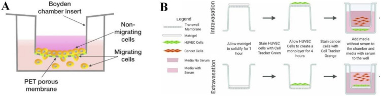
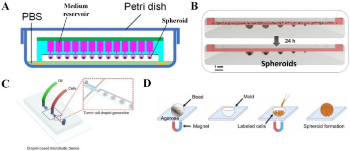
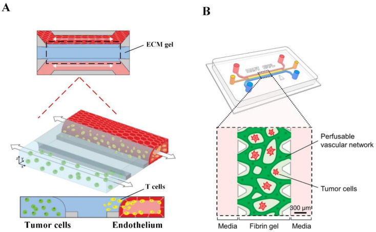
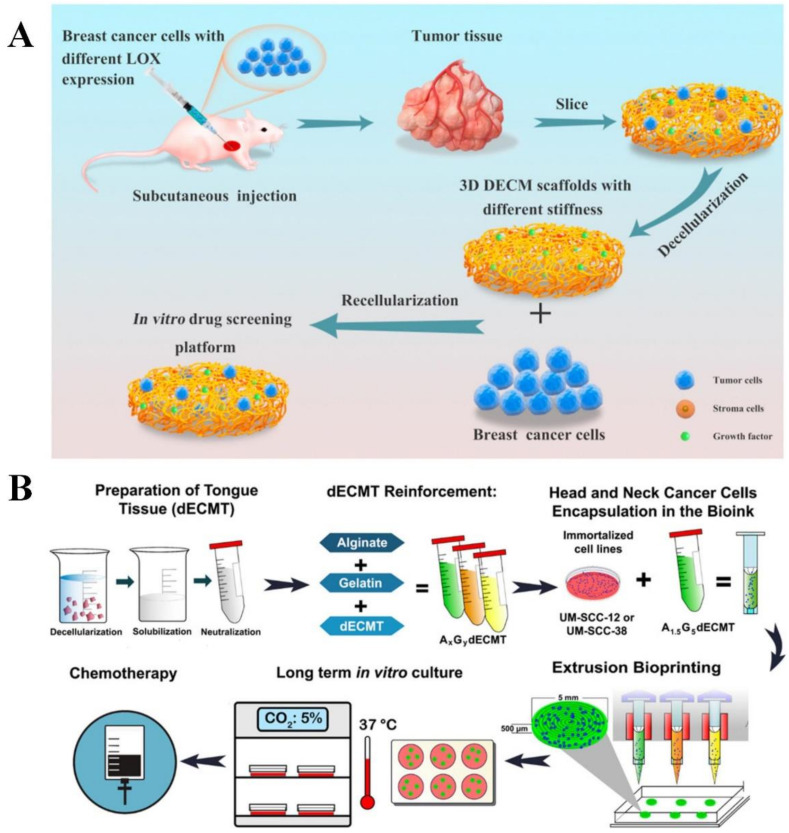
Cancer is one of the leading causes of death worldwide, and its incidence is steadily increasing. Although years of research have been conducted on cancer treatment, clinical treatment options for cancers are still limited. Animal cancer models have been widely used for studies of cancer therapeutics, but these models have been associated with many concerns, including inaccuracy in the representation of human cancers, high cost and ethical issues. Therefore, in vitro human cancer models are being developed quickly to fulfill the increasing demand for more relevant models in order to get a better knowledge of human cancers and to find novel treatments. This review summarizes the development of in vitro human cancer models for biomedical applications. We first review the latest development in the field by detailing various types of in vitro human cancer models, including transwell-based models, tumor spheroids, microfluidic tumor-microvascular systems and scaffold-based models. The advantages and limitations of each model, as well as their biomedical applications, are summarized, including therapeutic development, assessment of tumor cell migration, metastasis and invasion and discovery of key cancer markers. Finally, the existing challenges and future perspectives are briefly discussed.
Keywords: biomedical applications; cancer markers; human cancers; in vitro model; therapeutic development; tumor biology.
Conflict of interest statement
The authors declare no conflict of interest.
Figures
Similar articles
-
Nanotechnology: an evidence-based analysis.Ont Health Technol Assess Ser. 2006;6(19):1-43. Epub 2006 Nov 1. Ont Health Technol Assess Ser. 2006. PMID: 23074489 Free PMC article.
-
Microengineered 3D Tumor Models for Anti-Cancer Drug Discovery in Female-Related Cancers.Ann Biomed Eng. 2021 Aug;49(8):1943-1972. doi: 10.1007/s10439-020-02704-9. Epub 2021 Jan 5. Ann Biomed Eng. 2021. PMID: 33403451 Review.
-
Biomedical Applications of Non-Small Cell Lung Cancer Spheroids.Front Oncol. 2021 Dec 7;11:791069. doi: 10.3389/fonc.2021.791069. eCollection 2021. Front Oncol. 2021. PMID: 34950592 Free PMC article. Review.
-
The future of Cochrane Neonatal.Early Hum Dev. 2020 Nov;150:105191. doi: 10.1016/j.earlhumdev.2020.105191. Epub 2020 Sep 12. Early Hum Dev. 2020. PMID: 33036834
-
High-throughput screening approaches and combinatorial development of biomaterials using microfluidics.Acta Biomater. 2016 Apr 1;34:1-20. doi: 10.1016/j.actbio.2015.09.009. Epub 2015 Sep 8. Acta Biomater. 2016. PMID: 26361719 Review.
Cited by
-
Three-dimensional in vitro models in head and neck cancer: current trends and applications.Med Oncol. 2025 May 5;42(6):194. doi: 10.1007/s12032-025-02737-x. Med Oncol. 2025. PMID: 40320444 Review.
-
Experimental Murine Models for Colorectal Cancer Research.Cancers (Basel). 2023 Apr 30;15(9):2570. doi: 10.3390/cancers15092570. Cancers (Basel). 2023. PMID: 37174036 Free PMC article. Review.
-
Anticancer and Antioxidant Activities of the Root Extract of the Carnivorous Pitcher Plant Sarracenia purpurea.Plants (Basel). 2022 Jun 23;11(13):1668. doi: 10.3390/plants11131668. Plants (Basel). 2022. PMID: 35807620 Free PMC article.
-
Autophagy-related mechanisms for treatment of multiple myeloma.Cancer Drug Resist. 2023 Dec 25;6:838-857. doi: 10.20517/cdr.2023.108. eCollection 2023. Cancer Drug Resist. 2023. PMID: 38239705 Free PMC article. Review.
-
Cytotoxic, Antibacterial, and Antioxidant Activities of the Leaf Extract of Sinningia bullata.Plants (Basel). 2023 Feb 14;12(4):859. doi: 10.3390/plants12040859. Plants (Basel). 2023. PMID: 36840206 Free PMC article.
References
Publication types
LinkOut - more resources
Full Text Sources
