

Ultrasonographic Imaging Protocol and Sonoanatomy of the Lumbar Spine in Healthy Dogs
- PMID: 35565613
- PMCID: PMC9100366
- DOI: 10.3390/ani12091187
Ultrasonographic Imaging Protocol and Sonoanatomy of the Lumbar Spine in Healthy Dogs
Abstract
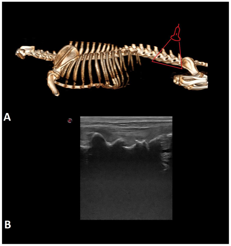
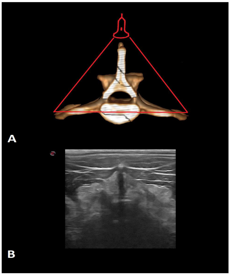
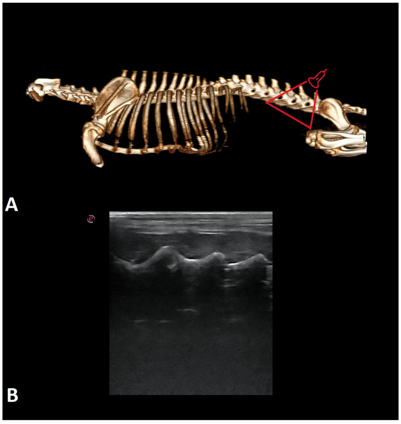
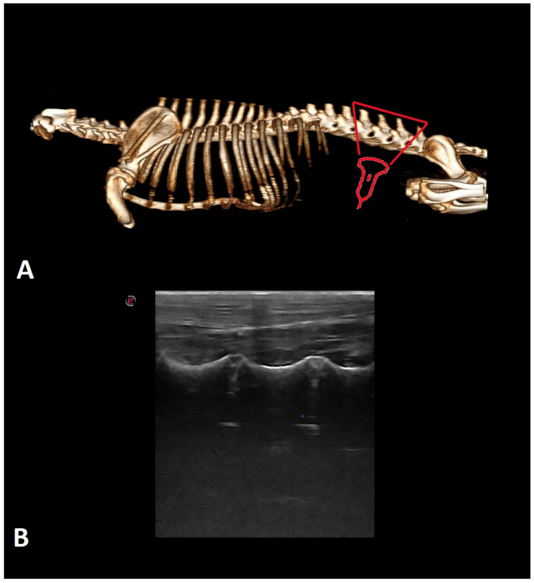
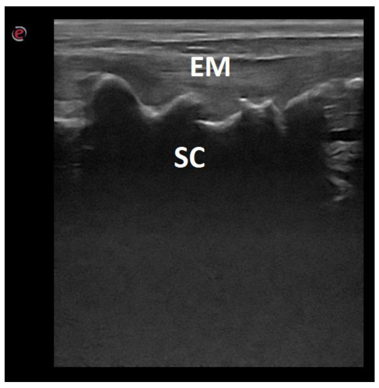
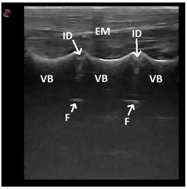
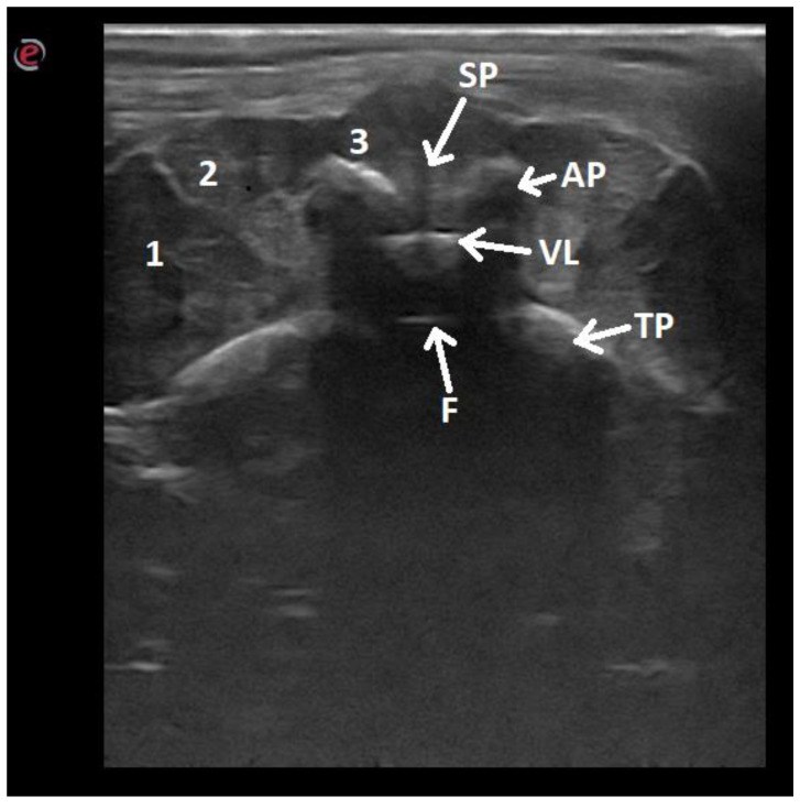
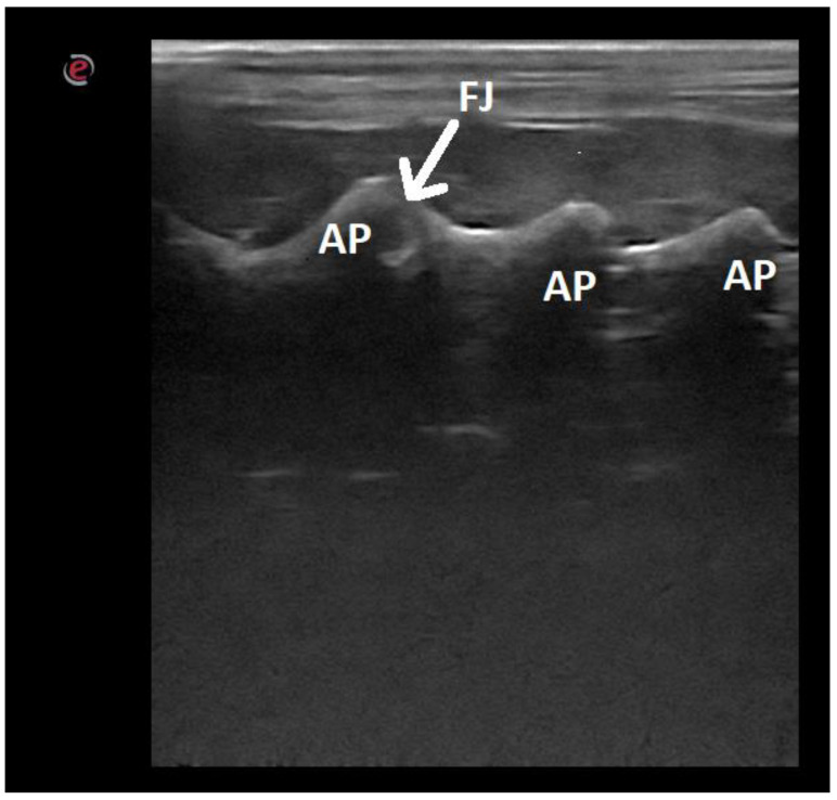
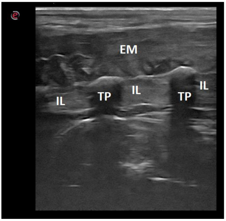
Ultrasound is an imaging technique commonly used in veterinary medicine. Ultrasound devices are widely available, their means of examination are relatively short and cheap, and they do not generate ionizing radiation. In addition, ultrasound generally does not need to be performed under general anesthesia. This study was performed on 23 canine cadavers with full clinical histories and with no confirmed pathological changes in the spine region. The imaging modalities were established in dogs in lateral recumbency, with the selected side being the uppermost angle, in a neutral position. All dogs were examined in the transverse and longitudinal planes. Sacral crest, intertransverse ligament, vertebral canal floor, vertebral body, and intervertebral discs were only visible in the longitudinal plane. Vertebral arch, supraspinal ligament, dorsal wall of the vertebral canal and muscles were visualized only in the transverse plane. This article provides a brief and relatively easy-to-perform protocol for ultrasound imaging of the lumbar spine of dogs. In addition, it presents a detailed description of the sonoanatomy of the area under investigation.
Keywords: anatomy; canine; spinal cord; ultrasound (USG).
Conflict of interest statement
The authors declare no conflict of interest.
Figures
Similar articles
-
Description and evaluation of four ultrasound-guided approaches to aid spinal canal puncture in dogs.Vet Anaesth Analg. 2016 Jul;43(4):444-52. doi: 10.1111/vaa.12324. Epub 2015 Dec 15. Vet Anaesth Analg. 2016. PMID: 26671565
-
Feasibility of ultrasound-guided epidural access at the lumbo-sacral space in dogs.Vet Radiol Ultrasound. 2015 Mar-Apr;56(2):220-8. doi: 10.1111/vru.12207. Epub 2014 Sep 3. Vet Radiol Ultrasound. 2015. PMID: 25187175
-
[Anatomical background of low back pain: variability and degeneration of the lumbar spinal canal and intervertebral disc].Schmerz. 2001 Dec;15(6):418-24. doi: 10.1007/s004820100026. Schmerz. 2001. PMID: 11793145 German.
-
Ultrasonographic identification of the dorsal atlantoaxial ligament in dogs.Vet Surg. 2017 Nov;46(8):1126-1130. doi: 10.1111/vsu.12702. Epub 2017 Aug 31. Vet Surg. 2017. PMID: 28858386
-
Ultrasonography of the lumbar spine: sonoanatomy and practical applications.Joint Bone Spine. 2014 Mar;81(2):130-6. doi: 10.1016/j.jbspin.2013.10.009. Epub 2014 Mar 4. Joint Bone Spine. 2014. PMID: 24618457 Review.
References
-
- Bonelli M., Tudury E., Santos C., Araújo B., Diogo C., Silva A., Costa F. Intraoperative ultrasonography of the vertebral canal in dogs. Arq. Bras. Med. Veterinária Zootec. 2015;67:655–663. doi: 10.1590/1678-4162-7494. - DOI
Grants and funding
LinkOut - more resources
Full Text Sources
