

The Mantle Transcriptome of Chamelea gallina (Mollusca: Bivalvia) and Shell Biomineralization
- PMID: 35565623
- PMCID: PMC9100110
- DOI: 10.3390/ani12091196
The Mantle Transcriptome of Chamelea gallina (Mollusca: Bivalvia) and Shell Biomineralization
Abstract
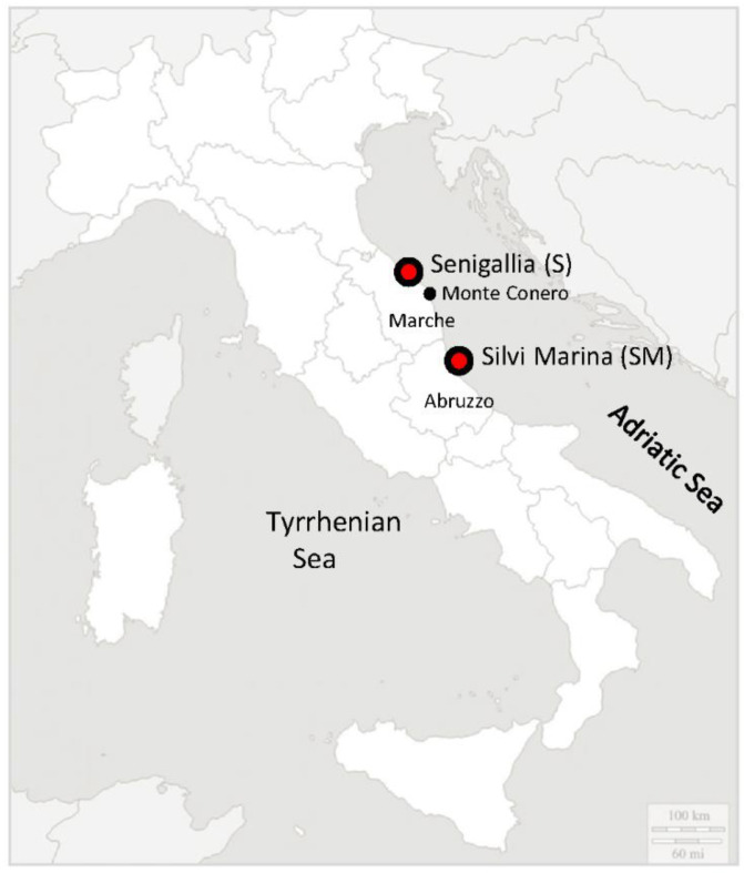
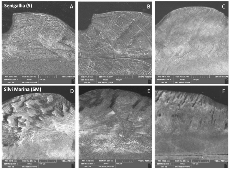
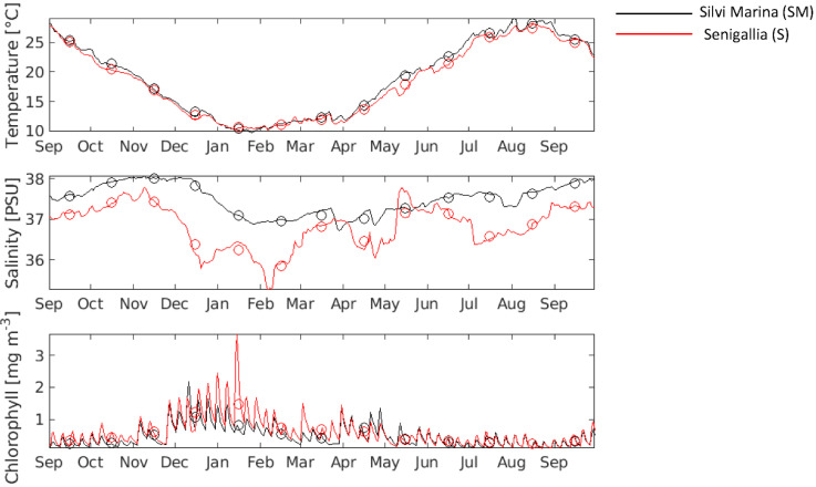
The striped venus Chamelea gallina is a bivalve mollusc that represents one of the most important fishery resources of the Adriatic Sea. In this work, we investigated for the first time the ability of this species to modulate the expression of genes encoding proteins involved in biomineralization process in response to biotic and abiotic factors. We provided the first comprehensive transcriptome from the mantle tissue of clams collected in two sampling sites located along the Italian Adriatic coast and characterized by different environmental features. Moreover, the assessment of environmental parameters, scanning electron microscopy (SEM), and X-ray diffraction (XRD) measurements on valves were conducted to better contextualize RNA sequencing (RNA-Seq) data. Functional annotation of differentially expressed genes (DEGs) and SEM observations highlighted a different shell mineralization behaviour in C. gallina clams collected from two selected sites characterized by diverse environmental parameters.
Keywords: biomineralization; gene expression analysis; mollusc; transcriptomics.
Conflict of interest statement
The authors declare no conflict of interest.
Figures
Similar articles
-
Shell properties of commercial clam Chamelea gallina are influenced by temperature and solar radiation along a wide latitudinal gradient.Sci Rep. 2016 Nov 2;6:36420. doi: 10.1038/srep36420. Sci Rep. 2016. PMID: 27805037 Free PMC article.
-
Omics approaches for conservation biology research on the bivalve Chamelea gallina.Sci Rep. 2020 Nov 5;10(1):19177. doi: 10.1038/s41598-020-75984-9. Sci Rep. 2020. PMID: 33154500 Free PMC article.
-
Sequencing and characterization of striped venus transcriptome expand resources for clam fishery genetics.PLoS One. 2012;7(9):e44185. doi: 10.1371/journal.pone.0044185. Epub 2012 Sep 18. PLoS One. 2012. PMID: 23028497 Free PMC article.
-
Deciphering mollusc shell production: the roles of genetic mechanisms through to ecology, aquaculture and biomimetics.Biol Rev Camb Philos Soc. 2020 Dec;95(6):1812-1837. doi: 10.1111/brv.12640. Epub 2020 Jul 31. Biol Rev Camb Philos Soc. 2020. PMID: 32737956 Review.
-
Sea shell diversity and rapidly evolving secretomes: insights into the evolution of biomineralization.Front Zool. 2016 Jun 7;13:23. doi: 10.1186/s12983-016-0155-z. eCollection 2016. Front Zool. 2016. PMID: 27279892 Free PMC article. Review.
Cited by
-
A review of the endangered mollusks transcriptome under the threatened species initiative of Korea.Genes Genomics. 2023 Aug;45(8):969-987. doi: 10.1007/s13258-023-01389-3. Epub 2023 Jul 5. Genes Genomics. 2023. PMID: 37405596 Review.
References
-
- Bieler R., Mikkelsen P.M., Collins T.M., Glover E.A., Gonzalez V.L., Graf D.L., Harper E.M., Healy J., Kawauchi G.Y., Sharma P.P., et al. Investigating the bivalve tree of life-an exemplar-based approach combining molecular and novel morphological characters. Invertebr. Syst. 2014;28:32–115. doi: 10.1071/IS13010. - DOI
-
- Popov S.V. Composite prismatic structure in bivalve shell. Acta Palaeontol. Pol. 1986;31:3–28.
-
- Checa A. Physical and biological determinants of the fabrication of molluscan shell microstructures. Front. Mar. Sci. 2018;5:353. doi: 10.3389/fmars.2018.00353. - DOI
Grants and funding
LinkOut - more resources
Full Text Sources
