. 2022 May 7;12(9):1209.
doi: 10.3390/ani12091209.

Anti-Müllerian Hormone Inhibits FSH-Induced Cumulus Oocyte Complex In Vitro Maturation and Cumulus Expansion in Mice

Affiliations

Anti-Müllerian Hormone Inhibits FSH-Induced Cumulus Oocyte Complex In Vitro Maturation and Cumulus Expansion in Mice

Xue Yu et al. Animals (Basel). .

Abstract

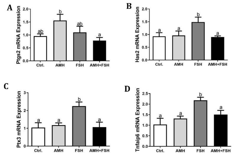
Anti-Müllerian hormone (AMH) is secreted by the ovaries of female animals and exerts its biological effects through the type II receptor (AMHR2). AMH regulates follicular growth by inhibiting the recruitment of primordial follicles and reducing the sensitivity of antral follicles to FSH. Despite the considerable research on the actions of AMH in granulosa cells, the effect of AMH on the in vitro maturation of oocytes remains largely unknown. In the current study, we showed that AMH is only expressed in cumulus cells, while AMHR2 is produced in both cumulus cells and oocytes. AMH had no significant effect on COCs nuclear maturation, whereas it inhibited the stimulatory effects of FSH on COCs maturation and cumulus expansion. Moreover, AMH treatment effectively inhibited the positive effect of FSH on the mRNA expressions of Hyaluronan synthase 2 (Has2), Pentraxin 3 (Ptx3), and TNF-alpha-induced protein 6 (Tnfaip 6) genes in COCs. In addition, AMH significantly decreased the FSH-stimulated progesterone production, but did not change estradiol levels. Taken together, our results suggest that AMH may inhibit the effects of FSH-induced COCs in vitro maturation and cumulus expansion. These findings increase our knowledge of the functional role of AMH in regulating folliculogenesis.

Keywords: anti-müllerian hormone; follicle-stimulating hormone; maturation; mice; oocyte.


Conflict of interest statement

The authors have no competing interest to declare.

Figures

Figure 1
Figure 1
Expression of Amh and Amhr in mouse cumulus cells and oocytes. PCR amplification product of representative agarose gel electrophoresis. RNA was isolated from cumulus cells (Lane 1), cumulus oocyte complexes (Lane 2), and cumulus-free oocytes (Lane 3). One-microliter amounts of cDNA were used as templates for Amh (A) and Amhr (B) amplification, ddH2O was used as negative control for PCR amplification (Lane 4).
Figure 2
Figure 2
Effect of rhAMH on COCs in vitro maturation. COCs were supplemented with AMH and FSH alone or in combination for 16 h, the first polar body extrusion, mRNA expression in cell lysate, cAMP and MPF levels in the cytoplasm were analyzed. (A), Polar body extrusion percentage; (BE), the relative mRNA expression of Fshr, Amhr2, Bmp15, and Gdf9; (F), cAMP levels; (G), MPF levels. Data were expressed as mean ± SEM from at least three independent experiments. Bars with different letters represent significant differences (p < 0.05).
Figure 3
Figure 3
Effect of rhAMH on cumulus cell expansion. COCs were supplemented with AMH and FSH alone or in combination for 16 h, cumulus cell expansion was analyzed by the cumulus expansion index (CEI). (A), representative images of different grades of cumulus cell expansion, a = 0, b = 1, c = 2, d = 3, e = 4, the scale bar represents 50 μm. (B), the representative cumulus cell expansion of each group, the scale bar represents 50 μm. (C), the CEI of each group. Data were from at least three independent experiments. Bars with different letters represent significant differences (p < 0.05).
Figure 4
Figure 4
Effect of rhAMH on expression of cumulus expansion related genes. COCs were supplemented with AMH and FSH alone or in combination for 16 h,relative expression of transcripts for Ptgs2 (A), Has2 (B), Ptx3 (C), and Tnfaip6 (D) genes in cumulus cells were detected by RT-qPCR. The results were evaluated as the relative ratio of the expression level of each mRNA to that of β-actin and were expressed as mean ± SEM from three independent experiments. Bars with different letters represent significant differences (p < 0.05).
Figure 5
Figure 5
Effect of rhAMH treatment on estradiol (A) and progesterone (B) productions in COCs. The data were expressed as mean ± SEM from three independent experiments. Bars with different letters represent significant differences (p < 0.05).

Similar articles

Cited by

References

    1. Cate R.L., Mattaliano R.J., Hession C., Tizard R., Farber N.M., Cheung A., Ninfa E.G., Frey A.Z., Gash D.J., Chow E.P., et al. Isolation of the bovine and human genes for müllerian inhibiting substance and expression of the human gene in animal cells. Cell. 1986;45:685–698. doi: 10.1016/0092-8674(86)90783-X. - DOI - PubMed
    1. MacLaughlin D.T., Donahoe P.K. Mullerian Inhibiting Substance: An Update. Adv. Exp. Med. Biol. 2002;511:25–38. doi: 10.1007/978-1-4615-0621-8_3. - DOI - PubMed
    1. Lee M.M., Donahoe P.K. Mullerian Inhibiting Substance: A Gonadal Hormone with Multiple Functions. Endocr. Rev. 1993;14:152–164. doi: 10.1210/edrv-14-2-152. - DOI - PubMed
    1. Moses M.M., Behringer R.R. A gene regulatory network for Müllerian duct regression. Environ. Epigenetics. 2019;5:dvz017. doi: 10.1093/eep/dvz017. - DOI - PMC - PubMed
    1. Fiçicioǧlu C., Kutlu T., Baglam E., Bakacak Z. Early follicular antimüllerian hormone as an indicator of ovarian reserve. Fertil. Steril. 2006;85:592–596. doi: 10.1016/j.fertnstert.2005.09.019. - DOI - PubMed

LinkOut - more resources