Review
. 2022 Apr 19;14(9):1690.
doi: 10.3390/nu14091690.

Comprehensive Analysis of the Structure and Function of Peptide:N-Glycanase 1 and Relationship with Congenital Disorder of Deglycosylation

Affiliations
Review

Comprehensive Analysis of the Structure and Function of Peptide:N-Glycanase 1 and Relationship with Congenital Disorder of Deglycosylation

Xiangguang Miao et al. Nutrients. .

Abstract

The cytosolic PNGase (peptide:N-glycanase), also known as peptide-N4-(N-acetyl-β-glucosaminyl)-asparagine amidase, is a well-conserved deglycosylation enzyme (EC 3.5.1.52) which catalyzes the non-lysosomal hydrolysis of an N(4)-(acetyl-β-d-glucosaminyl) asparagine residue (Asn, N) into a N-acetyl-β-d-glucosaminyl-amine and a peptide containing an aspartate residue (Asp, D). This enzyme (NGLY1) plays an essential role in the clearance of misfolded or unassembled glycoproteins through a process named ER-associated degradation (ERAD). Accumulating evidence also points out that NGLY1 deficiency can cause an autosomal recessive (AR) human genetic disorder associated with abnormal development and congenital disorder of deglycosylation. In addition, the loss of NGLY1 can affect multiple cellular pathways, including but not limited to NFE2L1 pathway, Creb1/Atf1-AQP pathway, BMP pathway, AMPK pathway, and SLC12A2 ion transporter, which might be the underlying reasons for a constellation of clinical phenotypes of NGLY1 deficiency. The current comprehensive review uncovers the NGLY1'ssdetailed structure and its important roles for participation in ERAD, involvement in CDDG and potential treatment for NGLY1 deficiency.

Keywords: ER associated degradation process; N-glycosylation; NFE2L1; NGLY1; congenital disorder of deglycosylation.


Conflict of interest statement

There were no competing interests between the authors and funding.

Figures

Figure 1
Figure 1
Brief history of the discovery of PNGase.
Figure 2
Figure 2
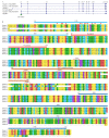
Predicted structures for non-animal Ngly1 and the core transglutaminase-like domain. (A) AlphaFold predicted structure of FmeNgly1; (B) AlphaFold predicted structure of SceNgly1; (C) AlphaFold predicted structure of AthNgly1; (D) Sequence alignment of the core transglutaminase-like domain. Fme, Flavobacterium meningosepticum; Sce, Saccharomyces cerevisiae; Ath, Arabidopsis thaliana; Osa, Oryza sativa Japonica; Zma, Zea mays; * represents conserved residuals; red * represents the three residuals of the catalytic triad.
Figure 3
Figure 3
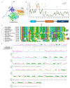
Structural analysis of human NGLY1. (A) Three-dimensional structure predicted by AlphaFold; (B) Intrinsically-disordered region of NGLY1; (C) Alignment of the intrinsically-disordered region; (D) Secondary structure of NGLY1.
Figure 4
Figure 4
Evolutionary relationships of NGLY1 in 33 taxa. (A) Evolutionary phylogeny; (B) Protein structures of Ngly1.
Figure 5
Figure 5
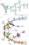
Glc3Man9GlcNAc2 trimming process and ER-associated degradation pathway (ERAD) involving NGLY1. (A) Glc3Man9GlcNAc2 trimming process; (B) ERAD under normal physiological state; (C) ERAD with NGLY1 deficiency.
Figure 6
Figure 6
Variants of NGLY1 generated by alternative splicing in humans. (A) Transcripts; (B) Protein alignments. Red triangles represent peptide binding sites; Red stars the Cys-His-Asp catalytic triad.
Figure 7
Figure 7
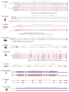
Alternative splicing patterns of NGLY1 in different species.
Figure 8
Figure 8
Protein network involving NLGY1 by STRING.
Figure 9
Figure 9
Characterization of mutations of NGLY1. (A) mRNA locations; (B) Amino acid locations; (C) Different types of mutations.

Similar articles

Cited by

References

    1. Hwang J., Qi L. Quality Control in the Endoplasmic Reticulum: Crosstalk between ERAD and UPR pathways. Trends Biochem. Sci. 2018;43:593–605. doi: 10.1016/j.tibs.2018.06.005. - DOI - PMC - PubMed
    1. Suzuki T., Park H., Lennarz W.J. Cytoplasmic peptide:N-glycanase (PNGase) in eukaryotic cells: Occurrence, primary structure, and potential functions. FASEB J. 2002;16:635–641. doi: 10.1096/fj.01-0889rev. - DOI - PubMed
    1. Need A.C., Shashi V., Hitomi Y., Schoch K., Shianna K.V., McDonald M.T., Meisler M.H., Goldstein D.B. Clinical application of exome sequencing in undiagnosed genetic conditions. J. Med. Genet. 2012;49:353–361. doi: 10.1136/jmedgenet-2012-100819. - DOI - PMC - PubMed
    1. Enns G.M., Shashi V., Bainbridge M., Gambello M.J., Zahir F.R., Bast T., Crimian R., Schoch K., Platt J., Cox R., et al. Mutations in NGLY1 cause an inherited disorder of the endoplasmic reticulum-associated degradation pathway. Genet. Med. 2014;16:751–758. doi: 10.1038/gim.2014.22. - DOI - PMC - PubMed
    1. Takahashi N. Demonstration of a new amidase acting on glycopeptides. Biochem. Biophys. Res. Commun. 1977;76:1194–1201. doi: 10.1016/0006-291X(77)90982-2. - DOI - PubMed

MeSH terms

Substances

Supplementary concepts