. 2022 Apr 29;14(9):1882.
doi: 10.3390/nu14091882.

Short-Chain Fatty Acids Ameliorate Depressive-like Behaviors of High Fructose-Fed Mice by Rescuing Hippocampal Neurogenesis Decline and Blood-Brain Barrier Damage

Affiliations

Short-Chain Fatty Acids Ameliorate Depressive-like Behaviors of High Fructose-Fed Mice by Rescuing Hippocampal Neurogenesis Decline and Blood-Brain Barrier Damage

Chuan-Feng Tang et al. Nutrients. .

Abstract

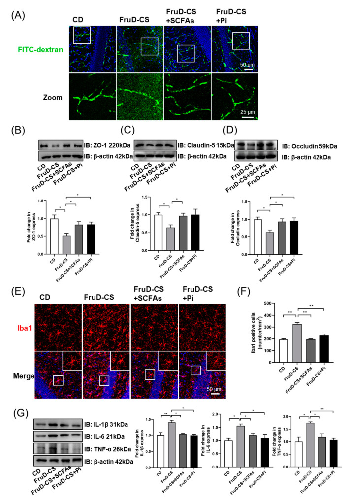
Excessive fructose intake is associated with the increased risk of mental illness, such as depression, but the underlying mechanisms are poorly understood. Our previous study found that high fructose diet (FruD)-fed mice exhibited neuroinflammation, hippocampal neurogenesis decline and blood-brain barrier (BBB) damage, accompanied by the reduction of gut microbiome-derived short-chain fatty acids (SCFAs). Here, we found that chronic stress aggravated these pathological changes and promoted the development of depressive-like behaviors in FruD mice. In detail, the decreased number of newborn neurons, mature neurons and neural stem cells (NSCs) in the hippocampus of FruD mice was worsened by chronic stress. Furthermore, chronic stress exacerbated the damage of BBB integrity with the decreased expression of zonula occludens-1 (ZO-1), claudin-5 and occludin in brain vasculature, overactivated microglia and increased neuroinflammation in FruD mice. These results suggest that high fructose intake combined with chronic stress leads to cumulative negative effects that promote the development of depressive-like behaviors in mice. Of note, SCFAs could rescue hippocampal neurogenesis decline, improve BBB damage and suppress microglia activation and neuroinflammation, thereby ameliorate depressive-like behaviors of FruD mice exposed to chronic stress. These results could be used to develop dietary interventions to prevent depression.

Keywords: blood-brain barrier; depressive-like behaviors; high-fructose diet; neurogenesis; short-chain fatty acids; stress resilience.


Conflict of interest statement

The authors declare no conflict of interest.

Figures

Figure 1
Figure 1
Chronic stress-induced depressive-like behaviors are aggravated in FruD mice. (A) Schematic diagram of mice dealing with high fructose diet and chronic stress. (B) Body weight of mice (* CD versus FruD group; # CD vs. CD-CS group; $ FruD vs. FruD-CS group). (C) Corticosterone level in serum of mice. (DJ) Behavioral test and representative state images of mice in OFT (D), SPT (E), FST (F,G) and TST (HJ). Data are expressed as mean ± SEM, * p < 0.05, ** p < 0.01; # p < 0.05; $$ p < 0.01, $$$ p < 0.001.
Figure 2
Figure 2
Chronic stress worsens the decline of hippocampal neurogenesis in FruD mice. (A) Representative confocal images labeled with DCX or NeuN. (B) Number of DCX positive newborn neurons was quantified. (C) Number of NeuN positive mature neurons was quantified. (D) Representative confocal images labeled with Nestin and GFAP. (E) Number of Nestin and GFAP positive NSCs was quantified. Data are expressed as mean ± SEM, * p< 0.05, ** p< 0.01.
Figure 3
Figure 3
Chronic stress exacerbates the impaired BBB integrity and activates microglia in FruD mice. (A) Representative confocal images for brain vessel visualization in mice injected with FITC-dextran. (BD) Representative immunoblot and quantification of ZO-1 (B), claudin-5 (C) and occludin (D) protein levels in the brain vessel. (E) Representative confocal images labeled with Iba1. (F) Number of Iba1 positive microglia was quantified. (G) Representative immunoblot and quantification of IL-1β, IL-6 and TNF-α protein levels in the hippocampus. Data are expressed as mean ± SEM, * p < 0.05, ** p < 0.01.
Figure 4
Figure 4
SCFAs and pioglitazone ameliorate depressive-like behaviors of FruD-CS mice. (A) Schematic diagram of mice dealing with SCFAs and pioglitazone treatment in FruD-CS mice. (B) Body weight of mice (* CD versus FruD-CS group). (C) Corticosterone level in serum of mice. (DJ) Behavioral test and representative state images of mice in OFT (D), SPT (E), FST (F,G) and TST (HJ). Data are expressed as mean ± SEM, * p < 0.05, ** p < 0.01, *** p < 0.01.
Figure 5
Figure 5
SCFAs and pioglitazone rescued the decline of hippocampal neurogenesis in FruD-CS mice. (A) Representative confocal images labeled with Nestin and GFAP. (B) Number of Nestin and GFAP positive NSCs was quantified. (C) Representative confocal images labeled with DCX or NeuN. (D) Number of DCX positive newborn neurons was quantified. (E) Number of NeuN positive mature neurons was quantified. Data are expressed as mean ± SEM, * p < 0.05, ** p < 0.01.
Figure 6
Figure 6
SCFAs and pioglitazone repaired BBB damage in FruD-CS mice. (A) Representative confocal images for brain vessel visualization in mice injected of FITC-dextran. (BD) Representative immunoblot and quantification of ZO-1 (B), claudin-5 (C) and occludin (D) protein levels in brain vessel. (E) Representative confocal images labeled with Iba1. (F) Number of Iba1 positive microglia was quantified. (G) Representative immunoblot and quantification of IL-1β, IL-6 and TNF-α protein levels in the hippocampus. Data are expressed as mean ± SEM, * p < 0.05, ** p < 0.01.

Similar articles

Cited by

References

    1. Gore F.M., Bloem P.J.N., Patton G.C., Ferguson J., Joseph V., Coffey C., Sawyer S.M., Mathers C.D. Global burden of disease in young people aged 10–24 years: A systematic analysis. Lancet. 2011;377:2093–2102. doi: 10.1016/S0140-6736(11)60512-6. - DOI - PubMed
    1. McCarron R.M., Shapiro B., Rawles J., Luo J. Depression. Ann. Intern. Med. 2021;174:ITC65–ITC80. doi: 10.7326/AITC202105180. - DOI - PubMed
    1. Osorio C., Probert T., Jones E., Young A.H., Robbins I. Adapting to Stress: Understanding the Neurobiology of Resilience. Behav. Med. 2017;43:307–322. doi: 10.1080/08964289.2016.1170661. - DOI - PubMed
    1. Santomauro D.F., Herrera A.M., Shadid J., Zheng P., Ashbaugh C., Pigott D.M., Abbafati C., Adolph C., Amlag J.O., Aravkin A.Y., et al. Global prevalence and burden of depressive and anxiety disorders in 204 countries and territories in 2020 due to the COVID-19 pandemic. Lancet. 2021;398:1700–1712. doi: 10.1016/S0140-6736(21)02143-7. - DOI - PMC - PubMed
    1. Li Y., Lv M.-R., Wei Y.-J., Sun L., Zhang J.-X., Zhang H.-G., Li B. Dietary patterns and depression risk: A meta-analysis. Psychiatry Res. 2017;253:373–382. doi: 10.1016/j.psychres.2017.04.020. - DOI - PubMed

LinkOut - more resources