

Bacillus amyloliquefaciens Enriched Camel Milk Attenuated Colitis Symptoms in Mice Model
- PMID: 35565934
- PMCID: PMC9101272
- DOI: 10.3390/nu14091967
Bacillus amyloliquefaciens Enriched Camel Milk Attenuated Colitis Symptoms in Mice Model
Abstract
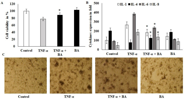
Fermented camel's milk has various health beneficial prebiotics and probiotics. This study aimed to evaluate the preventive efficacy of Bacillus amyloliquefaciens enriched camel milk (BEY) in 2-, 4- and 6-Trinitrobenzenesulfonic acid (TNBS)-induced colitis mice models. To this end, the immune modulatory effects of Bacillus amyloliquefaciens (BA) on TNF-α challenged HT29 colon cells were estimated using the cell proliferation and cytokines ELISA method. BEY was prepared using the incubation method and nutritional value was quantified by comparing it to commercial yogurt. Furthermore, TNBS-induced colitis was established and the level of disease index, pathological scores, and inflammatory markers of BEY-treated mice using macroscopic and microscopic examinations, qPCR and immunoblot were investigated. The results demonstrate that BA is non-toxic to HT29 colon cells and balanced the inflammatory cytokines. BEY reduced the colitis disease index, and improved the body weight and colon length of the TNBS-induced mice. Additionally, Myeloperoxidase (MPO) and pro-inflammatory cytokines (IL1β, IL6, IL8 and TNF-α) were attenuated by BEY treatment. Moreover, the inflammatory progress mRNA and protein markers nuclear factor kappa B (NFκB), phosphatase and tensin homolog (PTEN), proliferating cell nuclear antigen (PCNA), cyclooxygenase-2 (COX-2) and occludin were significantly down-regulated by BEY treatment. Interestingly, significant suppression of PCNA was observed in colonic tissues using the immunohistochemical examination. Treatment with BEY increased the epigenetic (microRNA217) interactions with PCNA. In conclusion, the BEY clearly alleviated the colitis symptoms and in the future could be used to formulate a probiotic-based diet for the host gut health and control the inflammatory bowel syndrome in mammals.
Keywords: Bacillus amyloliquefaciens; PCNA; TNF-α; inflammatory bowel disease; probiotics.
Conflict of interest statement
The authors declare no conflict of interest.
Figures


Similar articles
-
Bacillus amyloliquifaciens-Supplemented Camel Milk Suppresses Neuroinflammation of Autoimmune Encephalomyelitis in a Mouse Model by Regulating Inflammatory Markers.Nutrients. 2023 Jan 20;15(3):550. doi: 10.3390/nu15030550. Nutrients. 2023. PMID: 36771257 Free PMC article.
-
Camel's milk ameliorates TNBS-induced colitis in rats via downregulation of inflammatory cytokines and oxidative stress.Food Chem Toxicol. 2014 Jul;69:294-302. doi: 10.1016/j.fct.2014.04.032. Epub 2014 Apr 28. Food Chem Toxicol. 2014. PMID: 24788059
-
Probiotic-Fermented Camel Milk Attenuates Neurodegenerative Symptoms via SOX5/miR-218 Axis Orchestration in Mouse Models.Pharmaceuticals (Basel). 2023 Feb 25;16(3):357. doi: 10.3390/ph16030357. Pharmaceuticals (Basel). 2023. PMID: 36986457 Free PMC article.
-
Efficacy of thalidomide on trinitrobenzene sulfonate-induced colitis in young rats and its mechanism.Chin Med J (Engl). 2014;127(12):2368-75. Chin Med J (Engl). 2014. PMID: 24931258
-
Antoine-Barthélemy Clot, known as Clot-Bey (1793-1868), and spinitis in 1820.Rev Neurol (Paris). 2025 Mar;181(3):238-242. doi: 10.1016/j.neurol.2023.10.018. Epub 2024 Feb 8. Rev Neurol (Paris). 2025. PMID: 38336525 Review. No abstract available.
Cited by
-
Phenolic-Compound-Rich Opuntia littoralis Ethyl Acetate Extract Relaxes Arthritic Symptoms in Collagen-Induced Mice Model via Bone Morphogenic Markers.Nutrients. 2022 Dec 17;14(24):5366. doi: 10.3390/nu14245366. Nutrients. 2022. PMID: 36558525 Free PMC article.
-
The Halotolerant Probiotic Bacterium Enterococcus lactis ASF-2 from Al-Asfar Lake, Saudi Arabia, Reduces Inflammation in Carrageenan-Induced Paw Edema.Microorganisms. 2023 Sep 27;11(10):2415. doi: 10.3390/microorganisms11102415. Microorganisms. 2023. PMID: 37894072 Free PMC article.
-
Probiotics for the treatment of ulcerative colitis: a review of experimental research from 2018 to 2022.Front Microbiol. 2023 Jul 6;14:1211271. doi: 10.3389/fmicb.2023.1211271. eCollection 2023. Front Microbiol. 2023. PMID: 37485519 Free PMC article. Review.
-
Enterococcus faecium from chicken feces improves chicken immune response and alleviates Salmonella infections: a pilot study.J Anim Sci. 2023 Jan 3;101:skad016. doi: 10.1093/jas/skad016. J Anim Sci. 2023. PMID: 36651637 Free PMC article.
-
Bacillus amyloliquefaciens: Harnessing Its Potential for Industrial, Medical, and Agricultural Applications-A Comprehensive Review.Microorganisms. 2023 Aug 31;11(9):2215. doi: 10.3390/microorganisms11092215. Microorganisms. 2023. PMID: 37764059 Free PMC article. Review.
References
-
- Cordeiro B.F., Lemos L., Oliveira E.R., Silva S.H., Savassi B., Figueiroa A., Faria A.M.C., Ferreira E., Esmerino E.A., Rocha R.S., et al. Prato cheese containing Lactobacillus casei 01 fails to prevent dextran sodium sulphate-induced colitis. Int. Dairy J. 2019;99:104551. doi: 10.1016/j.idairyj.2019.104551. - DOI
-
- Rabah H., Do Carmo F.L.R., Carvalho R.D.d.O., Cordeiro B.F., da Silva S.H., Oliveira E.R., Lemos L., Cara D.C., Faria A.M.C., Garric G., et al. Beneficial propionibacteria within a probiotic emmental cheese: Impact on dextran sodium sulphate-induced colitis in mice. Microorganisms. 2020;8:380. doi: 10.3390/microorganisms8030380. - DOI - PMC - PubMed
MeSH terms
Substances
Grants and funding
LinkOut - more resources
Full Text Sources
Research Materials
Miscellaneous
