Review
. 2022 Apr 19;27(9):2625.
doi: 10.3390/molecules27092625.

Coordination Chemistry of Nucleotides and Antivirally Active Acyclic Nucleoside Phosphonates, including Mechanistic Considerations

Affiliations
Review

Coordination Chemistry of Nucleotides and Antivirally Active Acyclic Nucleoside Phosphonates, including Mechanistic Considerations

Astrid Sigel et al. Molecules. .

Abstract

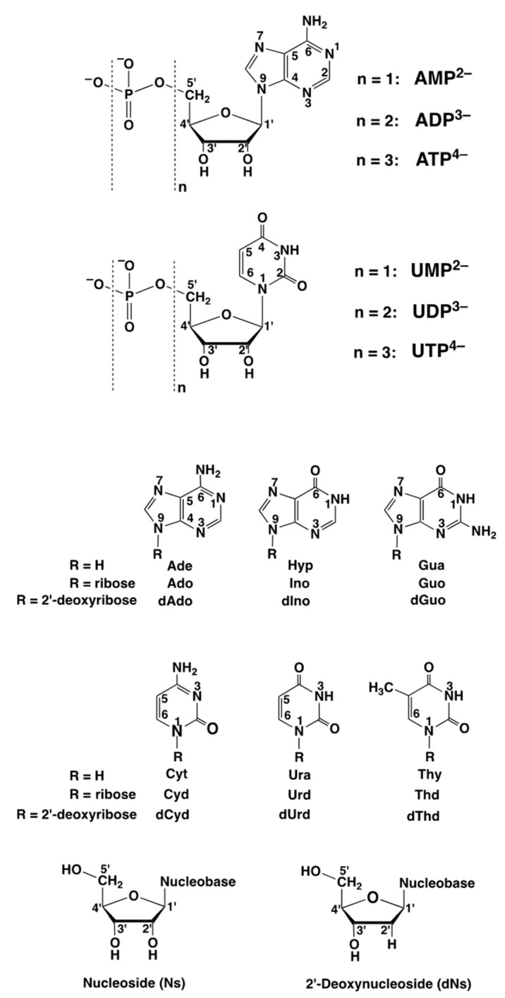
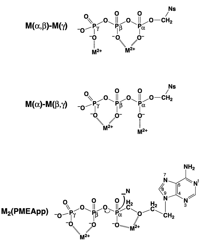
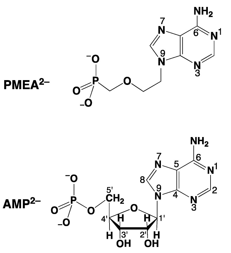
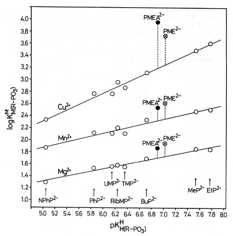
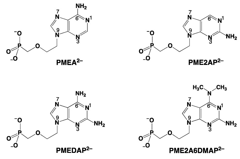
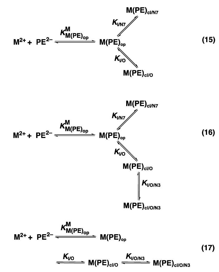
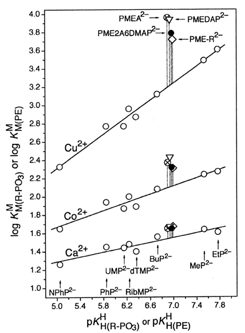
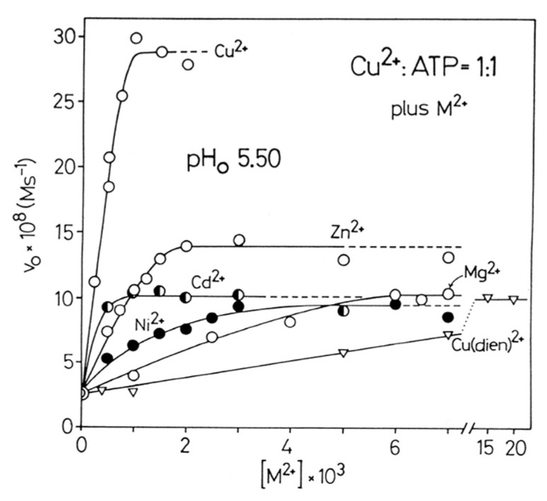
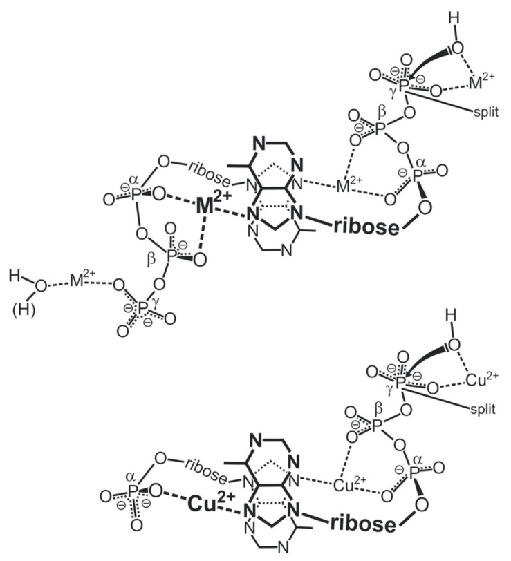
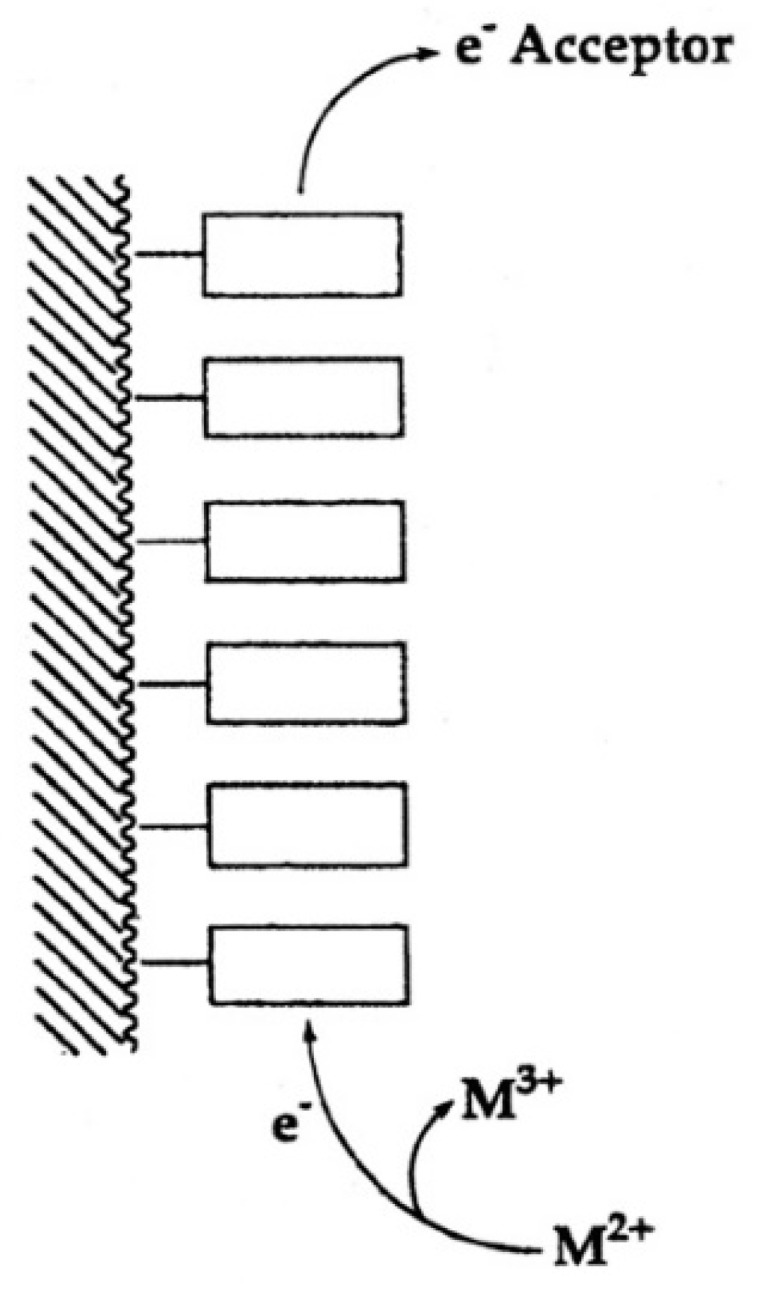
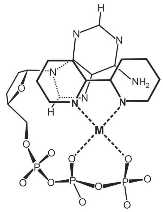
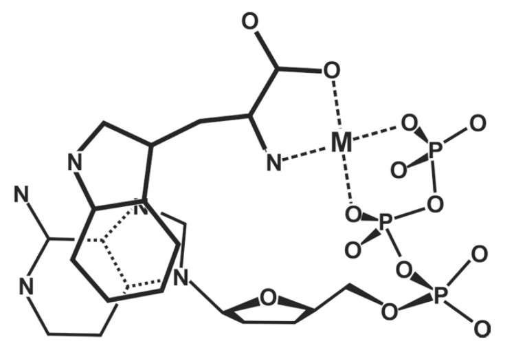
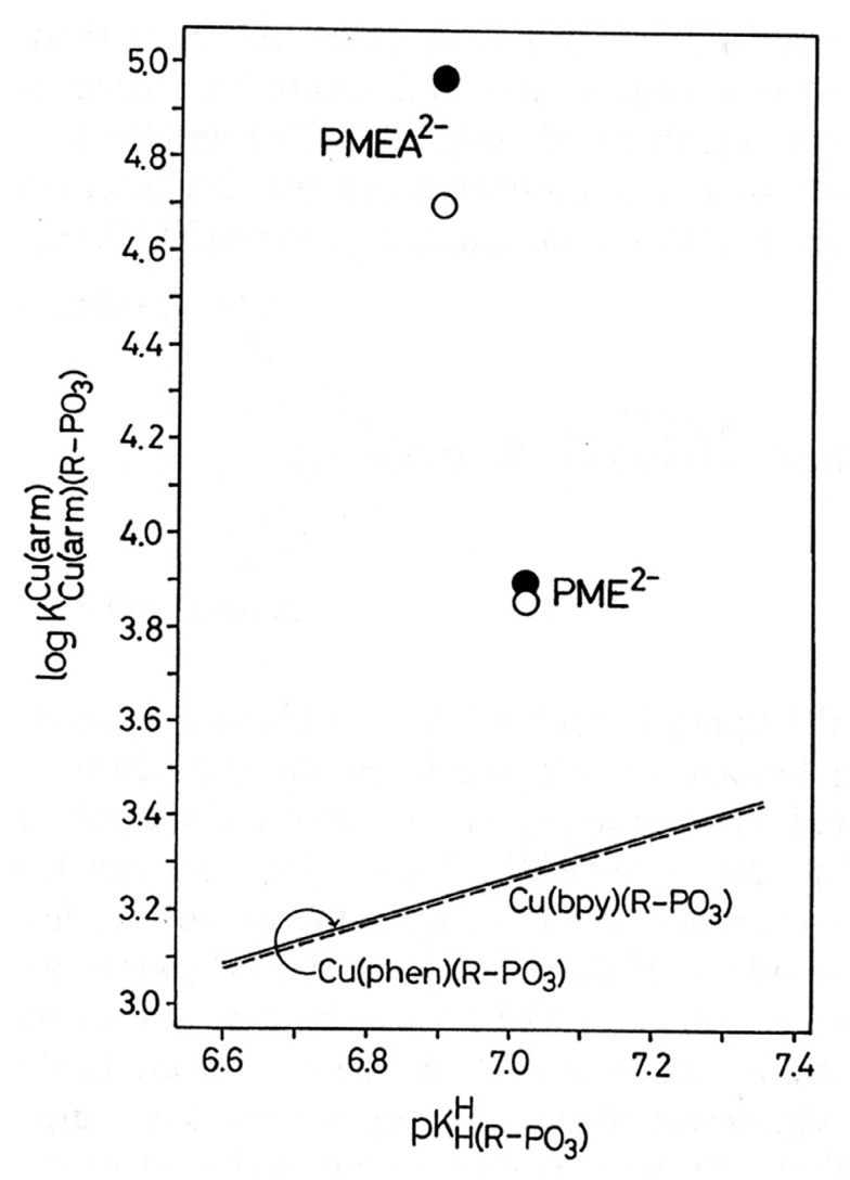
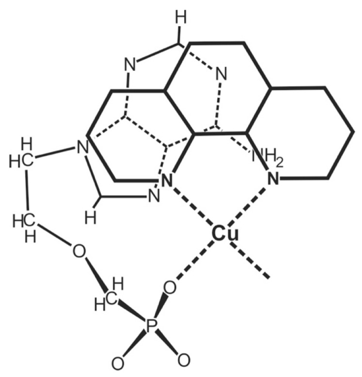
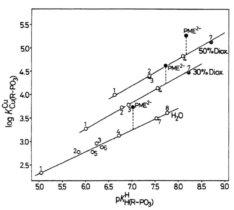
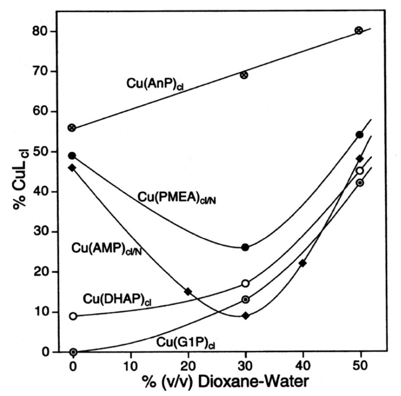
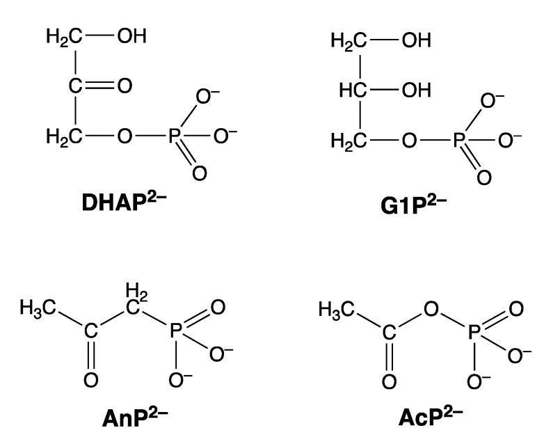
Considering that practically all reactions that involve nucleotides also involve metal ions, it is evident that the coordination chemistry of nucleotides and their derivatives is an essential corner stone of biological inorganic chemistry. Nucleotides are either directly or indirectly involved in all processes occurring in Nature. It is therefore no surprise that the constituents of nucleotides have been chemically altered-that is, at the nucleobase residue, the sugar moiety, and also at the phosphate group, often with the aim of discovering medically useful compounds. Among such derivatives are acyclic nucleoside phosphonates (ANPs), where the sugar moiety has been replaced by an aliphatic chain (often also containing an ether oxygen atom) and the phosphate group has been replaced by a phosphonate carrying a carbon-phosphorus bond to make the compounds less hydrolysis-sensitive. Several of these ANPs show antiviral activity, and some of them are nowadays used as drugs. The antiviral activity results from the incorporation of the ANPs into the growing nucleic acid chain-i.e., polymerases accept the ANPs as substrates, leading to chain termination because of the missing 3'-hydroxyl group. We have tried in this review to describe the coordination chemistry (mainly) of the adenine nucleotides AMP and ATP and whenever possible to compare it with that of the dianion of 9-[2-(phosphonomethoxy)ethyl]adenine (PMEA2- = adenine(N9)-CH2-CH2-O-CH2-PO32) [or its diphosphate (PMEApp4-)] as a representative of the ANPs. Why is PMEApp4- a better substrate for polymerases than ATP4-? There are three reasons: (i) PMEA2- with its anti-like conformation (like AMP2-) fits well into the active site of the enzyme. (ii) The phosphonate group has an enhanced metal ion affinity because of its increased basicity. (iii) The ether oxygen forms a 5-membered chelate with the neighboring phosphonate and favors thus coordination at the Pα group. Research on ANPs containing a purine residue revealed that the kind and position of the substituent at C2 or C6 has a significant influence on the biological activity. For example, the shift of the (C6)NH2 group in PMEA to the C2 position leads to 9-[2-(phosphonomethoxy)ethyl]-2-aminopurine (PME2AP), an isomer with only a moderate antiviral activity. Removal of (C6)NH2 favors N7 coordination, e.g., of Cu2+, whereas the ether O atom binding of Cu2+ in PMEA facilitates N3 coordination via adjacent 5- and 7-membered chelates, giving rise to a Cu(PMEA)cl/O/N3 isomer. If the metal ions (M2+) are M(α,β)-M(γ)-coordinated at a triphosphate chain, transphosphorylation occurs (kinases, etc.), whereas metal ion binding in a M(α)-M(β,γ)-type fashion is relevant for polymerases. It may be noted that with diphosphorylated PMEA, (PMEApp4-), the M(α)-M(β,γ) binding is favored because of the formation of the 5-membered chelate involving the ether O atom (see above). The self-association tendency of purines leads to the formation of dimeric [M2(ATP)]2(OH)- stacks, which occur in low concentration and where one half of the molecule undergoes the dephosphorylation reaction and the other half stabilizes the structure-i.e., acts as the "enzyme" by bridging the two ATPs. In accord herewith, one may enhance the reaction rate by adding AMP2- to the [Cu2(ATP)]2(OH)- solution, as this leads to the formation of mixed stacked Cu3(ATP)(AMP)(OH)- species, in which AMP2- takes over the structuring role, while the other "half" of the molecule undergoes dephosphorylation. It may be added that Cu3(ATP)(PMEA) or better Cu3(ATP)(PMEA)(OH)- is even a more reactive species than Cu3(ATP)(AMP)(OH)-. - The matrix-assisted self-association and its significance for cell organelles with high ATP concentrations is summarized and discussed, as is, e.g., the effect of tryptophanate (Trp-), which leads to the formation of intramolecular stacks in M(ATP)(Trp)3- complexes (formation degree about 75%). Furthermore, it is well-known that in the active-site cavities of enzymes the dielectric constant, compared with bulk water, is reduced; therefore, we have summarized and discussed the effect of a change in solvent polarity on the stability and structure of binary and ternary complexes: Opposite effects on charged O sites and neutral N sites are observed, and this leads to interesting insights.

Keywords: 9-[2-(phosphonomethoxy)ethyl]adenine (PMEA); acyclic nucleoside phosphonates; antivirals; cell organelles; competing solvent effects; complex stabilities; dephosphorylation; hydrolysis of ATP; intramolecular equilibria; isodesmic model; kinases; mechanistic considerations; metal ion complexes; mixed ligand complexes; nucleic acids; nucleotide analogues; polarity changes; polymerases; self-association; solvent effects; ternary complexes; triphosphate coordination modes.


Conflict of interest statement

The authors declare no conflict of interest.

Figures

Figure 1
Figure 1
In the upper part are shown the chemical structures of the adenosine and uridine 5′-phosphates—namely, adenosine 5′-monophosphate (AMP2−; n = 1), adenosine 5′-diphosphate (ADP3−; n = 2), and adenosine 5′-triphosphate (ATP4−; n = 3), as well as those of uridine 5′-monophosphate (UMP2−; n = 1), uridine 5′-diphosphate (UDP3−; n = 2), and uridine 5′-triphosphate (UTP4−; n = 3). The phosphate groups are named α, β, γ; the γ group being the terminal one. The nucleotides are depictured in their dominating anti conformation [4,5,6,7], which means that the adenine residue is pointing away from the ribose plane as is the (C2)O group in the uridine 5′-phosphates. It is obvious that the substitution with other nucleobases will also lead to the anti conformation. In the lower part of the figure, the structures are shown of the nucleobases (Nb), of the nucleosides (Ns), and of the 2′-deoxynucleosides (dNs). The Abbreviations employed (following the order given in the figure) are as follows: Ade = adenine, Ado = adenosine, and dAdo = 2′-deoxyadenosine; Hyp = hypoxanthine, Ino = inosine, and dIno = 2′-deoxyinosine; Gua = guanine, Guo = guanosine, and dGuo = 2′-deoxyguanosine; Cyt = cytosine, Cyd = cytidine, and dCyd = 2′-deoxycytidine; Ura = uracil, Urd = uridine, and dUrd = 2′-deoxyuridine; Thy = thymine; Thd = thymidine, and dThd = 2′-deoxythymidine = 2′-deoxy-5-methyluridine.
Figure 2
Figure 2
The upper and middle parts of the figure show structures of two M2(NTP) complexes, where NTP4− = nucleoside 5′-triphosphate: In one case, a M(α,β)-M(γ) coordination is depictured (upper part) indicating the structure relevant for transphosphorylations (kinase, etc.). In the other case, the metal ions are bound in a M(α)-M(β,γ)-type fashion (middle part) that is relevant for nucleic acid polymerases, which catalyze the transfer of a nucleotidyl unit. For the latter binding mode, the structure needs to be enforced by the enzyme; this means, the two metal ions need to be anchored [11,12,13,14] to amino acid side chains, often carboxylate groups of aspartate or glutamate residues of the enzyme [15,16,17,18,19]. The (lower part) shows a complex formed between two metal ions and diphosphorylated PMEA (9-[2-(phosphonomethoxy)ethyl]adenine)—that is, M2(PMEApp). Note, the M(α)-M(β,γ) binding mode, crucial for the polymerase reaction, is favored by the formation of a 5-membered chelate with the ether oxygen of the aliphatic chain (see Section 3). Of course, the adenine residue may be replaced by any other nucleobase moiety and the nucleophile (N) may in addition interact with the M2+ at the α-phosphonate group. Altered versions of similar situations are depictured in Refs [20,21,22,23].
Figure 3
Figure 3
Comparison of the chemical structure of the dianion of 9-[2-(phosphonomethoxy)ethyl]adenine (PMEA2−) with the one of adenosine 5′-monophosphate (AMP2−). The orientation of PMEA2− in solution [36] and in the solid state [37] resembles the anti conformation of AMP2− [4,5,6,38] and thus PMEA2− may be considered as an analogue of AMP2− or 2′-deoxy-AMP2−.
Figure 4
Figure 4
Evidence for enhanced stabilities of some M(PMEA) (●) complexes in comparison with those of the corresponding M(PME) (⊗) species, based on the relationship between log KM(R-PO3)M and pKH(R-PO3)H  for M(R-PO3) complexes of some simple phosphate monoester and phosphonate ligands (R-PO32 ) (◯): 4-nitrophenyl phosphate (NPhP2−), phenyl phosphate (PhP2−), uridine 5′-monophosphate (UMP2−), D-ribose 5-monophosphate (RibMP2−), thymidine [= 1-(2′-deoxy-β-D-ribofuranosyl)thymine] 5′-monophosphate (TMP2−), n-butyl phosphate (BuP2−), methanephosphonate (MeP2−), and ethanephosphonate (EtP2−) (from left to right). The least squares lines (Equation (3)) are drawn through the corresponding 8 data sets (◯) taken from Ref [48] for the phosphate monoesters and from Ref [44] for the phosphonates. The data due to the M2+/PMEA (●) and the M2+/PME (⊗) systems are from Ref [44]. The vertical broken lines emphasize the stability differences to the reference lines; they equal log Δ as defined in Equation (12) for the M(PME) complexes (for the M(PMEA) complexes the analogous formulation holds). All the plotted equilibrium constants refer to aqueous solutions at 25 °C and I = 0.1 M (NaNO3). Reproduced with permission from our publication in Coordination Chemistry Reviews [45]; copyright 1995, Elsevier Science S.A., Lausanne, Switzerland.
Figure 5
Figure 5
Chemical structures of the dianions of 9-[2-(phosphonomethoxy)ethyl]adenine (PMEA2−), 9-[2-(phosphonomethoxy)ethyl]-2-aminopurine (PME2AP2−), 9-[2-(phosphonomethoxy)ethyl]-2,6-diaminopurine (PMEDAP2−), and 9-[2-(phosphonomethoxy)ethyl]-2-amino-6-dimethylaminopurine (PME2A6DMAP2−). These compounds are abbreviated as PE2−. For the solution structure of PMEA2−, see legend of Figure 3. Replacement of the adenine residue in PMEA2− by a cytosine residue (Figure 1) gives PMEC2− = 1-[2-(phosphonomethoxy)ethyl]cytosine.
Figure 6
Figure 6
Equilibrium schemes involving various N sites in metal ion complexes of acyclic nucleoside phosphonates containing a purine moiety.
Figure 7
Figure 7
Evidence for enhanced stabilities of some M(PME2A6DMAP) (●) complexes in comparison with those of the corresponding M(PMEA) (⊗, M(PMEDAP) (▽), and M(PME-R) (◇) species, based on the relationship between log KM(R-PO3)M and pKH(R-PO3)H  for M(R-PO3) complexes of some simple phosphate monoester and phosphonate ligands (R-PO32) (◯) (see legend to Figure 4). The points due to the equilibrium constants for the M2+/PME2A6DMAP systems (●) are based on the values listed in Tables 1 and 2 of Ref [61]; those for the M2+/PMEA systems (⊗) are from Ref [44], for the M2+/PMEDAP systems (▽) from Ref [62], and those for the M2+/PME-R systems (◇) are based on pKH(PME-R)H = 6.99 ± 0.04 (average of the pKa values for H(PME) (7.02) and H(PMEC) (6.95) [63]) and the stability enhancements listed in Ref [63]. The vertical broken lines emphasize the stability differences to the reference lines; they equal log ΔM/PE, as defined in Equation (12) for the M(PE) complexes in general. All the plotted equilibrium constants refer to aqueous solutions at 25 °C and I = 0.1 M (NaNO3). Reproduced with permission from our publication in the Canadian Journal of Chemistry [61]; copyright 2014, NRC Research Press.
Figure 8
Figure 8
Dependence of the initial rate, v0 = d[PO4]/dt (M s−1), of the Cu2+-promoted dephosphorylation of ATP ([Cu2+]tot = [ATP]tot = 10−3 M) in aqueous solution on the addition of further divalent metal ions (empty circles, half-filled circles, full circles) or the Cu2+ 1:1 complex with diethylenetriamine (= dien = 1,4,7-triazaheptane (▽)) at pH0 5.50; I = 0.1 M, NaClO4; 50 °C. The broken line portions indicate uncertainty due to precipitation. The figure is reproduced with permission from our publication in Inorganica Chimica Acta [55]; copyright 1992, Elsevier Sequoia (see also [14]).
Figure 9
Figure 9
Variation in the proportions of ATP present in the monomer (1), dimer (2), trimer (3), …, and octamer (8) in D2O solutions in dependence on the total concentration of ATP4− (K = 1.3 M−1), Mg(ATP)2− (K = 4.0 M−1), and Zn(ATP)2− (KD* = 20 M−1 and Kst = 4 M−1) (see text in Section 6.2 and Equations (18)–(20)) (27 °C, I = 0.1 to ~2 M (NaNO3) in D2O). This figure is reproduced with permission from our publication in the Journal of the American Chemical Society [89]; copyright 1981, American Chemical Society (see also [53]).
Figure 10
Figure 10
TOP: Proposed structure of the reactive [M2(ATP)]2(OH) dimer, which occurs in low concentration during the metal ion-promoted dephosphorylation of ATP. The intramolecular attack of OH is indicated on the right-hand side, while the left-hand side is ready to transfer also into the reactive state by deprotonation of the coordinated water molecule or to undergo an intramolecular water attack, corresponding to the dimeric [M2(ATP)]2 species. BOTTOM: Probable structure of the reactive Cu3(ATP)(AMP)(OH) species. The intramolecular attack of OH is indicated on the right-hand side, while the left-hand side shows the metal ion bridge stabilizing the purine stack by M2+ coordination to the phosphate group of AMP2− and to N7 of ATP4− [14,80,94]. The structures are adapted from Ref [80].
Figure 11
Figure 11
Influence of AMP (◯) and PMEA (●) as well as of ε-AMP (Figure 12) on the initial rate v0 (M s−1) of the dephosphorylation of the Cu2+/ATP 2:1 system ([Cu2+]tot = 2 × 10⁻³ M and [ATP]tot = 10−3 M) in aqueous solution at pH 6.70 (I = 0.1 M, NaClO4; 50 °C). This figure is adapted with permission from Figure 3 of our publication in Chem. Commun. [94]; copyright 1998, The Royal Society of Chemistry.
Figure 12
Figure 12
Chemical structures of AMP2− analogues: tubercidin 5′-monophosphate (TuMP2− = 7-deaza-AMP2−), 1,N6-ethenoadenosine 5′-monophosphate (ε-AMP2−), and adenosine 5′-monophosphate N(1)-oxide (AMP·NO2−).
Figure 13
Figure 13
Simplified picture of a matrix-assisted ATP stack indicating an electron and thus an information transfer through this stack from one end to the other. This figure is reproduced with permission from our publication in Pure and Applied Chemistry [21]; copyright 2004, International Union of Pure and Applied Chemistry (IUPAC).
Figure 14
Figure 14
Probable schematic structure in solution of an intramolecular stack in the Cu(bpy)(ATP)2− complex [105]; adapted from similar structures for Cu(bpy)(AMP), as shown in Refs [124,125,126].
Figure 15
Figure 15
Tentative and schematic structure of the stacked isomer in solution of an M(ATP)(Trp)3− complex. Adapted from Ref [101].
Figure 16
Figure 16
Evidence for an enhanced stability of the Cu(arm)(PME) and Cu(arm)(PMEA) complexes based on the relationship between log KCu(arm)(R-PO3)Cu(arm) and pKH(R-PO3)H for the ternary. Cu(bpy)(PME) and Cu(bpy)(PMEA) (◯), as well as for the Cu(phen)(PME) and Cu(phen)(PMEA) (●) complexes in aqueous solution at I = 0.1 M (NaNO3) and 25 °C. The plotted data are from Table 6 in Section 8 of [141] (= Table 7 here). The two reference lines represent the log K versus pKa relationship for Cu(arm)(R-PO3) complexes; it should be emphasized that R-PO32 symbolizes here phosphonates (or phosphate monoesters) with an R group unable to undergo any kind of hydrophobic, stacking, or other type of interaction; the broken line holds for arm = bpy and the solid line for arm = phen. Both straight lines are calculated with the parameters listed in Table 5 of Ref [141], and they represent the situation for ternary complexes without an intramolecular ligand–ligand interaction. Redrawn in a slightly altered way from Ref [141] with permission of the Royal Society of Chemistry and then reproduced with permission from our publication in Coordination Chemistry Reviews [45]; copyright 1995, Elsevier Science S.A., Lausanne, Switzerland.
Figure 17
Figure 17
Tentative and simplified structure of a species with an intramolecular stack for Cu(phen)(PMEA) in solution [45,141]. Adapted from Ref [45].
Figure 18
Figure 18
Evidence for an enhanced stability of the Cu(PME) (●) complex in 1,4-dioxane-water mixtures as solvents, based on the relationship between log KCu(R-PO3)Cu and pKH(R-PO3)H for the Cu2+ 1:1 complexes of 4-nitrophenyl phosphate (1), phenyl phosphate (2), D-ribose 5-monophosphate (3), n-butyl phosphate (4),uridine 5′-monophosphate (5), thymidine [= 1-(2′-deoxy-β-D-ribofuranosyl)thymine] 5′-monophosphate (6), methanephosphonate (7), and ethanephosphonate (8) in water and in water containing 30% or 50% (v/v) 1,4-dioxane. The least-squares lines are drawn in each case through the data sets shown [44,150]; the equations for these reference lines are given in Ref [149]. The data points due to the methanephosphonate system in the mixed solvents (⊗,7) (see Ref [149]) are shown to prove that simple phosphonates fit within the experimental error limits on the reference lines established with phosphate monoester systems (see also Ref [149). The points due to the Cu2+ 1:1 complexes formed with PME2− (●) in the three mentioned solvents demonstrate the enhanced complex stabilities. The vertical broken lines emphasize the stability differences to the reference lines; these differences equal log ΔCu(R-PO3), as defined in Equation (12). All the plotted equilibrium constants refer to 25 °C and I = 0.1 M (NaNO3). This figure is adapted with permission from our publication in Inorganic Chemistry [149]; copyright 1993, American Chemical Society (see also [45]).
Figure 19
Figure 19
Formation degrees of the chelated isomers, CuLcl, in the Cu(AMP) and Cu(PMEA) systems (Table 11) as well as for comparison in the Cu(DHAP) [47], Cu(G1P) [47], and Cu(AnP) [41,47,154] complex systems (Figure 20) as a function of the percentage of 1,4-dioxane added to an aqueous reagent mixture. All the plotted equilibrium constants refer to 25 °C and I = 0.1 M (NaNO3). This figure is reproduced with permission from our publication in Coordination Chemistry Reviews [47]; copyright 2000, Elsevier, Science S.A., Lausanne, Switzerland).
Figure 20
Figure 20
Chemical structures of dihydroxyacetone phosphate (DHAP2−) and glycerol 1-phosphate (G1P)2−. Acetonylphosphonate (AnP2−) is often employed as a model compound for acetyl phosphate (AcP2−), which is biologically very relevant but difficult to study because it is very hydrolysis-sensitive. It may be added that in the so-called [155] α-glycerophosphate shuttle, DHAP2− and G1P2− are interconverted into each other [155,156].
Figure 21
Figure 21
Irving–Williams sequence-type plots [158] for the stability constants log KM(N)M (N = nucleotide) of the 1:1 complexes of Ba2+ through Cd2+ formed with mono- (R-MP2−), di- (R-DP3−), and triphosphate monoesters (R-TP4−) (◯), as well as of those for AMP2−, ADP3−, and ATP4− (●). The plotted data of the phosphate ligands are from Table 13 of [23]; they also represent the stability constants for the M2+ complexes of the pyrimidine-nucleoside 5′-mono-, di- or triphosphates (except for Cu(CTP)2− [74]). The log stability constants for the M2+ complexes of AMP2− and ADP3− are from Table 4 of [153]; those for ATP4− are from Table II of [74] (25 °C; I = 0.1 M, NaNO3) ([23,74,163,164] as well as pp. 364–365 in [165]).

Similar articles

Cited by

References

    1. Sigel A., Sigel H., editors. Metal Ions in Biological Systems. Volume 32. Marcel Dekker, Inc.; New York, NY, USA: Basel, Switzerland: Hong Kong, China: 1996. Interactions of Metal Ions with Nucleotides, Nucleid Acids, and Their Constituents; pp. 1–814.
    1. Sigel A., Sigel H., Sigel R.K.O., editors. Metal Ions in Life Sciences. Volume 9. Royal Society of Chemistry; Cambridge, UK: 2011. Structural and Catalytic Roles of Metal Ions in RNA; pp. 1–391. - PubMed
    1. Sigel A., Sigel H., Sigel R.K.O., editors. Metal Ions in Life Sciences. Volume 10. Springer; Dordrecht, The Netherlands: 2012. Interplay Between Metal Ions and Nucleic Acids; pp. 1–351.
    1. Aoki K. General Conclusions from Solid State Studies of Nucleotide-Metal Ion Complexes. Met. Ions Biol. Syst. 1996;32:91–134.
    1. Martin R.B., Mariam Y.H. Interactions between Metal Ions and Nucleic Bases, Nucleosides, and Nucleotides in Solution. Met. Ions Biol. Syst. 1979;8:57–124.

LinkOut - more resources